
Exodeconus maritimus
|
publication ID |
https://doi.org/ 10.5281/zenodo.4605860 |
|
persistent identifier |
https://treatment.plazi.org/id/6B72879E-FF95-FFC6-FFBB-FBCFA38AFA17 |
|
treatment provided by |
Carolina |
|
scientific name |
Exodeconus maritimus |
| status |
|
Exodeconus maritimus View in CoL
Calyx
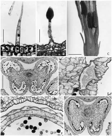
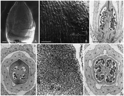
The sepals are triangular, overlaping up to half of their length, 5.5-6.5 mm long and 1.5-1.8 mm wide. Trichomes vary considerably: eglandular, simple, pluricellular, and glandular with spherical unicellular or elipsoidal head ( Fig. 1A,B View Fig ), and are located over the external face of the sepals. Anatomically, the calyx is formed by two epidermises, with a thick cuticle, and five to six layers of parenchymatous tissue ( Fig. 2D View Fig ). These cells are isodiametric in cross sections leaving reduced spaces among them ( Fig. 2D View Fig ). Chloroplasts and starch are usually observed in the parenchyma cells.
Corolla
The shape is tubular-infundibuliform; it is white, with a violet throat, 17-18 mm long and 4.5-6.5 mm wide. The anatomical structure is very similar to the calyx, with slight differences: the petals’ epidermis is covered by a thin cuticle, and the parenchyma cells possess a large lumen. — Fig. 2D,F View Fig .
Androecium
The filaments are pubescent at the base with simple eglandular trichomes ( Fig. 1A,C View Fig ), and are composed of an epidermis, parenchymatic tissue, and a single vascular strand. The length of filaments is distinctive; three filaments are shorter than the other two ( Fig. 1C View Fig ). The anthers are small (3.2-3.5 mm), glabrous, and formed of a simple epidermis and an endothecium constituted of two cell strata with fibrous thickenings ( Fig. 1D,F View Fig ). Dehiscence occurs at the stomium ( Fig. 1D,E View Fig ), which is formed by a layer of seven to ten cells with crystalline sand. After dehiscence, the walls disorganize, and their contents are leaked into the lysigenous cavity they form ( Fig. 1D,E,G View Fig ). Andro-sterility is observed in E. maritimus , where the flowers display two or three sterile anthers ( Fig. 1G View Fig ). The pollen sacs possess normal endothecium, collapsed tapetum and empty pollen grains ( Fig. 1G View Fig ).
Gynoecium
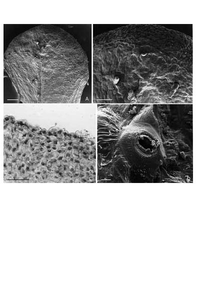
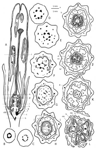
The stigma is depressed and enlarged toward its flanks, giving it a fan-like shape ( Fig. 3A View Fig ). The receptive surface is covered by unicellular papillae enlarged at the base and rounded towards the apex ( Fig. 3B,C View Fig ). The style bears eglandular, simple, pluricellular trichomes and stomata shared on the epidermis ( Fig. 3B,D View Fig ). The ovary is ringshaped in cross section ( Fig. 2D,F View Fig ), bicarpellar, with 4 locules at the base due to the false septa ( Fig. 2D View Fig ). This characteristic disappears at the apex of the ovary ( Fig. 2F View Fig ). Crystalline sand is frequently found in the parenchymatic tissue of the ovary. The nectary, located at the base of the ovary ( Fig. 2A View Fig ), has the shape of a ring with two swellings at opposite sides and at the level of the septa ( Fig. 2A View Fig ,C-E). The secretory tissue is constituted by 5-6 layers of isodiametric cells with large nuclei and vacuolated cytoplasm ( Fig. 2D View Fig ). Also, there are 12-15 layers of cells ( Fig. 2D,E View Fig ) and 20 ± 3 stomata concentrated at the upper part of the nectary swellings ( Fig. 2B View Fig ). The secre- tory tissue is supplied by two dorsal and several lateral vascular bundles that ascend to the external wall of the ovary ( Fig. 4A,H View Fig ).
Vascularization
The peduncle shows a siphonostele, which divides into 10 vascular bundles forming a circle ( Fig. 4 View Fig A-C). Five traces split to constitute the main sepal bundles, which give rise to lateral ramifications ( Fig. 4A View Fig ,D-F). The five remaining vascular bundles are reorganized, acquiring a star shape ( Fig. 4A,E,F View Fig ). Five main corolla bundles are immediately formed taking an external position (each one produces two or three lateral ramifications). The five staminal bundles acquire an internal position ( Fig. 4A,G View Fig ). After these divisions, the remaining vascular tissue is reordered in a circle, entering the gynoecium ( Fig. 4A,G,H View Fig ). The ovary has 2 or 4 ventral vascular bundles in the central region ( Fig. 4A,H View Fig ); when 4, they then decrease to only 2 by fusion at the level of the apical portion of the ovary ( Fig. 4A,I,J View Fig ). The dorsal vascular bundles enter the style, reaching the base of the stigma without ramifications ( Fig. 4A,K,L View Fig ).
No known copyright restrictions apply. See Agosti, D., Egloff, W., 2009. Taxonomic information exchange and copyright: the Plazi approach. BMC Research Notes 2009, 2:53 for further explanation.
|
Kingdom |
|
|
Phylum |
|
|
Class |
|
|
Order |
|
|
Family |
|
|
Genus |