
Foldilecanium multisetosus Kondo, 2011
|
publication ID |
https://doi.org/ 10.5281/zenodo.5160587 |
|
publication LSID |
lsid:zoobank.org:pub:583BAAAE-8AED-4E4F-8E5C-1B42027DD8D9 |
|
DOI |
https://doi.org/10.5281/zenodo.5164475 |
|
persistent identifier |
https://treatment.plazi.org/id/20E0EB21-D69F-41EC-ADF2-6B0D45E134E5 |
|
taxon LSID |
lsid:zoobank.org:act:20E0EB21-D69F-41EC-ADF2-6B0D45E134E5 |
|
treatment provided by |
Felipe |
|
scientific name |
Foldilecanium multisetosus Kondo |
| status |
sp. nov. |
Foldilecanium multisetosus Kondo sp. nov.
( Fig. 2-3 View Figure 2 View Figure 3 )
Proposed common names. Spanish: Escama blanda multisetosa; English: Multisetose soft scale.
Type material examined. Holotype, adult female. Colombia, Valle del Cauca, Cali, 03 o 18’08.7"N, 76 o 32’06.7"W, 1005 m a.s.l., 1.iii.2006, coll. Takumasa Kondo, ex twigs of Cananga odorata (Lam.) Hook. f. and Thomson, inside ant shelters of Azteca sp. , 1(1) ( USNM). Paratypes, same data, 19 (22 specimens: 17 adult females + 2 third-instar nymphs + 1 second-instar nymph + 2 first-instar nymphs) ( USNM).
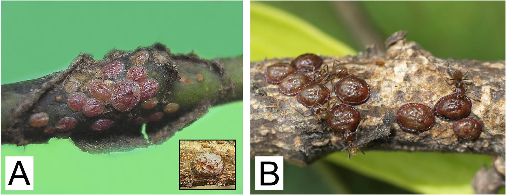
Unmounted material ( Fig. 2 View Figure 2 ). Adult female in life about 3.0 mm long, 2.8 mm wide, oval, moderately convex. Scale covered by a thin layer of a flaky glassy wax (see inset on Fig. 2A View Figure 2 ). Young adult females ( Fig. 2A View Figure 2 ) sligthly convex, orange to purplish red in color, often with mottlings of a darker color. Older specimens ( Fig. 2B View Figure 2 ) becoming heavily sclerotized, brown in color, with thick and dark color elevated rim around body margins.
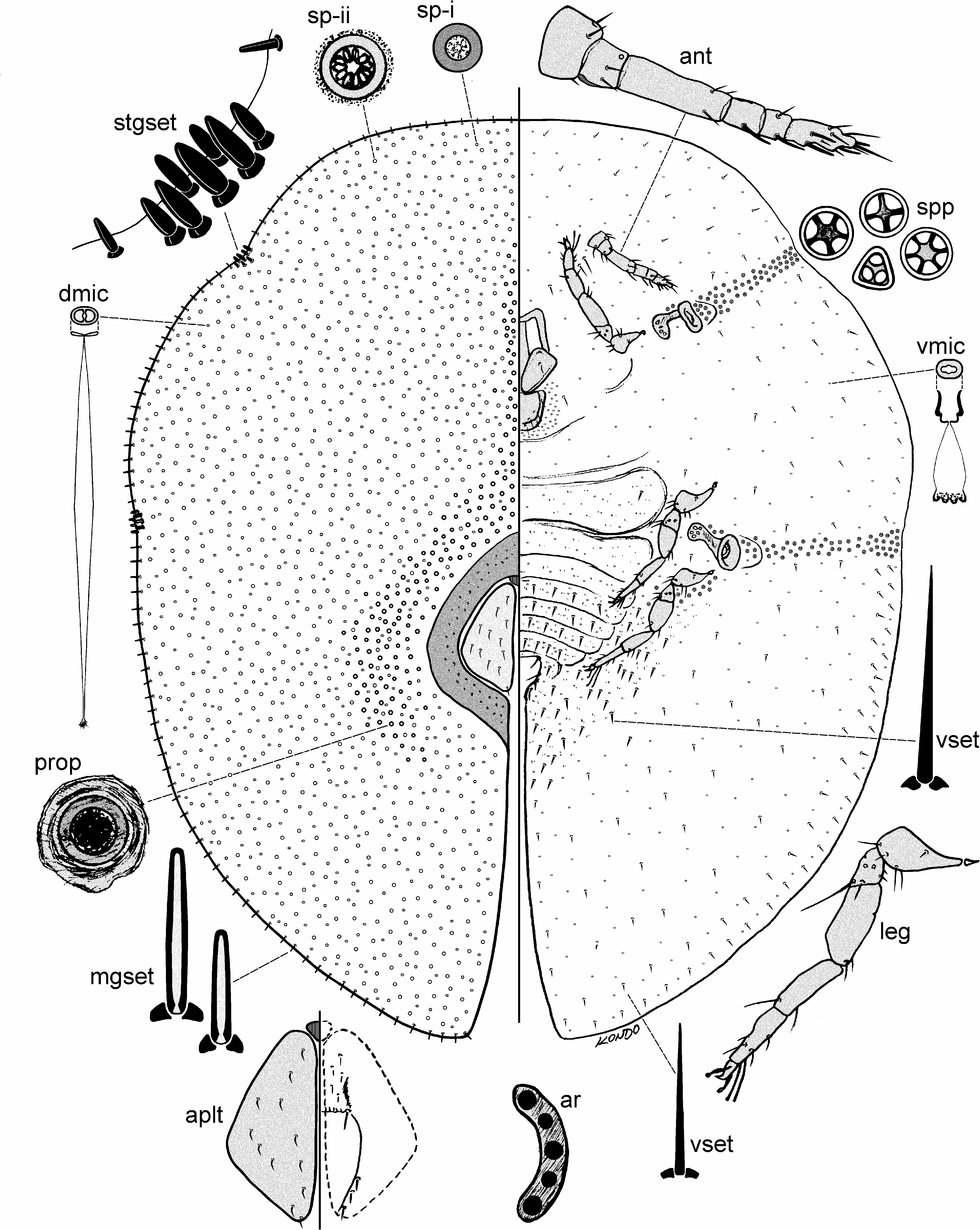
Slide-mounted material ( Fig. 3 View Figure 3 ). Slide mounted specimens1.8-3.0 mm long, 1.5-2.6 mm wide, body oval in shape.
Description. Adult female (measurements based on n=18).
Dorsum. Derm membranous, becoming sclerotized in older specimens. Dorsal setae completely absent. Dorsal microducts (dmic) each about 2.0 µm wide, scattered over dorsum. Simple pores (sp) of 2 types: type (i) pore small, with a thick sclerotized rim and a small central opening, each 2.5-3.0 µm wide, scattered over dorsum; type (ii) pore median in size, each 4.0-5.0 µm wide, with a thick rim, and a larger central opening, under high magnification appearing as having about 10 loculi, scattered over dorsum and intermixed with type (i) pores. Preopercular pores somewhat similar to type (ii) pore, each 4.0-6.0 µm wide, surrounded by a sclerotized rim, present around anal plates and extending in a narrow mid-dorsal line anteriorly up to area dorsad to antennae. Dorsal tubular ducts, dorsal tubercles and pocket-like sclerotizations absent. Anal plates (aplt) together broadly pyriform, with smooth rounded outer angles, plates located about mid-dorsum, dorsad to area between just posterior to metathoracic legs, each plate 210-265 µm long, 100-125 µm wide, anterolateral margin 160-200 µm long, posterolateral margin 135-150 µm long, with 9-14 setose setae on dorsal surface, plus 1 pair of fringe setae anteriorly, ventral subapical setae 3 pairs, and hypopygial setae about 5 pairs. Anal ring (ar) with 10 setae. A well-developed sclerotic area present around anal plates in mature specimens.
Margin. Marginal setae (mset) bluntly spinose, straight, with parallel sides, each 10-15 µm long, arranged in an irregular single row, with about 20-30 on each side between anterior and posterior stigmatic areas. Stigmatic clefts shallow to deep, each with 5-7 stigmatic seta (stgset) per stigmatic area, each conical, with shorter and longer setae, each 20-40 µm long. Eyes not detected.
Venter. Derm entirely membranous. Spiracular pores (spp) each 4.0-5.0 µm wide, with 3-6 (mostly 5) loculi, present in a narrow band as wide as peritreme (about 2-4 pores wide), with band of pores extending laterally from each spiracle to body margin. Ventral microducts (vmic) scattered evenly throughout, each about 2.5-3.0 µm wide. Ventral tubular ducts absent. Ventral submarginal setae slender, straight or slightly bent, present in a single row; ventral setae each 7.5-28.0 µm long, with longest setae present across abdominal segments. Anterior spiracular peritremes each 70-78 µm wide, posterior peritremes each 73-83 µm wide. Legs well developed, but small, each coxa 50-95 µm long, trochanter + femur 108-133 µm long; tibia + tarsus 118-155 µm long, without tibio-tarsal scleroses; claw 18.0-23.0 µm long, without a denticle. Antennae (ant) each 182-200 µm long, 6 segmented, with fleshy setae present on last three antennal segments. With 3 pairs of interantennal setae, each interantennal setae 7.5-30.0 µm long. Clypeolabral shield 163-213 µm wide; labium 1 segmented, with 4 pairs of labial setae.
Diagnosis. The adult female of F. multisetosus can be diagnosed by the combination of the following features: (1) dorsal setae completely absent; (2) simple pores of 2 types; (3) preopercular pores present around anal plates and extending medially towards head region; (4) anal plates together broadly pyriform, with 9-14 setae on dorsal surface; (5) sclerotic area present around anal plates; (6) marginal setae bluntly spinose, with parallel sides, with 20-30 setae on each side between anterior and posterior stigmatic areas; (7) stigmatic clefts shallow, each with 5-7 stigmatic setae; (8) eyespots not detected; (9) spiracular pores with 3-6 (mostly 5) loculi, (10) dense concentration of ventral microducts around labium; (11) ventral setae across abdominal segments, sharply spinose, setose elsewhere; (12) legs well developed, but small, claw without a denticle; (13) antennae 6 segmented. Foldilecanium multisetosus comes closest to its congener F. amazonensis , however, the two can be easily separated by the combination of features given in the diagnosis section of F. amazonensis (see also Notes under F. amazonensis ).
Etymology. The species epithet “ multisetosus ” is named after the many (5-7) stigmatic setae of the species. Gender male.
Biology. The soft scales were abundant on the twigs of its host and tended by Azteca sp. ants inside ant cartons. Colonies were composed of different growth stages.
Distribution. Neotropical region. Colombia.
| USNM |
Smithsonian Institution, National Museum of Natural History |
No known copyright restrictions apply. See Agosti, D., Egloff, W., 2009. Taxonomic information exchange and copyright: the Plazi approach. BMC Research Notes 2009, 2:53 for further explanation.
|
Kingdom |
|
|
Phylum |
|
|
Class |
|
|
Order |
|
|
Family |
|
|
Genus |