
Geckobia estherae, Bertrand & Pfliegler & Sciberras, 2012
|
publication ID |
https://doi.org/ 10.1051/acarologia/20122073 |
|
persistent identifier |
https://treatment.plazi.org/id/03C587FA-F821-7D0B-FEEF-E2A4FE03FCB3 |
|
treatment provided by |
Marcus |
|
scientific name |
Geckobia estherae |
| status |
sp. nov. |
Geckobia estherae n. sp.
Parasitic on Maltese wall geckos Tarentola mauritanica (Linnaeus, 1758) .
Holotype female. Locus typicus: Wied Encita, near Attard, Malta. The habitat was mostly abandoned farmland surrounded with rubble walls and with species among others, Ceratonia siliqua , Opuntia ficus-indica , Hedysarum coronarium , Ricinus communis , Nicotiana glauca , Arundo donax and Olea europea .
Deposit : in the MusØum National d’Histoire Naturelle de Paris ( MNHN) Collection of Arthropoda. Holotype: female from sample Sciberras 20.X.2010, Malta. Paratypes. 4 females in MNHN Paris. No male was found .
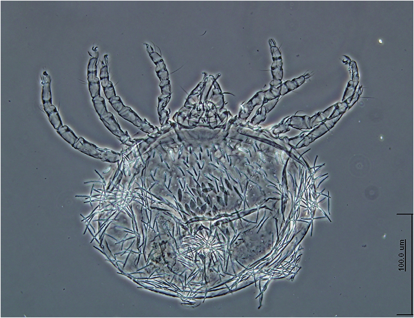
Female — Twelve specimens. Body roughly triangular in shape, red colored, wider than long, rather small in size, length and width of animals preserved in ethanol: 260 – 290 long, 480 – 490 wide. Soft cuticle, covered by long and scarcely dense setae on the dorsum ranging from 25 µm to 75 – 80 µm in length.
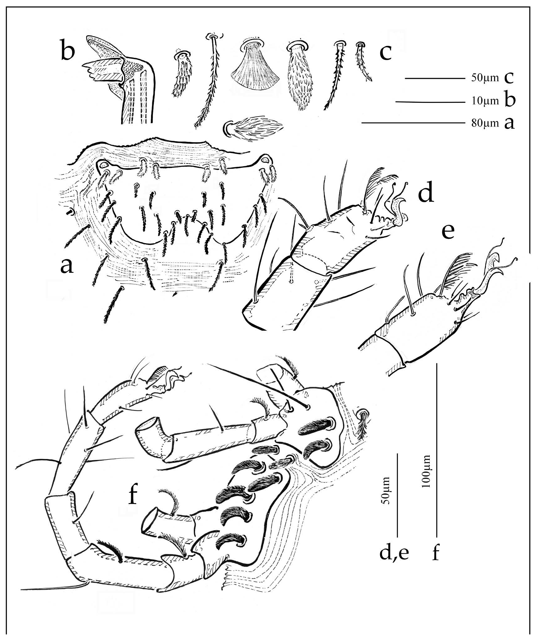
Dorsal view ( Fig. 1a View FIGURE ) — Dorsum: cuticle with striation, covered by pilose setae (ca. 100 dorsal without scutal and lateral setae) longer on the central and peripheral parts than on the anterior. The scutum ( Figs. 1a View FIGURE , 2a View FIGURE ) is symmetric, clearly wider than long, generally with incised backward arbelos shape, centrally concave backwards. The scutum does not reach the slightly concave anterior edge of the body. Posterior to the frontal edge of the scutum, a double pair of setae is present. Ocular lenses are situated on a small latero-anterior extension of the scutum. Three paired lateral scutal setae, placed in rows all along the lateral limit of the scutum, four pairs of mediolateral setae and four pairs of setae along the concavity of the posterior limit of the scutum are present.
Dorsal setae, (more than 60 excluding peripheral and scutal setae) fairly elongated, are arranged in files symmetrically on each side of the body with a four setae file (each 60 – 75 µm long) in the median part of the dorsum. Medially the anogenital fields are visible with three short anal setae (30 – 45 µm) and three longer perigenital setae (75 µm in length)( Fig. 1d View FIGURE ).
Ventral view ( Fig. 1b View FIGURE ) — Epimeral plates: Coxae are developed, gathered in two groups with setation from I to IV: 4(=2-2)-5(=3-2). Anterior epimeral plate (coxae I & II): one thin and long setae (coxa I) (>40 µm), the external seta shorter, two robust paraxial stout setae (29 – 32). Posterior epimeral plate (coxae III & IV) with five long and large epimeral brush-like setae (>30 µm), subequal in length. The anterior and posterior epimeral plates are well separated by striated cuticle bearing two or three robust setae (22 µm).
Ventral surface is covered by several differently shaped types of setae ( Figs. 2c View FIGURE , 3 View FIGURE ):
i) in anterior position, at the same level than the first pair of coxae and between the right and left coxae and near the posterior end of the infracapitulum a row of four robust brush-like setae,
ii) a row of 14 – 18 setae, shorter (20 µm) and briefly ended, and at least some longer setae (25 – 34 µm),
iii) the scale-like setae occupying the major surface of the central part of the venter, arranged in tiles, softly sharpened at the extremity and with irregular edges.
iv) usual setae, barbed and briefly ended distally.
Gnathosoma — Infracapitulum: In ventral view ( Fig. 1c View FIGURE ), the infracapitulum is subquadrangular in shape with one pair of long setae (= gnathobasal setae) slightly longer than the palp that can reach 100 µm length. In dorsal view, the pedipalps (ca. 100 µm long), the chelicerae (>120 µm) and the lips tube are visible, flanked by the peritremes, directed upward. Chelicerae are 100 – 120 µm long. The proximal end (30 – 40 µm) is subquadrangular in shape; it continues in a long slender branch ended by the mobile digit abruptly ended by 1 – 4 little teeth protected by an incomplete hood ( Fig. 2b View FIGURE ). Palpi: femora are as long as wide, the femoral dorsal setae are ciliate, shorter than the dorsal seta of genua that is simple. Palpal chaetotaxy is 1-1-2-4 + ω. Dorsal tibial seta is rather long compared to other species, but distinctly shorter than femoral and genual ones. The two longest setae of tarsus are 25 – 30 µm long. The terminal claw is rather long, embedded and protected by a developed cuticle fold.
Legs — The shorter legs are the leg I and II (140 – 160 µm long) the longer are III and IV (200 – 250 µm long). Separations between coxae I and II and coxae III and IV are not visible. Only the setae on coxae I are simple and long (40 – 60 µm). On some specimens, the distal seta on coxa I was not observed. Other setae are stout and short (ca 25 µm long). Coxal chaetotaxy= 4 or 3(1 or 2+2), 5(2+3). Anterior and posterior epimera are well separated and a file of two setae similar to the stout setae of coxae II and IV are situated on this inter-epimeric space.
Tibial, genual, and femoral setae are simple except on the fourth pair of legs; this ventral femoral seta is plumose. All trochanteral setae are plumose and strong.
Leg chaetotaxy remarkable by the long and ciliated ventro-lateral seta on each trochanter (20 – 30 µm). Chaetotaxy from trochanter to tibia is (1-1- 1-1) (3-2-2-2) (1-0-0-1) (5-5-5-5). This pattern corresponds to the Group I defined by Jack (1964). Femora I, II, III, IV with simple setae with the exception of the ventral seta of femur IV which is ciliate. Dorsal macrochetae (41 – 42, 62 – 69) of tibia I and IV are located behind the lateral setae (paraxial and antiaxial), dorsal setae of tibia II and III are in front of nearest and not too much longer (27 – 36, 34 – 36). Tarsi I and II ( Figs. 2d,e View FIGURE ) are short (50 µm), snub, whereas tarsus IV is a bit elongated. ( Fig. 2f View FIGURE ) The dorsal solenidia ω 2 of tarsi II visible. On tarsi I, two solenidia: the short solenidion ω 1 (7 – 8 µm) with the companion seta alm (28 – 29 µm long). Tarsus I with usual setation: md, (bv), (td), (tld), (tlv) and characteristic (tdf) well visible, only the ventral setae are ciliate. Setation of tarsus II: (bv), (td), (tld), (lvt) (tdf) + ω 2. The claws are projected forward on elongated ends of the tarsi. Tenent hairs with flattened ends present, two by unguis, the external branch being the longest.
Male — no male was collected.
Etymology — Species named in honor of Mrs. Esther Sciberras who assisted in the collection of the specimens and data during the field works and supported the herpetological works of Mr. Sciberras for years.
With the species here described, a second species was found on the Maltese wall-geckoes determined as G. latastei MØgnin, 1878 , a common parasite of Tarentola spp. The latter species was already recorded from several countries ( Spain, Balearic Islands, Morocco, Italy or France...) ( Girot 1969, Haitlinger 2004, Hirst 1926, Willmann 1955, Zapatero-Ramos et al. 1989).

| MNHN |
Museum National d'Histoire Naturelle |
No known copyright restrictions apply. See Agosti, D., Egloff, W., 2009. Taxonomic information exchange and copyright: the Plazi approach. BMC Research Notes 2009, 2:53 for further explanation.
|
Kingdom |
|
|
Phylum |
|
|
Class |
|
|
Order |
|
|
Family |
|
|
Genus |