
Gomphocythere besni, Külköylüoğlu, Okan, Yavuzatmaca, Mehmet, Cabral, Maria Cristina & Colin, Jean-Paul, 2015
|
publication ID |
https://doi.org/ 10.11646/zootaxa.3937.3.2 |
|
publication LSID |
lsid:zoobank.org:pub:73B69F58-99BC-4A9B-A714-8CE9FD5AEFCA |
|
DOI |
https://doi.org/10.5281/zenodo.6115770 |
|
persistent identifier |
https://treatment.plazi.org/id/03A1E50E-CC7B-FFBA-FF69-A00632A67AB6 |
|
treatment provided by |
Plazi |
|
scientific name |
Gomphocythere besni |
| status |
sp. nov. |
Gomphocythere besni View in CoL n. sp.
( Figs. 2–7 View FIGURE 2 View FIGURE 3 View FIGURE 4 View FIGURE 5 View FIGURE 6 View FIGURE 7 )
Etymology. named after the type locality, Besni , where the species was found.
Type locality. Tavaş pool (37°33'373''N, 37°48'596''E), Besni town, Adıyaman, Turkey. Note that the pool is used for human activities (e.g., swimming, irrigation). Most parts of the pool were covered by macrophytes but our samples were taken from the parts of fine pebble and sandy bottom.
Material. a total of 39 individuals, from which 9 are paratypes, all dissected (14 females, 7 males, 3 juveniles, 13 empty carapaces and 1 valve), collected on 19 July 2012. Leg. Mehmet Yavuzatmaca and Ozan Yılmaz.
Type material. Holotype: One dissected female, Tavaş pool, Besni , Adıyaman, Turkey, collected by Mehmet Yavuzatmaca and Ozan Yılmaz. (no: OK-AD-2012: 01).
Allotype: one dissected male from the type locality (no: OK-AD-2012: 02). Soft body parts dissected in lactophenol (no: OK-AD-2012: 02); empty valves of the allotype kept in micropalaeontological slides (no: OK- AD-2012: 03).
Paratypes (all dissected): four females and one juvenile from the type locality (no: OK-AD-2012: 04-08), and two females and two males used for SEM photography kept in the laboratory of the University of Lisbon, Department of Geology, Portugal (no: OK-AD-2012: 09-12).
Measurements. Female (RV-LV): L = 0.75– 0.67 mm (n = 7), H = 0.35– 0.32 mm (n = 7), W = 0.23 mm (n = 3) ( Figs. 2 View FIGURE 2 A–H, 3C). Male (RV-LV): L = 0.66–0.64 (n = 3), H = 0.34 mm, W = 0.30 mm. Juveniles: L = 0.53, H (RV) = 0.29.
Diagnosis. A species of Gomphocythere with weakly inflated posterior part in the female carapace. Valves externally reticulated, with around eight fossae arranged approximately in a circle in each mesh of the reticulum. Presence of a shallow ventral ridge along all the ventral margin and lower part of the posterior one. Main body of the hemipenis with a short and straight upper ramus of clasping organ. Copulatory organ of hemipenis short and distally curved.
Description of female. Carapace sub-rectangular in lateral view, pyriform in dorsal view, with a weak median sub-vertical sulcus. RV larger than the LV, slightly overlapping it ( Fig. 3 View FIGURE 3 A, B). Greatest height situated at anterior cardinal angle. Dorsal margin straight to slightly concave, sloping posteriorly; ventral margin slightly concave; anterior margin flattened and asymmetrically rounded; posterior margin rounded. External surface reticulated, with ca. eight fossae roughly arranged in a circle, in each mesh of the reticulum. A distinct shallow ventral ridge, more prominent posteriorly, running parallel to the margin ( Fig. 3 View FIGURE 3 –D). A few sparse pore conuli ( Fig. 3 View FIGURE 3 H, I) visible along the anterior and posterior margins. Over the whole surface, rounded or elongated (in the margins) sieve plates and separate single pores with a sensillum present, the latter being relatively rare.
Hinge “reversed lophodont with smooth teeth separated by a smooth groove in the LV. Both valves with marked selvage inwardly displaced along the anterior and posterior margins and with narrow calcified inner lamellae. Muscle scars: four elongated adductor muscle scars in a sub-vertical row, one rounded frontal scar and two rounded to elongated mandibular scars.
Sexual dimorphism well marked: females larger and swollen posteriorly; males with almost parallel sides weakly converging posteriorly in dorsal view.
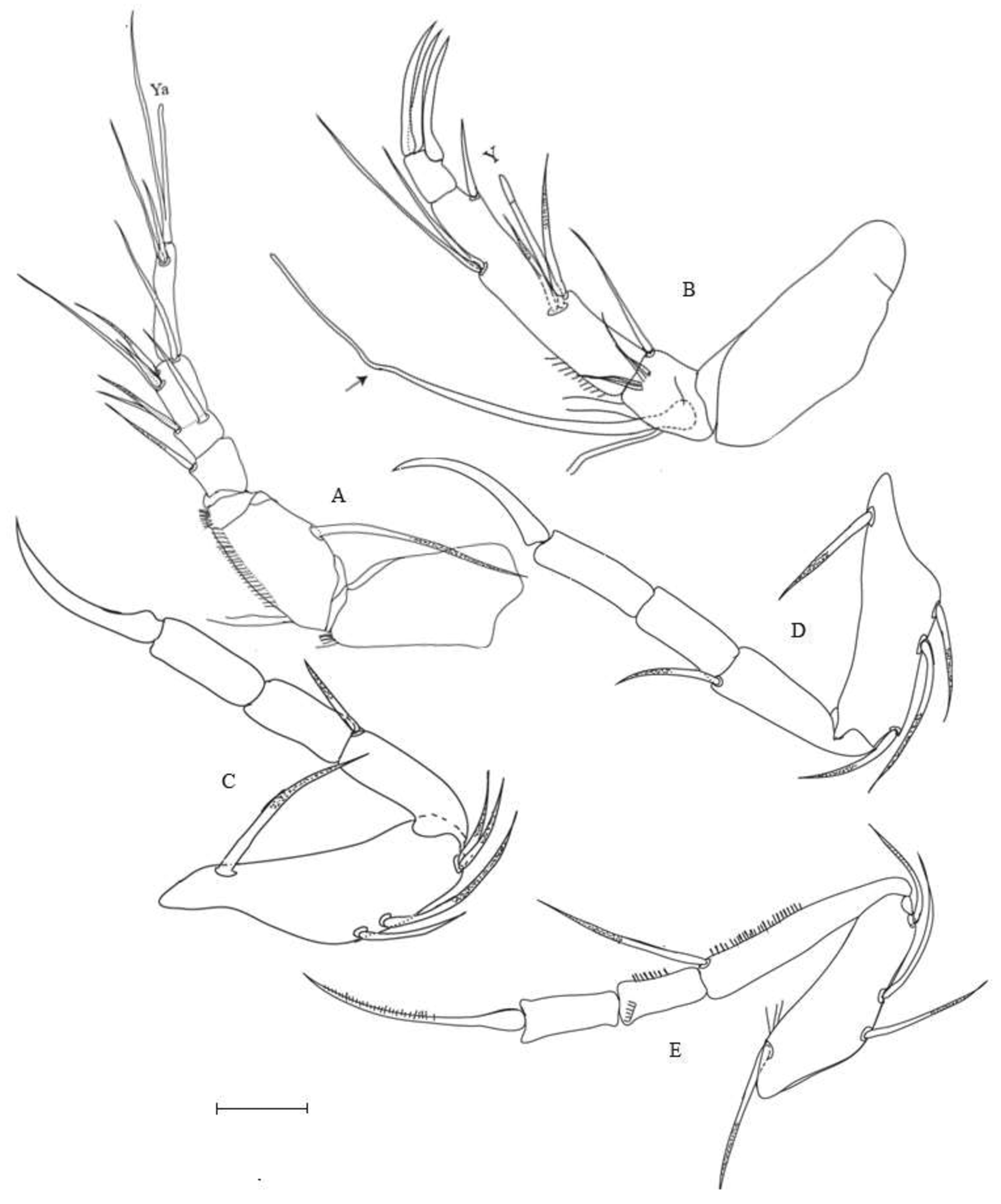
Female Soft parts: Antennula (A1) ( Fig. 4 View FIGURE 4 A): Six segmented without setae on protopodite (first segment). Second segment with one long (ca. 1.5 x of the length of 2nd segment) medio-lateral seta posteriorly and a group of tiny setules anteriorly. Rome organ absent. Third segment, subquadrate with one medium (bluntly plumose) dorsal seta (ca. 2 x of length of this segment). Fourth segment with three apical setae: two dorsal (plumose) and one ventral smooth setae. Fifth segment with four apical smooth setae, three dorsal (one shorter than the half of the others) and one long seta (extending the length of terminal segment). Terminal segment elongated (length more than 3 times width), with four setae apically, one long and one medium claw-like setae, and one seta fused at the base with aesthetasc Ya (fused seta longer than Ya).
Second antenna (A2) ( Fig. 4 View FIGURE 4 B) without no seta on protopodite. First segment of endopodite subquadrate with long (ca. of second segment) apical ventral seta. Second segment long (ca. at least of the A2), mid-ventrally with two long plumose setae and aesthetasc Y (slightly shorter than accompanying setae), mid-dorsally with two subequal setae (both extending the tip of the terminal segment); one stout, claw-like medium size apical seta (ca. reaching the tip of the terminal segment). Terminal segment with three (two apically and one subapically located) smooth and stout claws, almost equal in length. Exopodite long, extending to the tips of terminal claws, not divided but a node present almost in the middle.
Mandibula (Md) ( Fig. 5 View FIGURE 5 A): with a masticatory process and a four-segmented palp. Masticatory process with tiny setules and 8 strong teeth, the last three in one group. First segment of palp with a vibratory plate consisting of three well developed setae only (two long, one short plumose setae) and with two subequal ventral setae. Second segment short and subquadrate, bearing three apical setae. Third segment of similar shape, with five apical setae, three dorsally (length of these setae almost reaching tips of terminal claws) and two located ventrally. Terminal (fourth segment) pyramidal, with two claws (more than twice the length of this segment).
Maxillula (Mxl) ( Fig. 5 View FIGURE 5 B): with 3 endites of normal shape, ending with 3, 4, and 4 slightly plumosed short setae in 1, 2, and 3rd endites, respectively. Maxillular palp with two segments fused, as typical of the subfamily, ending with 3 long sclerotized claw-like lateral and 2 (one additional short seta bluntly seen) apical setae. Vibratory plate with 14 well developed and plumose whorls.
T1-T3 ( Figs. 4 View FIGURE 4 C-D-E) all well-developed walking legs, with T1 the shortest and T3 the longest. First segment (in all legs) with 2 long dorsal and one ventral seta and with two (T1) and one (T2, T3) knee-setae. Second segment with one claw-like antero-ventral seta in all legs. Terminal claw-like seta smooth (T1-T2) and weakly sclerotized (T3).
Sternum as in Fig. 5 View FIGURE 5 C.
Abdomen ( Fig. 5 View FIGURE 5 D): Posterior part with 3 hirsute lobes (crl1-3) equal in size, ending with a short filamentous tip. First seta of caudal ramus (crs1) short (ca. 1/2 of crs2) whip-like. Second seta (crs2) long, inverse L-shaped. Caudal seta large and claw like. Forked organ (FO) short, stout spine-like seta. Genital operculum (GeO) large, rounded.
Labium or upper lip ( Fig. 5 View FIGURE 5 E): with two lobes (hlo1, hlo2), anterior (hlo1) lobe at least twice as long as posterior (hlo1) one.
Male soft parts. Antennula (A1) ( Fig. 6 View FIGURE 6 A): Five-segmented (3rd and 4th segments fused) (also see discussion). A1 morphology and chaetotaxy are identical in both sexes, apart of the division between segments 3 and 4 (present in males, missing in females).
Second antenna (A2) ( Fig. 6 View FIGURE 6 B): Exopodite long extending to the tips of terminal claws, not divided but a node present almost in about 2/3 of the length.
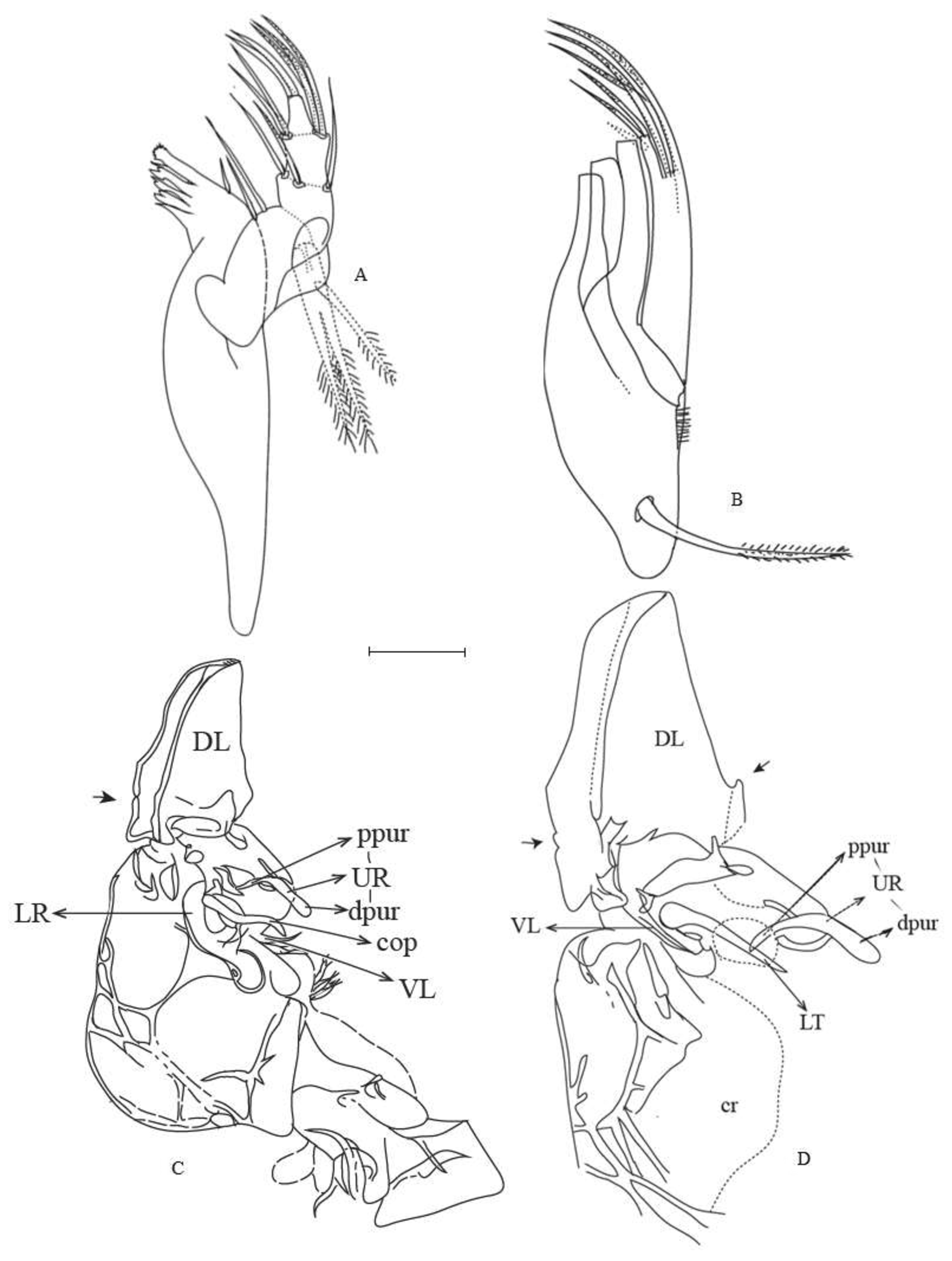
Mandibula (Md) ( Fig. 7 View FIGURE 7 A): Similar as in female.
Maxillula (Mxl) ( Fig. 7 View FIGURE 7 B): Similar as in female.
T1-T3 ( Figs. 6 View FIGURE 6 C-D-E): All walking legs as in female.
Hemipenis ( Figs. 7 View FIGURE 7 C–D): Typical of the genus with a large, well developed muscular base and a prominent, distal lobe (DL) without setae. No comb-like structure on the blunt tip of the main body of DL. Main body with a short and straight ur (upper ramus of clasping organ of hemipenis); copulatory organ of hemipenis (cop) short and distally curved. Caudal ramus short and straight.
Remarks. The closest species, regarding the carapace morphology, are G. geareyi (Holocene) and G. ortali (Recent) , both from Middle-East (see “Discussion for comparisons), G. h u w i Martens, 2003 and G. l i s a e Martens, 2003, both living species in Lake Nyassa/ Malawi, East Africa. Gomphocythere huwi is smaller than G. b e s n i n. sp. with a more pronounced median sulcus, different reticulation with absence of ventral ridge and a selvage inwardly displaced only along the anterior margin. Gomphocythere lisae is also smaller, with a more pronounced median sulcus, different reticulation with absence of ventral ridge and a posterior margin less uniformly rounded.
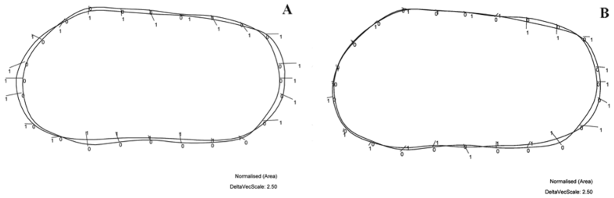
Due to the geographical proximity of G. b e s n i n. sp., G. geareyi and G. ortali and the fact that they all are very close in morphological characteristics of the carapace, we compared the area deviations between the superposed normalized outlines of the following couple of species: G. b e s n i n. sp. versus G. geareyi ( Fig. 8 View FIGURE 8 A) and G. b e s n i n. sp. versus G. ortali ( Fig. 8 View FIGURE 8 B) (cf. for this algorithm and its utilization Neubauer & Linhart 2008 and Minati et al. 2008).
The outline of G. b e s n i n. sp. compared to the one of G. g e a re yi appears more anteriorly rounded and with a more important height displayed at the anterior and posterior cardinal angles. G. g e a rey i is more elongated than its pair species.
The outline of G. b e s n i n. sp. compared to the one of G. ortali , when superposed and standardized to equal surface, shows less shape differences than the species pair G. b e s n i n. sp. - G. geareyi . This becomes particularly apparent along the anterior margin. Significant differences exist along the dorsal and posterior margins. These latter differences are due to the oblique position of the dorsal intercardinal segment and to a lower posterior height of G. ortali . Therefore, the geometric morphometric data, point to three distinct morphotypes which complemented with the differences for limb traits allow to decide for separation of the new Gomphocythere taxon, G. b es n i n. sp., beside the two already known species G. ortali and G. geareyi of the Eastern Mediterranean area, more precisely from Israel ( Martens 1993) and Western Turkey ( Boomer & Gearey 2010).
Ecology. So far, the individuals of the species Gomphocythere besni n. sp. have been found on sediments with a mixture of sand and gravel in the type locality only. Tavaş pool, Besni , Adıyaman has relatively cool (16.6°C), medium oxygenated (dissolved oxygen = 7.36 mg /L, percent dissolved oxygen = 75 %), slightly alkaline (pH = 7.53) and brackish to fresh waters (specific electrical conductivity = 609 µS/cm, salinity = 0.3 ppt). Air temperature (38.5°C), air moisture (25.5 %) and atmospheric pressure (701 mm Hg) were also measured in situ before sampling. Accompanying species included Ilyocypris inermis Kaufmann, 1900 (20 individuals) and Psychrodromus olivaceus (Brady & Norman, 1889) (4 individuals).


No known copyright restrictions apply. See Agosti, D., Egloff, W., 2009. Taxonomic information exchange and copyright: the Plazi approach. BMC Research Notes 2009, 2:53 for further explanation.