
Intrepidocythere ibipora, Pinto, Ricardo L., Rocha, Carlos E. F. & Martens, Koen, 2008
|
publication ID |
https://doi.org/ 10.5281/zenodo.183142 |
|
DOI |
https://doi.org/10.5281/zenodo.6229821 |
|
persistent identifier |
https://treatment.plazi.org/id/827887B2-FF80-FF84-FF6B-D2B0FAFFFE2D |
|
treatment provided by |
Plazi |
|
scientific name |
Intrepidocythere ibipora |
| status |
gen. nov. |
Intrepidocythere ibipora View in CoL n.gen. n.sp.
( Figs. 1–4 View FIGURE 1 View FIGURE 2 View FIGURE 3 View FIGURE 4 )
Type material. Holotype: a dissected male, with valves stored dry in a micropalaeontological slide and soft parts mounted in a permanent slide with CMC-9AF mounting media ( MZUSP 18479).
Allotype: an ovigerous female, dissected and stored like the holotype ( MZUSP 18480).
Paratypes: a male, dissected and stored like the holotype ( MZUSP 18483); a male ( MZUSP 18482) and a female ( MZUSP 18481), both dissected, with soft parts mounted in permanent slides with CMC-9AF and coated valves stored in micropalaeontological slides; soft parts of a male ( MZUSP 18487) mounted in a permanent slide with CMC-9AF; left valve of a male, coated for scanning electron microscopy and stored in a micropalaeontological slide ( MZUSP 18484); a male, dried and coated for scanning electron microscopy ( MZUSP 18486); a female, dried and coated for scanning electron microscopy ( MZUSP 18485); 7 females and 4 males kept whole in 70% ethanol ( MZUSP 18488).
Instars occurred in the population, but were not picked from the samples.
Type locality. Parque Estadual da Serra do Mar Núcleo Cunha /Indaiá, Municipality of Cunha, São Paulo State, Brazil. Approximate GPS coordinates: 23º14’03.3”S, 45º01’23.2”W.
The entrance to the State Park Serra do Mar Núcleo Cunha /Indaiá can be reached from highway Paulo Virgínio—SP171, by accessing (at Km 56) the municipal road Paraibuna and driving along 20 Km on nonpaved road. The Park is managed and controlled by the Instituto Florestal, which belongs to the São Paulo State Government. Information about the Park can be obtained from the web page of Instituto Florestal (http:/ /www.iflorestal.sp.gov.br/unidades_conservacao/busca.asp).
Altitude inside the Park ranges from 950m to 1561m, and temperature averages 18ºC and 22ºC, in winter and summer, respectively. The area consists of secondary vegetation typical of the Atlantic Ombrophilous Dense Forest, which is composed predominantly of evergreens that form a continuous canopy. Specimens used in the present description were retrieved from forest leaf litter collected alongside a track that runs from the base of the Park towards the river Paraibuna.
Material collected on 05 March 2004 by Marco Aurélio de Sena.
Etymology. The word ibipora comes from the Tupi-Guarani indigenous language and means ‘terrestrial’ or ‘pertaining to land’. From ibi, land and pora, pertaining to. The name of this species thus makes reference to the habitat in which it occurs, i.e. terrestrial.
Diagnosis. Distal lobe on hemipenis triangular and slender; copulatory process a projection from a large hook-like structure; the latter, in turn, articulating with an intricate sequence of irregular and folded pieces; inner edge of muscular body with a small bifid lobe midway from the copulatory process to the caudal ramus. Males subquadrate in lateral view, females somewhat rounded; in dorsal and ventral views, male carapace with greatest width located at midlength, female with greatest width in posterior part.
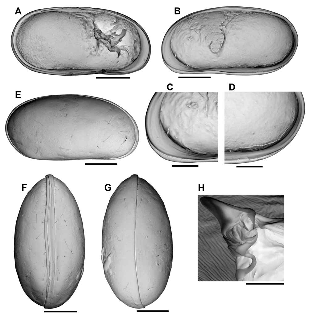
Description of male. Carapace ( Fig. 1 View FIGURE 1 E–G). Relatively small (426 μm in length), with brownish smooth surface and sparse sensillae; subquadrate in lateral view, relatively elongate (length/height=1.96); left valve overlapping right valve along posterior, dorsal, anterior and anteroventral margins; ventral margin straight; dorsal margin slightly arched; posterior and anterior margins rounded, the anterior margin slightly produced towards the ventral side; oval shaped in ventral and dorsal views, maximum width located at midlength; ventrally with well marked marginal ridges.
Left valve ( Fig.1 View FIGURE 1 A). In internal view, posterior margin broadly rounded; anterior margin rounded but slightly produced towards the ventral side; ventral margin nearly straight; dorsal margin slightly arched, running parallel to ventral margin along part of its length and sloping more pronouncedly near the anterior margin; vestibule weakly developed in anterior margin and absent in posterior margin; central muscle scars consisting of 4 spots arranged in a vertical row.
Right valve ( Fig. 1 View FIGURE 1 B–D). In internal view, posterior margin broadly rounded, slightly produced towards the dorsal side; anterior margin broadly rounded, slightly produced towards the ventral side; ventral margin nearly straight; dorsal margin arched; selvage well developed and sinuous, forming a continuous flange along ventral margin; vestibule weakly developed in anterior margin and absent in posterior margin; central muscle scars consisting of 4 spots arranged in a vertical row.
Hinge ( Fig.1 View FIGURE 1 A–B). Smooth medial ridge (c. 1/3 of the valve length) and posterior socket in left valve, and the complementary smooth medial groove and posterior tooth in right valve.
Small pigmented naupliar eye present.
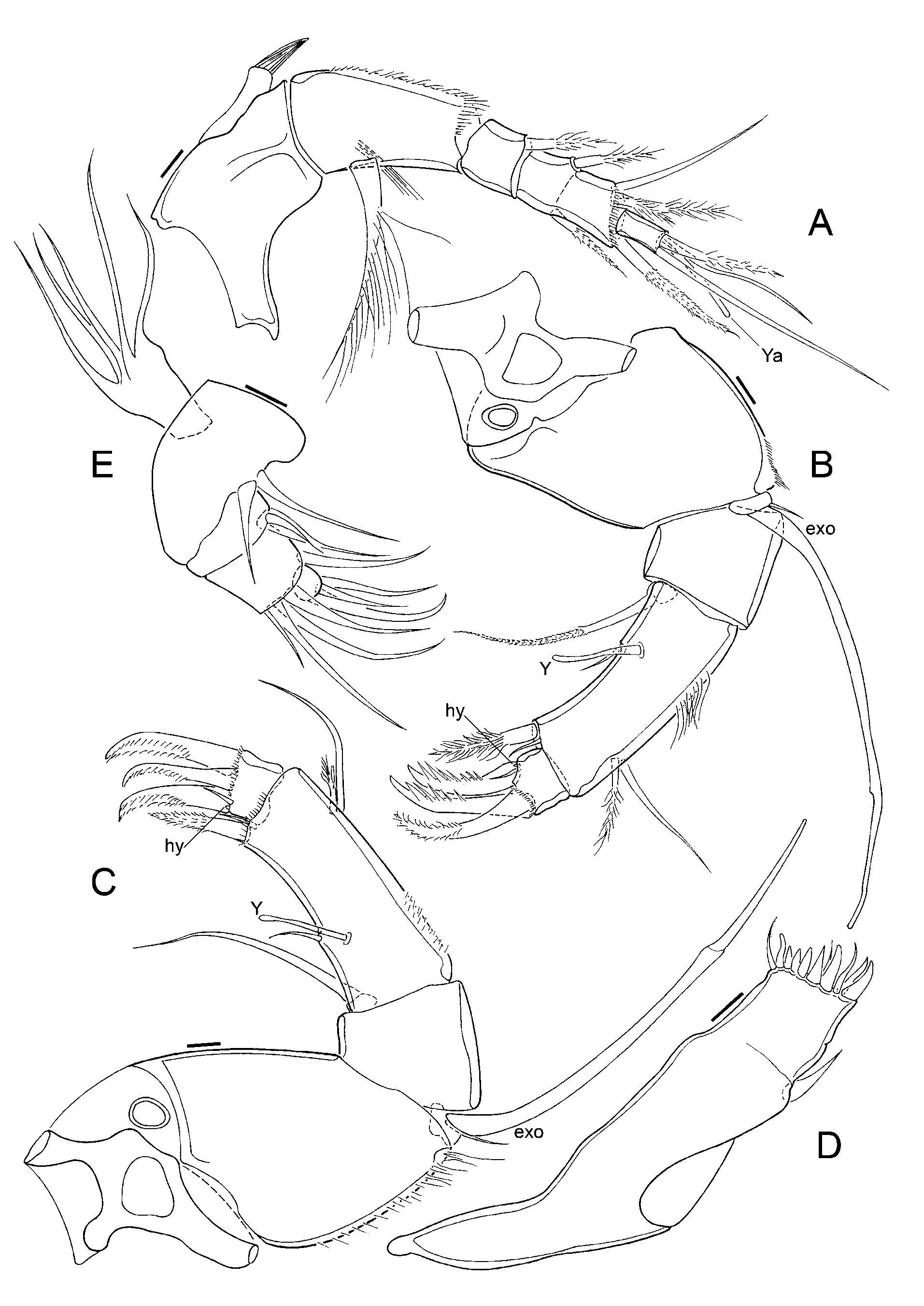
Antennula ( Fig. 2 View FIGURE 2 A). Five functional articles; first article relatively large, bearing, on the dorsal margin, a subapical expansion with a tuft of tiny setules; second article the longest, with a ventroproximal long, thick seta; third article small, with a dorsoapical seta; fourth article partially divided, with a dorsal and a ventral setae implanted at this nonfunctional joint, as well as a dorsoapical and a ventroapical setae; fifth (terminal) article with two long and one short setae and a short aesthetasc Ya.
Antenna ( Fig. 2 View FIGURE 2 B). Protopodite 2-segmented, the first one very short and the second one long and wide; endopodite 3-articulated; first segment short, bearing a ventroapical seta; second segment very long and narrow, dorsally with two subapical setae, one half as long as the other, ventrally with a short medial seta and aesthetasc Y and two ventroapical setae, one large and one minute; last segment small, with three large claws, two of them strongly serrated and the other one bearing two rows of fine setules, and a minute seta and a tiny lobe (hyaline formation); exopodite with a very small seta and a long spinneret seta reaching the tip of the penultimate endopodal segment.
Mandibula ( Fig. 2 View FIGURE 2 D–E). Coxa with 7 strong teeth and 4 setae on inner edge and a seta on outer edge (near the articulation with the palp); palp 4-segmented (basis + 3-segmented endopodite); basis with respiratory plate (exopodite) consisting of 5 rays and with two unequal internal apical setae; first endopodal segment with two apical internal setae, one half as long as the other; second endopodal segment with an internal apical seta and 4 external apical setae, one short and 3 long; terminal endopodal segment with 3 setae of different length.
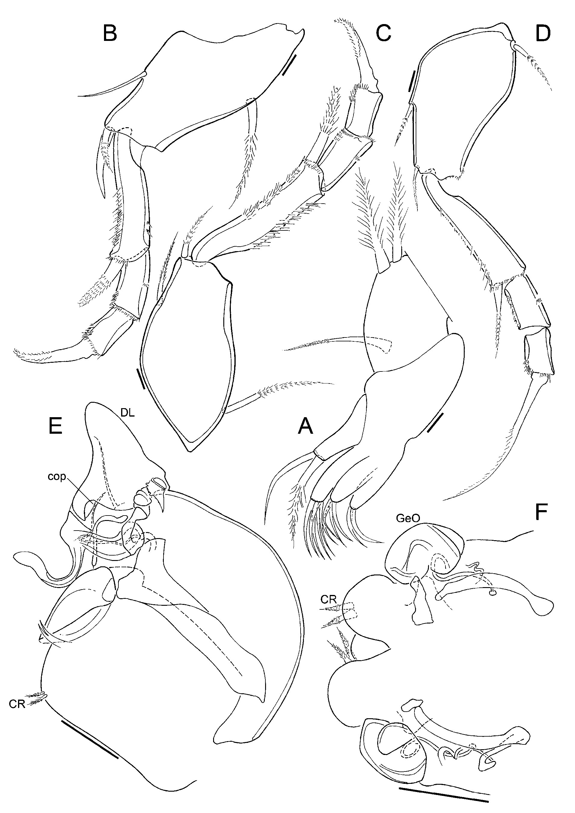
Maxillula ( Fig. 3 View FIGURE 3 A). Internally with three endites, first one with 2 setae, second one with 4 setae and third one with approximately 5 setae; palp not segmented, tapering, with 2 apical setae; respiratory plate well developed, carrying a reflexed seta (i.e. reversed towards the front) and about 16 long rays.
First thoracopod ( Fig. 3 View FIGURE 3 B). Four-segmented; first segment with a long mediodorsal seta, a short medioventral seta and two stout ventroapical setae, one half as long as the other; second segment quite long, with a strong ventroapical seta; third segment devoid of setae; terminal segment with an apical claw.
Second thoracopod ( Fig. 3 View FIGURE 3 C). Four-segmented; first segment with a long mediodorsal seta, a medioventral seta and a ventroapical seta; second segment long, with a very strong ventroapical seta reaching tip of terminal segment; third segment devoid of setae; terminal segment with an apical claw.
Third thoracopod ( Fig. 3 View FIGURE 3 D). Four-segmented, quite slender; first segment with a proximal dorsal seta, a medioventral seta and a ventroapical seta; second segment very long, with a ventroapical seta; third segment devoid of setae; terminal segment with a long apical claw.
Hemipenis ( Fig. 1 View FIGURE 1 H, 3E). Consisting of a large rounded muscular body and a triangular articulating distal lobe; this lobe quite slender, carrying a dorsal seta; copulatory process a projection from a large hook-like structure; the latter, in turn, articulating with an intricate sequence of irregular and folded pieces; inner edge of muscular body with a small bifid lobe midway from the copulatory process to the caudal ramus; caudal ramus a pair of short setae.
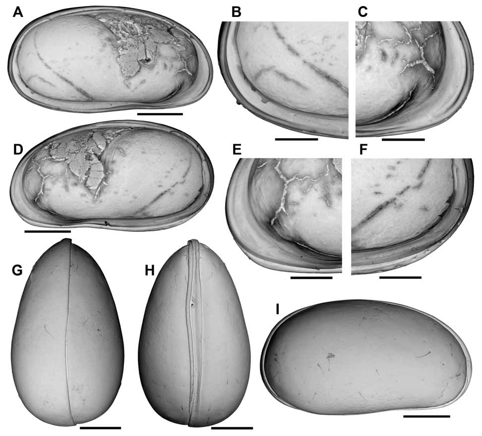
Additional description of female. Carapace ( Fig.4 View FIGURE 4 G–I). Relatively small (468 μm in length), with brownish smooth surface and sparse sensillae; left valve overlapping right valve along posterior, dorsal, anterior and anteroventral margins; ventral margin nearly straight; dorsal margin arched; posterior and anterior margins broadly rounded, the latter slightly produced towards the ventral side; oval shaped in dorsal and ventral views, with posterior part expanded, forming a brood pouch that carries, on average, between 4 and 5 offspring simultaneously (Mean = 4.57; SD = 1.51; N = 7; see also discussion below); maximum width displaced posteriorly (c. 2/3 of the length); ventrally with well marked marginal ridges.
Left valve (Fig. A–C). In internal view, posterior margin broadly rounded; anterior margin broadly rounded, slightly produced towards the ventral margin; ventral margin slightly sinuous; dorsal margin arched; vestibule weakly developed in anterior margin and absent in posterior margin; central muscle scars consisting of 4 spots arranged in a vertical row.
Right valve (Fig. D–F). In internal view, posterior and anterior margin broadly rounded, the latter slightly produced towards the ventral side; ventral margin sinuous; dorsal margin arched; selvage well developed and sinuous, forming a continuous flange along ventral margin; vestibule weakly developed in anterior margin and absent in posterior margin; central muscle scars consisting of 4 spots in a vertical row.
Hinge as in the male. Antenna ( Fig. 2 View FIGURE 2 C). Protopodite 2-segmented, with first segment very short and the second one long and wide; endopodite 3-articulated; first segment short, bearing a ventroapical seta; second segment very long and narrow, dorsally with 2 subapical setae, one short and one long, ventrally with a short medial seta and aesthetasc Y and two ventroapical setae, one large and one minute; last segment small, with three large claws, all of them with a double row of fine setules, and a minute seta and a tiny lobe (hyaline formation); exopodite with a very small seta and a long spinneret seta reaching the tip of the penultimate endopodal segment.
Female abdomen ( Fig. 3 View FIGURE 3 F). Genital operculum rounded, connected by tubes to a large trabecule; caudal ramus a pair of short setae.
Measurements
Males
Carapace: Length = 426 µm, Height = 217 µm, Width = 234 ìm (specimen MZUSP 18485).
Left valve: Length = 428 µm, Height = 218 µm (specimen MZUSP 18484).
Right valve: Length = 434 µm, Height = 227 µm (specimen MZUSP 18482).
Females
Carapace: Length = 468 µm, Height = 264 µm, Width = 288 µm (specimen MZUSP 18485).
Left valve: Length = 463 µm, Height = 252 µm (specimen MZUSP 18481).
Right valve: Length = 440 µm, Height = 241 µm (specimen MZUSP 18481).
Accompanying fauna. Intrepidocythere ibipora n. gen. n. sp. coexists with at least five other species of terrestrial ostracods: Caaporacandona shornikovi Pinto et al., 2005 , Callistocypris mckenziei Pinto et al., 2005, Callistocypris rossettii Pinto et al., 2005, Penthesilenula brasiliensis Pinto & Kotzian, 1961 and Terrestricypris wurdigae Pinto et al., 2005. Harpacticoid copepods were also present in the samples.
| MZUSP |
Museu de Zoologia da Universidade de Sao Paulo |
No known copyright restrictions apply. See Agosti, D., Egloff, W., 2009. Taxonomic information exchange and copyright: the Plazi approach. BMC Research Notes 2009, 2:53 for further explanation.