
Jorottui ipuanai, Moreno-González & Gutierrez-Estrada & Prendini, 2023
|
publication ID |
https://doi.org/ 10.1206/4000.1 |
|
persistent identifier |
https://treatment.plazi.org/id/043487D1-2C45-B647-FE0E-C193275F4AD4 |
|
treatment provided by |
Felipe |
|
scientific name |
Jorottui ipuanai |
| status |
sp. nov. |
Jorottui ipuanai , sp. nov.
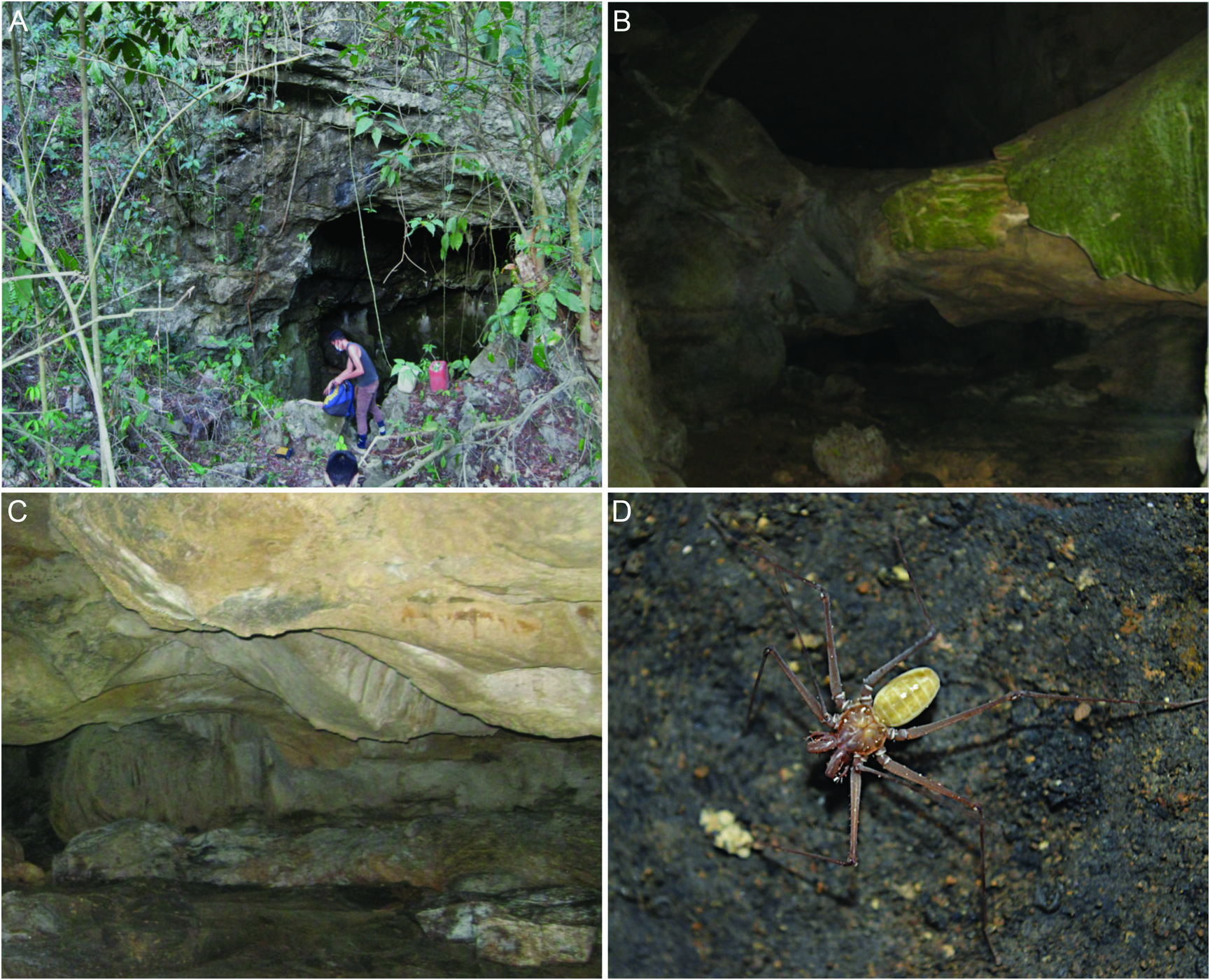
Figures 1–3 View FIGURE 1 View FIGURE 2 View FIGURE 3 , 4A, B View FIGURE 4 , 5A, B View FIGURE 5 , 6A, B, 7A–D, 8A–D, 9, 10B, 11B, 12, 13C–F, 14C– F, 15C–F, 16C–F, 17C–F, 18, 19B, C, 20; table 1
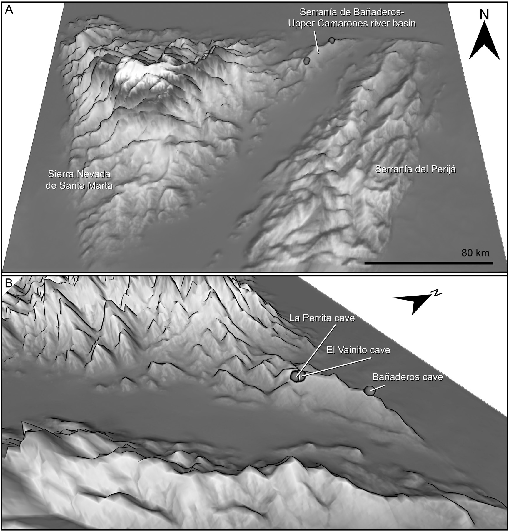
TYPE MATERIAL: COLOMBIA: La Guajira Department: Serranía de Bañaderos, upper basin of Camarones River: Holotype ♀ (ICN-Am 180), Hatonuevo , Bañaderos sidewalk, cave 100 m from Bañaderos Cave , 980 m, iv.2022, M. Gutierrez-Estrada. Paratypes: 1 ♀ ( AMNH IZC 357368 , AMCC [ LP 18072 ]), same data as holotype; 1 ♀ ( AMNH IZC 357367 , AMCC [ LP
19086]), same locality as the holotype, 28.x.2022, M. Gutirrez-Estrada; 1 ♀ [poorly sclerotized] (ICN-Am 181), Hatonuevo , Bañaderos Sidewalk , Bañaderos Cave , 11°07′51.5″N 72°47′23.9″W, 1005 m, x.2015, M. Gutierrez-Estrada; 8 ƋƋ (ICN-Am 182–188, AMNH IZC 357366 ), Fonseca , Los Chorros sidewalk, La Perrita Cave , 11°00′22″N 72°55′00.4″W, 968 m, 11.x.2015, M. Gutierrez-Estrada; 1 Ƌ (ICN-Am 189), Fonseca , Los Chorros sidewalk, La Perrita Cave , 11°00′22″N 72°55′00.4″W, 968 m, 13.i.2017, M. Gutierrez-Estrada; 1 Ƌ (ICN-Am 190), Barrancas, El Vainito Cave, 11°01′04.2″N 72°54′45.1″W, 998 m, x.2021 M. Gutierrez-Estrada GoogleMaps .
DIAGNOSIS: As for genus.
ETYMOLOGY: This new species is named after Ramón Paz Ipuana (1937–1992), a Venezuelan Wayuu educator, researcher, linguist, poet and writer, who devoted his life to studying Wayuu culture and promoting the rights and traditions of Wayuu people.
DESCRIPTION: Based on adult male and female (fig. 4A, B). Measurements (mm) in table 1.
Coloration: Carapace and opisthosoma light reddish brown; chelicerae, pedipalps, and legs reddish brown; tibiae and tarsi of legs, light brown (fig. 1D).
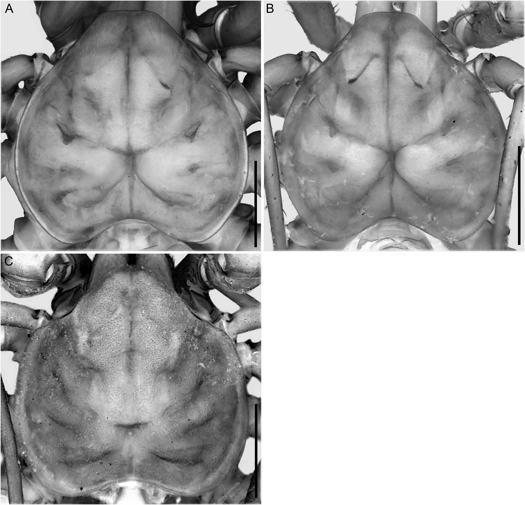
Carapace: Shape cordate (fig. 5A, B), as wide as long, without bumpy area anteromedially; anterior margin truncate, markedly projecting, with lateral margins linear, width of projection half width of carapace; posterior margin with deep median notch. Two median and four pairs of dorsosubmedian transverse sulci, all distinct; anteromedian sulcus extending from anterior margin to 0.6 of carapace length, connected to fovea; posteromedian sulcus extending from posterior margin to fovea; in anterior-posterior axis, first and third dorsosubmedian pairs of sulci not connected to fovea, second and fourth pairs connected to fovea. Median ocular tubercle, and median and lateral ocelli absent (fig. 5A, B). Frontal process projecting and recurved, 1.4× wider than long (fig. 6A, B). Cuticle feebly sclerotized, translucent, apodemes clearly visible.
Sterna: Tetra segmented (fig. 7A–D). Tritosternum vestigial, not projecting, consisting of single, flat, suboval plate, with single anteromedian seta and two pairs of lateral microsetae. Tetrasternum comprising single plate with rounded (Ƌ) or bifurcate (♀) apex bearing two setae (fig. 7B, D), and with two setae anteriorly and two setae posteriorly on surface of plate. Pentasternum comprising single plate, projecting slightly ventrally and terminating in rounded apex bearing two setae, with posteromedian seta. Metasternum comprising single subtriangular plate, anterior margin narrow, posterior margin wider, with two ventrosubmedian setae in posterior half.
Chelicerae: Basal segment retrolateral margin without teeth but with vestigial eminence (fig. 8B, D); prolateral margin with four teeth (fig. 8A, C), first tooth (ventralmost) ca. 1.5× longer and broader than other teeth, second, third, and fourth teeth similar in size, fourth tooth (dorsalmost) bicuspid, dorsal cusp smaller than ventral cusp (fig. 9C); prolateral surface densely covered with minute spicules, probably stridulatory (fig. 9E) and with transverse row of ca. 12 setae in basal third; proventral and retroventral margins densely covered with fringe of setae (figs. 8A–D, 9A, D). Movable finger (claw) with seven denticles on ventral margin (fig. 9C); prolateral surface densely covered with fringe of setae (figs. 8A, C, 9A, B).
Pedipalps: Oriented perpendicular to body in vertical plane (figs. 10B, 11B, 12A, B). Trochanter with extremely elongated anteroventral apophysis (AVA), ca. 2× length of trochanter, terminating in small spine (fig. 13C, E); prolateral margin of AVA with row of large dorsal setiferous tubercles, row of small ventral setiferous tubercles, and row of microsetae ventral to small setiferous tubercles; two dorsal primary spines (Tr1 = Tr2; fig. 13D, F), small setiferous tubercle distal to Tr1, two small setiferous tubercles between Tr1 and Tr2, and three small setiferous tubercles proximal to Tr2. Femur with four ventral primary spines (FII> FI> FIII> FIV; fig. 14C, E), large setiferous tubercle proximal to FI, and one (Ƌ; fig. 14E) small accessory spine distal to FIV; femur ca. 3× longer than FII, FII ca. 1.3× longer than FI, ca. 4.6× (Ƌ; fig. 14E) to 4.8× (♀; fig. 14C) longer than FIII, and ca. 8× longer than FIV; one dorsal primary spine (F1; fig. 14D, F) and two
(Ƌ; fig. 14D) to four (♀; fig. 14F) small, setiferous tubercles proximal to F1; femur ca. 6.3× (♀) to 7.6× (Ƌ) longer than F1. Patella with four ventral primary spines (PII> PI> PIII> PIV; fig. 15C, E), one (Ƌ; fig. 15E) small accessory spine proximal to PIV, one between PIII and PII, and two distal to PI; patella ca. 2.1× longer than PII, PII ca. 2× (Ƌ; fig. 15C) to 2.4× (♀; fig. 15E) longer than PI, ca. 2.2× (Ƌ) to 2.5× (♀) longer than PIII, and ca. 7× longer than PIV; three dorsal primary spines (P2> P3> P1; fig. 15D, F), small setiferous tubercle proximal to P3, two accessory spines between P3 and P2, one between P2 and P1, and one distal to P1; patella ca. 2.3× (Ƌ; fig. 15F) to 2.5× (♀; fig. 15D) longer than P2, P2 ca. 1.9× (Ƌ) to 1.76× (♀) longer than P1 and ca. 1.3× (Ƌ) to 1.4× (♀) longer than P3. Tibia with five ventral primary spines (TiI> TiII> TiIII> TiIV> TiV; fig. 16C, E), tibia ca. 1.3× (Ƌ; fig. 16E) to 1.4× (♀; fig. 16C) longer than TiI, TiI ca.
2.1× (Ƌ) to 2.2× (♀) longer than TiII, ca. 2.3× (Ƌ) to 2.4× (♀) longer than TiIII, ca. 5× (Ƌ) to 7.3× (♀) longer than TiIV, and ca. 14× (Ƌ) to 24× (♀) longer than TiV; five dorsal primary spines (Ti1> Ti2> Ti3> Ti4> Ti5; fig. 16D, F), tibia ca. 2× length of Ti1, Ti1 ca. 1.7× longer than Ti2, ca. 2.1× (♀; fig. 16D) to 2.5× (Ƌ; fig. 16F) longer than Ti3, ca. 3.8× longer than Ti4, and ca. 7.8× (♀) to 8× (Ƌ) longer than Ti5. Tarsus with one ventral primary spine (TaI; fig. 17C, E); tarsus ca. 1.1× (♀; fig. 17C) to 1.7× (Ƌ; fig. 17E) longer than TaI; three dorsal primary spines (Ta1> Ta2> Ta3; fig. 17D, F), tarsus ca. 1.1× (Ƌ; fig. 17F) to 1.2× (♀; fig. 17D) longer than Ta1, Ta1 ca. 1.4× (♀) to 1.5× (Ƌ) longer than Ta2, ca. 3.6× (♀) to 5.4× (Ƌ) longer than Ta3. Cleaning brush organ (fig. 18C–F) situated along distal two-thirds of tarsus (fig. 17C, E); ventral setal row comprising imbricated, sickle-shaped (falcate) setae (fig. 18D), each with concave anterior margin; dorsal setal row comprising curved setae (fig. 18E), each covered with numerous small, lanceolate projections basally and dorsally; granular area densely covered with rectangular squamiform structures (fig. 18F) each with ca. 12 to>21 digitiform projections, with several lipped, major glands; clavate setae, situated near cleaning organ, long and lanceolate (fig. 18C). Claw ca. 1.3× (Ƌ; fig. 17E) to 1.6× (♀; fig. 17C) longer than tarsus, curved, terminating in truncate apex (fig. 18A, B).
Legs: Legs II–IV femora with rounded process projecting distally. Leg I tibia comprising 16 articles; tarsus comprising 31 (holotype ♀) to 33 (paratype Ƌ) very elongate articles. Leg IV basitibia comprising two articles, distal third of second article with bt trichobothrium (fig. 19B, C); distitibia with 10 trichobothria: single proximal bf trichobothrium near articulation with basitibia, three trichobothria in sc series, four trichobothria in sf series, and single tc and tf trichobothria, all situated distally, near articulation with tarsus.
Opisthosoma: Translucent, ca. 1.7× longer than wide (fig. 4A, B); each tergite with pair of dorsosubmedian depressions (internal apodemes) in anterior half.
Male Genitalia: LoL1 and LoL2 similar in length and shape, both lobes becoming progressively narrower apically (fig. 20C–E); LoL1 2× width of LoL2 (fig. 20C–E); both lobes with distal half markedly curved and projecting; LoL1 and LoL2 densely covered with small digitiform projections,
each with basalmost surface markedly rounded and becoming slenderer towards distal region of lobe. LoD vestigial, separated from LoL1 and LoL2 by deep groove. LaM with corrugated texture, densely covered with small digitiform projections (fig. 20E) and projecting dorsally, almost reaching ventral region of LoD. PI shorter than LaM, large, with rounded apex, visible in anterior aspect.
Female Genitalia: Gonopods oval, globose, cushionlike, situated near posterior margin of genital operculum (fig. 20B); each with soft projections, forming unsclerotized, membranous flaps covering atrial opening; lateral flap 2× width of median flap, without projections on anterolateral surface, inner margin mostly covered by median flap, and posterior terminus rounded; inner margin of lateral flap with few visible spiniform projections anteriorly and posteriorly near areas covered by median flap; inner margin of median flap forming inconspicuous lobe medially, covering lateral flap; atrial opening barely visible in dorsal aspect.
VARIATION: Total length: Ƌ, 7.2–10.4 mm (n = 10), ♀, 10–11 mm (n = 4); Carapace L: W ratio: Ƌ, 0.8–1.2 (n = 10), ♀, 0.9–1.1 (n = 4); 16–22 tibial segments and 31–44 tarsal segments (table 1). Pedipalp, femur, ventral primary spines: Ƌ, 3–4 (n = 20 pedipalps), ♀, 4–6 (n = 8); patella, ventral accessory spines: Ƌ, 4–6 (n = 20 pedipalps), ♀, 4–6 (n = 7), dorsal accessory spines: Ƌ, 2–6 (n = 20 pedipalps), ♀, 4–5 (n = 7); tibia, ventral primary spines: Ƌ, 4–6 (n = 19 pedipalps), ♀, 5–6 (n = 7), dorsal primary spines, Ƌ, 4–5 (n = 19 pedipalps), ♀, 5 (n = 7).
DISTRIBUTION: Jorottui ipuanai , sp. nov., is currently known from only four caves (Bañaderos Cave and an unnamed cave 100 m away from it, El Vainito Cave, and La Perrita Cave) in the Serranía de Bañaderos, a mountain range in the upper basin of the Camarones River in the La Guajira Department of northeastern Colombia (figs. 1A–C, 2, 3).
NATURAL HISTORY: Jorottui ipuanai , sp. nov., is an obligate troglobite that exhibits several troglomorphic characters such as depigmentation, thin cuticle, lack of ocelli, and elongated appendages. Several other arachnid taxa inhabit the same caves, e.g., the agoristenid harvestman, Avima wayuunaiki García et al., 2022 , the sicariid spider, Loxosceles guajira Cala-Riquelme et al., 2015 ( Cala-Riquelme et al., 2015; García et al., 2022), and an undescribed species of Charinus Simon, 1892 , whip spider. The caves in which J. ipuanai has been collected have not been thoroughly documented or included in the national cave record of Colombia ( Instituto Alexander von Humboldt, 1998). However, the area in which they are situated was recently incorporated into a protected area.


No known copyright restrictions apply. See Agosti, D., Egloff, W., 2009. Taxonomic information exchange and copyright: the Plazi approach. BMC Research Notes 2009, 2:53 for further explanation.
|
Kingdom |
|
|
Phylum |
|
|
Class |
|
|
Order |
|
|
Family |
|
|
Genus |