
Lacronia serripes (Mello-Leitão, 1923)
|
publication ID |
https://doi.org/ 10.5852/ejt.2023.859.2043 |
|
publication LSID |
lsid:zoobank.org:pub:8110A1B3-C4A4-4495-8AFD-1FED3D11D0A4 |
|
DOI |
https://doi.org/10.5281/zenodo.7643373 |
|
persistent identifier |
https://treatment.plazi.org/id/6A2C87B3-8F7D-5A0F-FB5F-FDC0FEC5DB01 |
|
treatment provided by |
Felipe |
|
scientific name |
Lacronia serripes (Mello-Leitão, 1923) |
| status |
|
Lacronia serripes (Mello-Leitão, 1923) View in CoL
Figs 4D View Fig , 9C View Fig , 16–18 View Fig View Fig View Fig ; Tables 6–7 View Table 6 View Table 7
Luederwaldtia serripes Mello-Leitão, 1923a: 519 View in CoL View Cited Treatment , fig. 5.
Luederwaldtia serripes View in CoL – Roewer 1929: 218. — Mello-Leitão 1932: 166. — B. Soares 1946: 520. — Soares & Soares 1954: 270. — H. Soares 1966: 284, figs 7–10. — Muñoz-Cuevas 1973: 226.
Lacronia serripes View in CoL – Strand 1942: 397. — Kury 2003a: 174; 2003b: 30. — Kury & Orrico 2006: 148.
Diagnosis
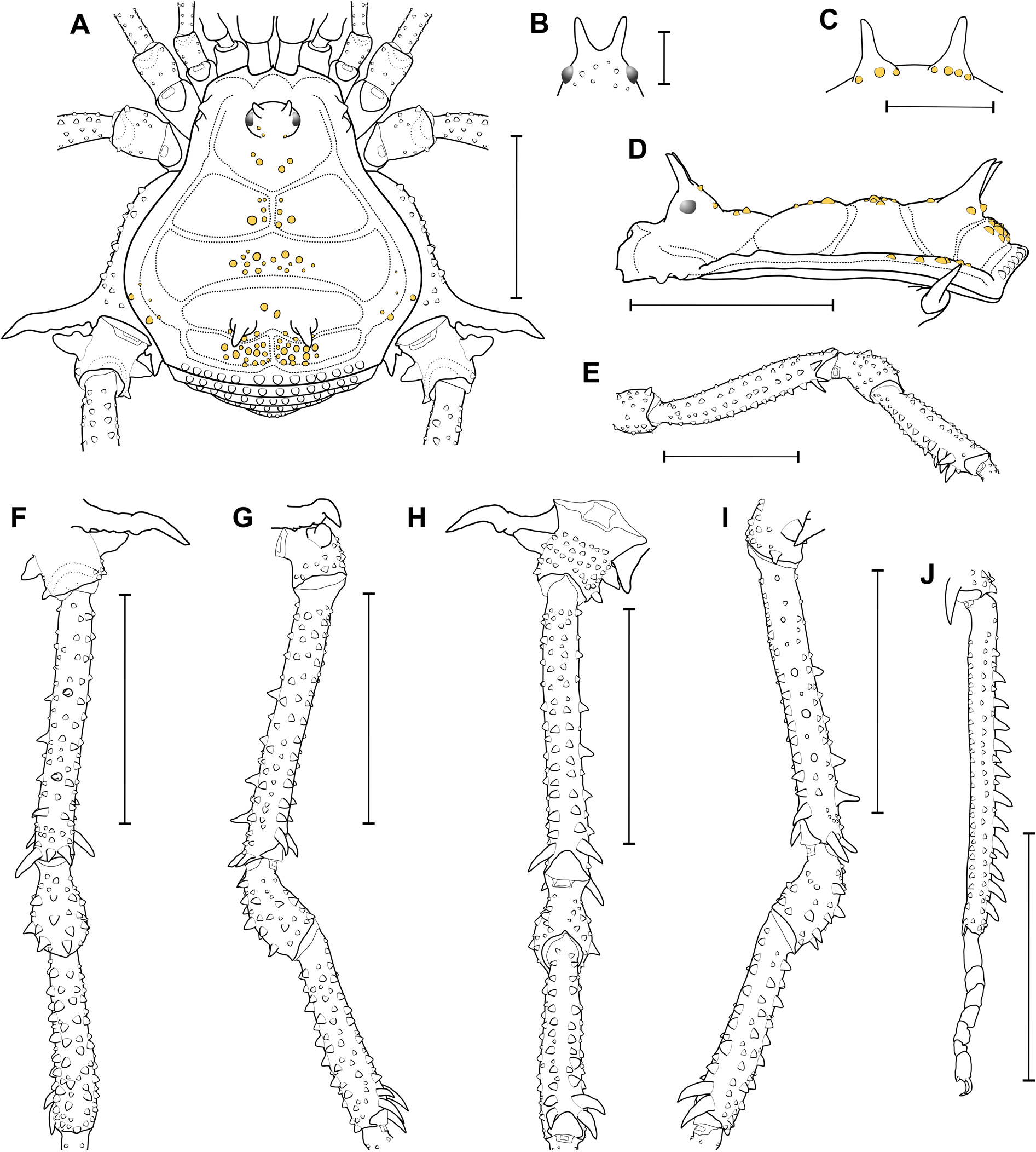
Lacronia serripes can be differentiated from the other species of the genus by the following combination of characters: 1) mesotergum areas I–IV with light-colored tubercles contrasting with its background (without areolated spots surrounding the tubercles) ( Figs 16A, C View Fig , 17A, D View Fig ); 2) mesotergum area II with two paramedian pairs of conspicuous tubercles ( Fig. 17A View Fig ); 3) Ti III proventral face with a comb of three spines (iII) on the distal third, the larger ones touching each others’ tip ( Fig. 17E View Fig ); 4) Tr IV prolateral proximal/central portion with an isosceles triangle-shaped apophysis with a prodorsal protuberance ( Fig. 17A, G View Fig ); 5) Tr IV prodorsal and prolateral distal portions only with ordinary tubercles ( Fig. 16F– G View Fig ); 6) Mt IV with a dorsal row of conical spines (absent only on the proximal a fifth) ( Fig. 17J View Fig ); 7) subapical portion of the stylus covered by tiny spines on lateral faces ( Fig. 18B, D View Fig ); 8) sub-apical portion of the stylus swollen ( Fig. 18B, D View Fig ); 9) ventral process flabellum scallop-shaped, straight (not bent ventrad) ( Fig. 18A–D View Fig );
Type material
Holotype BRAZIL • ♂; State of São Paulo, Ilha dos Alcatrazes ; MZSP 550 View Materials (examined).
Additional material examined
BRAZIL – State of Rio de Janeiro • 7 ♂♂, 2 ♀♀, 1 juv; Angra dos Reis, Ilha Grande ; 14 Dec. 1985; R.L.C. Baptista leg.; MNRJ 6100ꜝ . – State of São Paulo • 1 ♂; Ilhabela, Ilha São Sebastião ; 8–10 Feb. 1948; H. Urban leg.; MNRJ 9286ꜝ • 3 ♂♂, 1 ♀; Salesópolis ; MZSP 9973 View Materials • 10 ♂♂, 8 ♀♀; Estação Biológica de Boracéia ; 26 Nov. 1968; E.X. Rabello leg.; MZSP 14343 View Materials • 3 ♂♂, 3 ♀♀; same collection data as for preceding; 6 Nov. 1968; MZSP 18308ꜝ • 1 ♀; São Sebastião, Ilha dos Alcatrazes ; Oct. 1921; MZSP 13590 View Materials .
Redescription
Male
MZSP 14343 for the external body illustrations and description; MZSP 9973 for genitalic illustrations. MEASUREMENTS. DS: CW 2.0, CL 1.5, AW 3.7, AL 2.4; legs I–IV measurements in the Table 6 View Table 6 ; right / left tarsal (distitarsal) counts: 5(3) / 4(2) - 11(3) / 11(3) - 7 / 7 - 7 / 7.
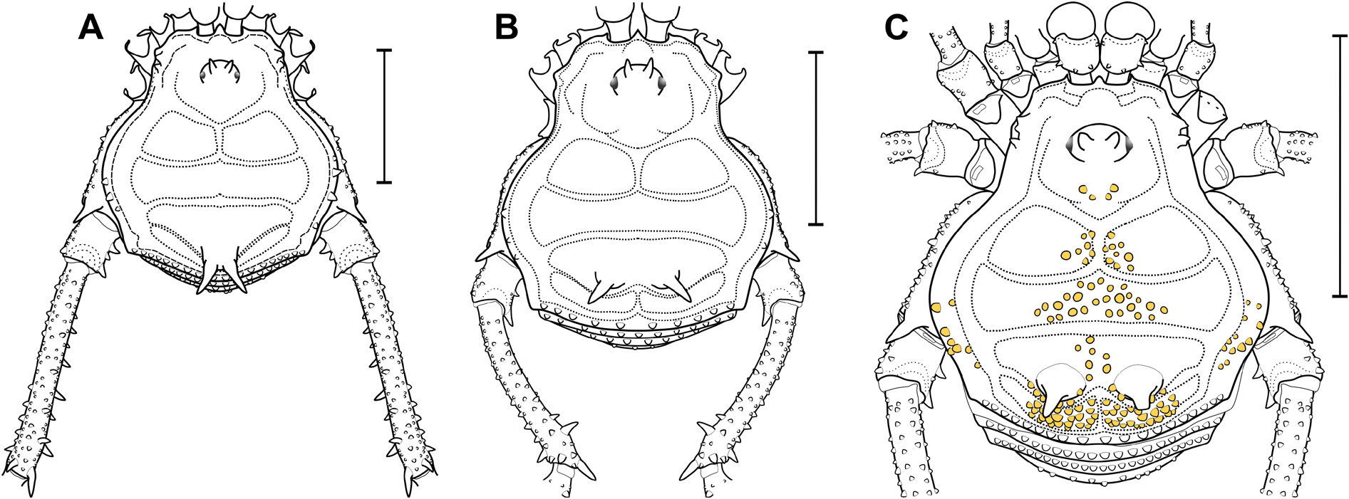
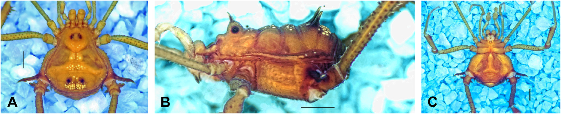
DORSUM. DS gamma-pyriform, longer than wide, with lateral borders of the AS convex, widest and thickest at mesotergum area II, with a slightly convex posterior border ( Figs 16A View Fig , 17A View Fig ). DS anterior border with a pair of shallow cheliceral sockets divided by a smallcentral projection ( Fig. 17A View Fig ). Carapace with one or two pair(s) of prominent tubercles posterior ro ocularium ( Figs 16A View Fig , 17A View Fig ). Ocularium elliptical (in dorsal view), high (ca 3 × the eye diameter), almost forming a 90º angle to the DS, placed in the anterior portion of the carapace ( Figs 16A–B View Fig , 17A–B, D View Fig ). Ocularium with a pair of divergent spines (ca 3 × the eye diameter), slightly inclined frontwards ( Figs 16A–B View Fig , 17A–B, D View Fig ). Mesotergum divided in four clearly defined areas ( Fig. 17A, D View Fig ). AS lateral borders with two yellowish prominent tubercles contrasting with the background ( Figs 16A–B View Fig , 17A View Fig ). Mesotergum areas I and IV divided into left and right halves by a longitudinal median groove ( Fig. 17A View Fig ). All areas tuberculate on the center, with all yellowish tubercles contrasting with the background ( Figs 16A–B View Fig , 17A, D View Fig ). Mesotergum area I with a pair of prominent tubercles ( Figs 16A View Fig , 17A View Fig ). Mesotergum area II with two pairs of prominent tubercles ( Figs 16A View Fig , 17A View Fig ). Mesotergum area III with a pair of paramedian outstanding spines (ca 1.5 × the ocularium spines) ( Fig. 17A, C–D View Fig ). Mesotergum area IV with transversal rows of four to five prominent tubercles ( Fig. 17A View Fig ). DS posterior border with a transversal row of prominent tubercles (same color as the background) ( Figs 16A–B View Fig , 17A, D View Fig ). Free tergites III with a transversal row of prominent tubercles ( Fig. 17A View Fig ). Free tergite III with a transversal row of ordinary tubercles ( Fig. 17A View Fig ). Anal operculum covered by ordinary tubercles.
VENTER. Cx I–III sub-parallel to each other ( Fig. 16C View Fig ), each with ventral longitudinal rows of setiferous tubercles (Cx I rows with higher and sharper tubercles than the others). Cx II and III with a retro-ventral distal row of acuminate tubercles. Cx IV much larger than the others, directed obliquely ( Figs 16C View Fig , 17A View Fig ). Intercoxal bridges well-marked ( Fig. 16C View Fig ). Stigmatic area Y-inverted-shaped, clearly sunken in relation to the Cx IV distal part ( Fig. 16C View Fig ). Cx IV covered by ordinary tubercles. Cx IV posterior border and stigmatic area each with a transversal row of ordinary tubercles. Stigmata visible ( Fig. 16C View Fig ). Free sternites each with a transverse row of ordinary tubercles.
CHELICERA. Basichelicerite elongate, bulla well-marked, with one setiferous tubercle on the ectal margin; hand not swollen.
PEDIPALPS. Tr with two geminate ventral setiferous tubercles. Fe with a ventral basal and a mesal apical setiferous tubercle. Pa unarmed. Ti with two rows (ventro-mesal and ventro-ectal) of four spines (IiIi). Ta with two rows of spies: three (iII) ventro-mesal and three (III) ventro-ectal.
LEGS. All the unmentioned podomeres are unarmed or without relevant armature. Tr I–III each with several ventral tubercles. Fe I sub-straight; Fe II straight; Fe III sinuous ( Fig. 17E View Fig ). Fe and Ti I–III all faces with rows of small tubercles ( Fig. 17E View Fig ). Fe II–III with an apical retro-dorsal spur. Fe III with an apical prodorsal spur (reduced when compared to the retro-dorsal spur). Fe III and Ti III with proventral and retro-ventral rows of small acuminate tubercles with spines on the distal third [four proventral (iiII); three retro-ventral (iII), the largest ones touching each other’s tip] ( Fig. 17E View Fig ). Pa I–III covered dorsally by acuminated tubercles ( Fig. 17E View Fig ). Ti III mace-shaped ( Fig. 17E View Fig ). Cx IV reaching as far as mesotergum areas III–IV ( Fig. 17A View Fig ). Cx IV tuberculate between prodorsal and ventral faces ( Fig. 17A View Fig ). Cx IV with a thick prolateral distal conical apophysis, posteriorly crenated, with apical portion slightly curved backward ( Fig. 17A, F–H View Fig ). Cx IV with a retro-lateral distal spiniform apophysis, fused with a small secondary branch ( Fig. 17A, H–I View Fig ). Tr IV square-shaped in dorsal view ( Fig. 17A, F, H View Fig ). Tr IV with a prolateral proximal/central apophysis that is isosceles triangle-shaped, with a secondary prolateral medial protuberance ( Fig. 17A, F–H View Fig ). Tr IV with a prolateral distal sub-conical tubercle ( Fig. 17A, F–G View Fig ). Tr IV with a retro-lateral proximal conical apophysis ( Fig. 17A, F, H–I View Fig ). Tr IV with a retro-lateral distal spine ( Fig. 17A, F, H–I View Fig ). Tr IV ventral face tuberculate ( Fig. 17G–I View Fig ). Fe IV almost straight (in dorsal view), slightly arched on the central portion towards dorsal face ( Fig. 17F–I View Fig ). Fe IV with all the faces covered by longitudinal rows of acuminated tubercles (largest in size on the proventral and retroventral distal half) ( Fig. 17G–I View Fig ). Fe IV with three dorsal conical spines (two on central portion, one on apex) ( Fig. 17F–G, I View Fig ). Fe IV retro-lateral face with two to three central spines (iIi) ( Fig. 17F, H–I View Fig ). Fe IV retro-dorsal face with one conical spine (I) on the distal ¼ ( Fig. 17F, I View Fig ). Fe IV prodorsal and retrodorsal faces with an outstanding spur on distal apex ( Fig. 17F–G, I View Fig ). Fe IV proventral and retro-ventral faces with an outstanding spine on distal portion ( Fig. 17F–I View Fig ). Pa IV dorsally covered by spines and acuminated prominent tubercles ( Fig. 17F–G, I View Fig ). Pa IV proventral face with a row of four spines (iiii) ( Fig. 17G–H View Fig ). Pa IV retro-ventral face with two spines (ii) ( Fig. 17H–I View Fig ). Ti IV with all the faces (except ventral) covered by longitudinal rows of acuminated tubercles (retro-lateral and retro-dorsal faces with largest ones) ( Fig. 17F–I View Fig ). Ti IV prolateral face with two spines (iI) on the distal ¼ ( Fig. 17F–H View Fig ). Ti IV retro-lateral face with two outstanding spines (II) on the distal ¼ ( Fig. 17F, H–I View Fig ). Mt IV with a dorsal row of spines (absent on the proximal a fifth) ( Fig. 17J View Fig ).
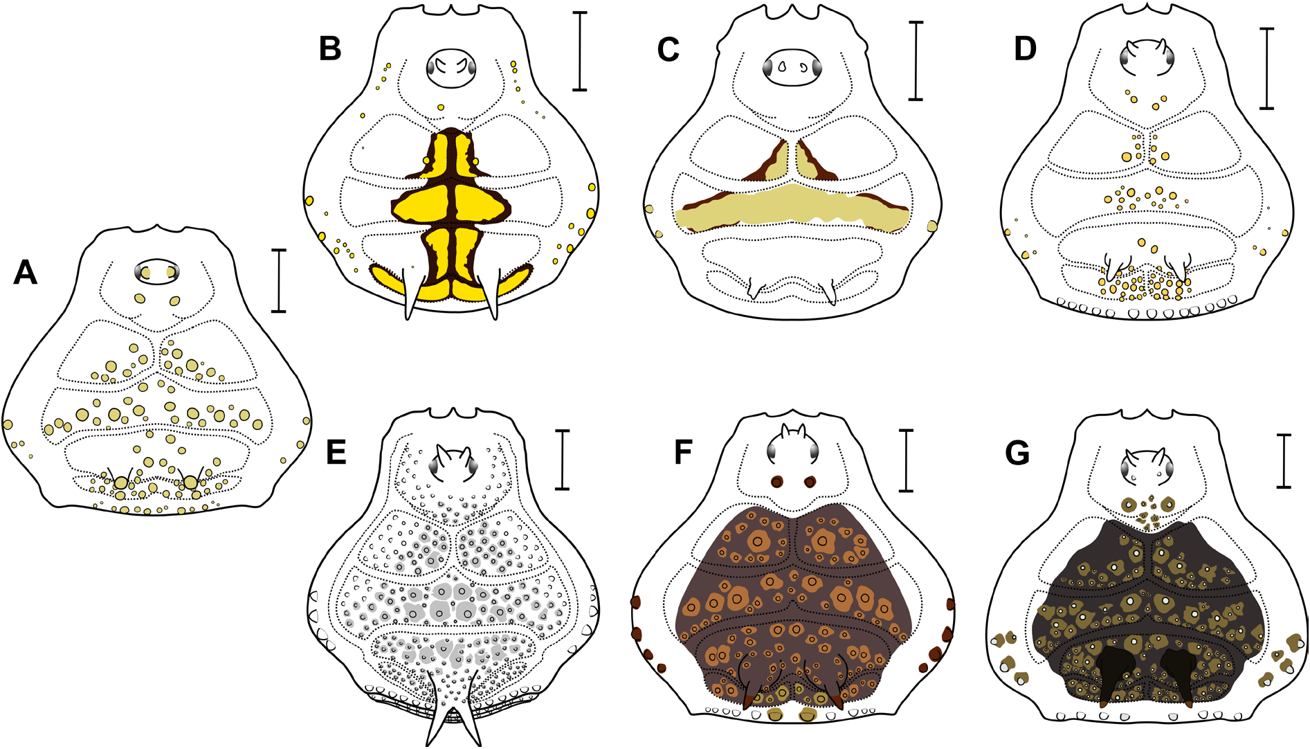
COLOR (in alcohol) ( Fig. 16A–C View Fig ). Ocularium, DS background and Cx IV Strong Orange (50). Spines on ocularium Deep Brown (56). Tubercles on DS Brilliant Yellow (83). Spines of the mesotergum area III and Cx IV prodorsal distal apophysis Dark Violet (212). Ch and Pp background Strong Yellow (84). Legs –III background Moderate Greenish Yellow (102). Tr IV background Dark Orange Yellow (72) with apophyses Dark Grayish Red (20). Fe–Mt IV background Dark Yellow (88). Tubercles and spines on legs IIV Deep Brown (56).
MALE GENITALIA. VP slightly divided into a distal trapezoidal half (widest at base, with latero-apical flaps), and a proximal half elliptical ( Fig. 18A, C View Fig ). VP ventral surface entirely covered with microsetae of type 1. All macrosetae inserted on the ventro-laterals of VP. MS A1–A3 cylindrical, thick, and acuminate, forming a triangle in lateral view (A1 on the basal portion of the distal part, A2–A3 on the proximal part, A2 dorsalmost) ( Fig. 18A–B, D View Fig ). MS B1 small, inserted ventrally, ventral and close to A3 ( Fig. 18B–D View Fig ). MS C1–C3 longer than the MS A, forming a row (C3 dorsalmost) on the distal ¼ of VP ( Fig. 18A–D View Fig ). MS D1 small, close to C3 ( Fig. 18A–D View Fig ). MS E1–E2 small, on the laterodistal flange of VP – E1 more ventrally placed between MS C1–C2, E2 same, below C3 ( Fig. 18D View Fig ). Glans sac arising
from the middle bulge on the podium, not extended as a dorsal process ( Fig. 18A–B, D View Fig ). Stylus and its ventral process axis fused basally, forming a prominent trapezoidal-shaped pedestal ( Fig. 18A–B, D View Fig ). Stylus cylindrical, almost straight, inserted on the pedestal forming a 25º angle, without conspicuous head (slightly swollen at subapical portion), with subdistal tiny spines ( Fig. 18A–B, D View Fig ). Ventral process is ¾ of the stylus length, slightly bent dorsad, with an apical flabellum ( Fig. 18B, D View Fig ). Flabellum not curved ventrally, scallop-shaped with serrulations and, with approximately 50% of the ventral process stem length ( Fig. 18B–D View Fig ).
Female (MZSP 9973) ( Fig. 9C View Fig )
DS, measurements: CW 2.2, CL 1.6, AW 3.9, AL 2.6; legs I–IV measurements in the Table 7 View Table 7 ; right / left tarsal (distitarsal) counts: 5(3) / 5(3) - 10(3) / 10(3) - 7 / 7 - 7 / 7.
DS gamma type. Cx IV narrower than in the males, with the prodorsal distal apophysis reduced to a single spine and without the retro-lateral distal apophysis. Tr IV prolateral face with a row of acuminated tubercles (without apophyses). Tr IV retro-lateral face with a prominent proximal spine and a distal one.
Fe IV thinner than in the male and unarmed on the distal portion. Mt IV dorsally covered by ordinary tubercles.
Intraspecific variation
In the minor morph males (compared to major morph): DS narrower; Cx IV with reduced prolateral and retro-lateral distal apophyses; Fe IV thinner, with reduced armature size. It was not found intraspecific variation among the major morph males or among females.
Records
BRAZIL, state of São Paulo: [Ilhabela]: Ilha dos Búzios; Ilha da Vitória (H. Soares 1966).
Geographic distribution (new records with an asterisk)
BRAZIL: state of Rio de Janeiro: Angra dos Reis*. State of São Paulo: Ilhabela, São Sebastião, Salesópolis*.

No known copyright restrictions apply. See Agosti, D., Egloff, W., 2009. Taxonomic information exchange and copyright: the Plazi approach. BMC Research Notes 2009, 2:53 for further explanation.
|
Kingdom |
|
|
Phylum |
|
|
Class |
|
|
Order |
|
|
Family |
|
|
Genus |
Lacronia serripes (Mello-Leitão, 1923)
| Carvalho, Rafael N. & Kury, Adriano B. 2023 |
Lacronia serripes
| Kury & Orrico 2006: 148 |
| Kury 2003: 284 |
| Kury 2003: 30 |
| Strand 1942: 397 |
Luederwaldtia serripes
| Muñoz-Cuevas 1973: 226 |
| H. Soares 1966: 284 |
| Soares & Soares 1954: 270 |
| Soares B. A. M. 1946: 520 |
| Mello-Leitao C. F. 1932: 166 |
| Roewer C. F. 1929: 218 |