
Lutzomyia (Lutzomyia) renei (Martins, Falcão & Silva), Martins, Falcao & Silva
|
publication ID |
https://doi.org/ 10.11646/zootaxa.3999.4.9 |
|
publication LSID |
lsid:zoobank.org:pub:8F59A37D-B14D-404B-AA37-249B097DA99D |
|
DOI |
https://doi.org/10.5281/zenodo.6095555 |
|
persistent identifier |
https://treatment.plazi.org/id/7B550535-E450-FFB5-FF69-4868FD008F7B |
|
treatment provided by |
Plazi |
|
scientific name |
Lutzomyia (Lutzomyia) renei (Martins, Falcão & Silva) |
| status |
|
Lutzomyia (Lutzomyia) renei (Martins, Falcão & Silva) View in CoL
( Figs 1–4 View FIGURE 1 View FIGURE 2 View FIGURE 3 View FIGURE 4 )
Phlebotomus renei Martins, Falcão & Silva, 1957: 321 View in CoL . Type series: six males “cotypes”: Lapinha Cave, Lagoa Santa municipality, Minas Gerais state, Brazil, July/ September1957, F. R. Bastos coll. (Revista Brasileira de Malariologia e Doenças Tropicais); Sherlock 1957: 547 (description of female and immature forms); Coelho 1962: 102 (exp. Leishmania infection); Coelho & Falcão 1962: 220 (exp. Leishmania braziliensis infection); Sherlock & Pessôa 1964: 332 (sample methods).
Lutzomyia renei Barretto1962: 92 View in CoL (cat., comb.); Theodor 1965: 181 (cat., male fig.); Coelho et al. 1967a – d (exp. Leishmania infection); Forattini 1971: 100; 1973: 266 (cat., figs, tax.); Christensen et al. 1972: 55 (exp. Leishmania infection); Lewis et al. 1977: 325 (cat.); Martins et al. 1978: 25 (cat., distribution); Galati et al. 1985: 266 (tax. male); Killick-Kendrick 1986: 135 (cat., Leishmania infection); Gontijo et al. 1987: 445 (exp. Leishmania infection); Artemiev 1991: 73 (cat.); Young & Duncan 1994: 53 (cat., figs, tax., distribution); Galati & Nunes 1999; 280 (tax.); Alves et al. 2003: 121 (biological cycle); Galati 2003: 14 (cat., tax., distr.).
Diagnosis. Both sexes: Preapical papilla on flagellomeres I, II and III, ascoids without long posterior spur; external ascoid inserted on level more apical than the internal one. Newstead’s sensilla dispersed on third palpal segment, labial suture forming a fork. Male: gonostyle with five spines: two apical, the upper external one implanted on the apical third, the lower external one more basal than the internal and this latter implanted just before the middle of the gonostyle. The dorsal margin of the paramere slightly concave and presenting in its middle two bristles with hooked apex. Gonocoxite with a basal cluster (tuft) presenting four bristles, three fine and one semi-foliaceous, implanted directly on its surface. Female: cibarium with sclerotized complete arch and absence of strong sclerotization below the posterior teeth. Two pairs of posterior teeth and several anterior teeth lateralized. 8th tergum with two to five bristles and 10th sternite with three to five apical bristles. Spermathecae with rings of equivalent length; individual spermathecal duct more than four times the length of the spermatheca and a short common spermathecal duct. Cercus ca. 2.0 times longer than its width.
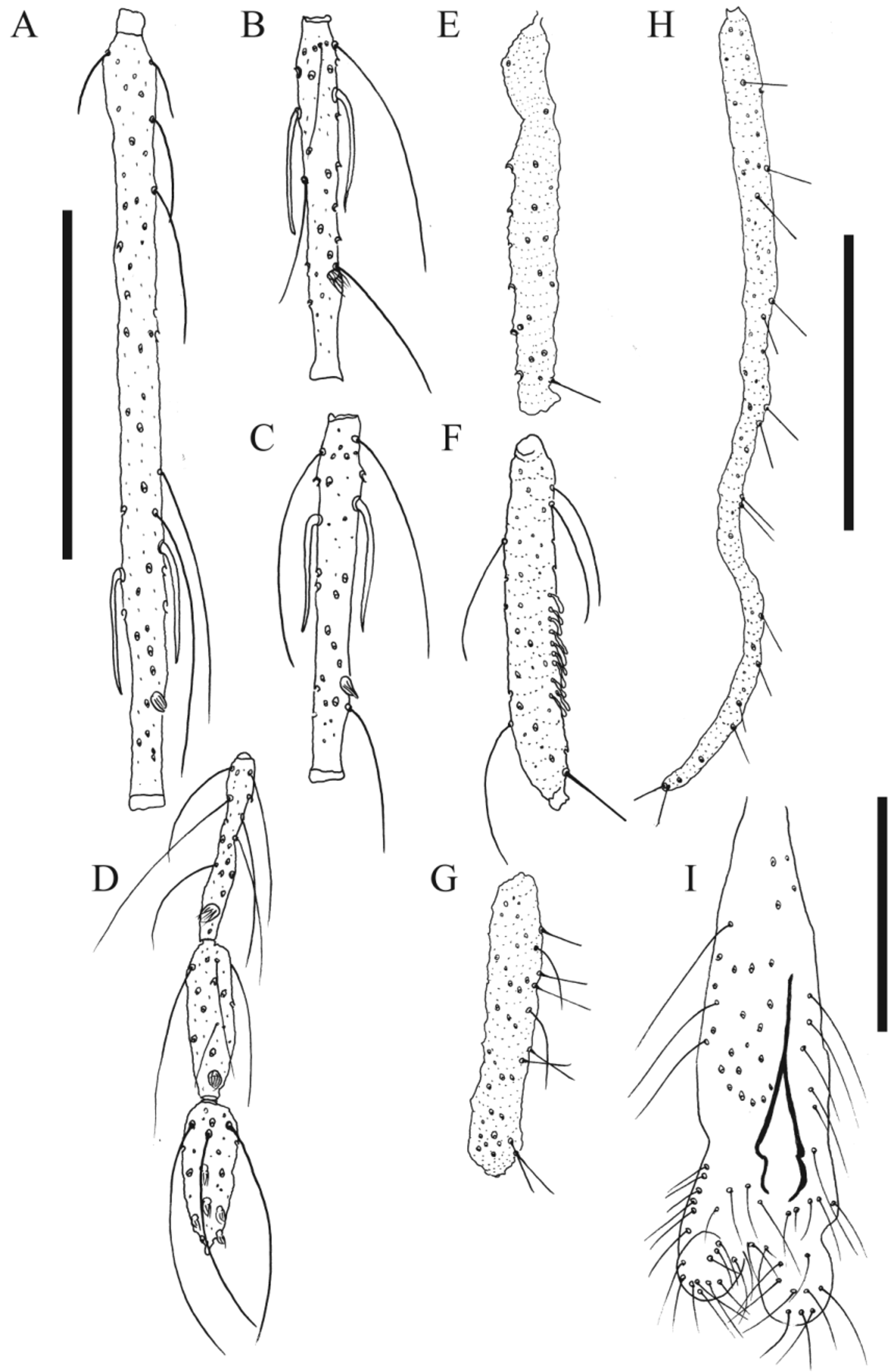
Redescription. Male. Head 380 (370) in length. Clypeus 156 (161 and 143) long. Eyes 174 (166) long. Cibarium without teeth. Labrum-epipharynx 230 (210 and 230). Antenna ( Figs 1 View FIGURE 1 A–D): flagellomere (F) length: F1 280 (280 and 280), FII 140 (140 and 130), FIII 140 (130), FXIII (60), FXIV (57). Only (FI–FVII) and (FI–FVIII) are present in the lectotype; paralectotypes: one of them only with FI and FII and the other with one full antenna (FI–FXIV). Ascoids: long anterior spur almost reaches the level of the preapical papilla and absence of posterior spur; external ascoid inserted on level more apical than the internal one ( Figs 1 View FIGURE 1 A–C); antennal formula FI–FXIII 2, FXIV 0. Papilla implanted in the preapical region on FI–FIII ( Figs 1 View FIGURE 1 A–C); presence of papillae on FXII–FXIV. Presence of simple setae on FII–FXIV. Palpus ( Figs 1 View FIGURE 1 E–H): palpal segment (P) length: PI 42 (42 and 42), PII 133 (143 and 138), PIII 166 (161 and 179), P IV 133 (127 and 122), PV 385 (307 and 374). Palpal formula: 1-(4-2)-3-5 and 1-4-2-3-5; PII without Newstead’s sensilla, PIII with several Newstead’s sensilla dispersed on the middle region ( Fig. 1 View FIGURE 1 F). Labial suture forming a fork ( Fig. 1 View FIGURE 1 I).
Cervix. Ventro-cervical sensilla present. Cervical sclerites bearing a pair of spiniform sensilla.
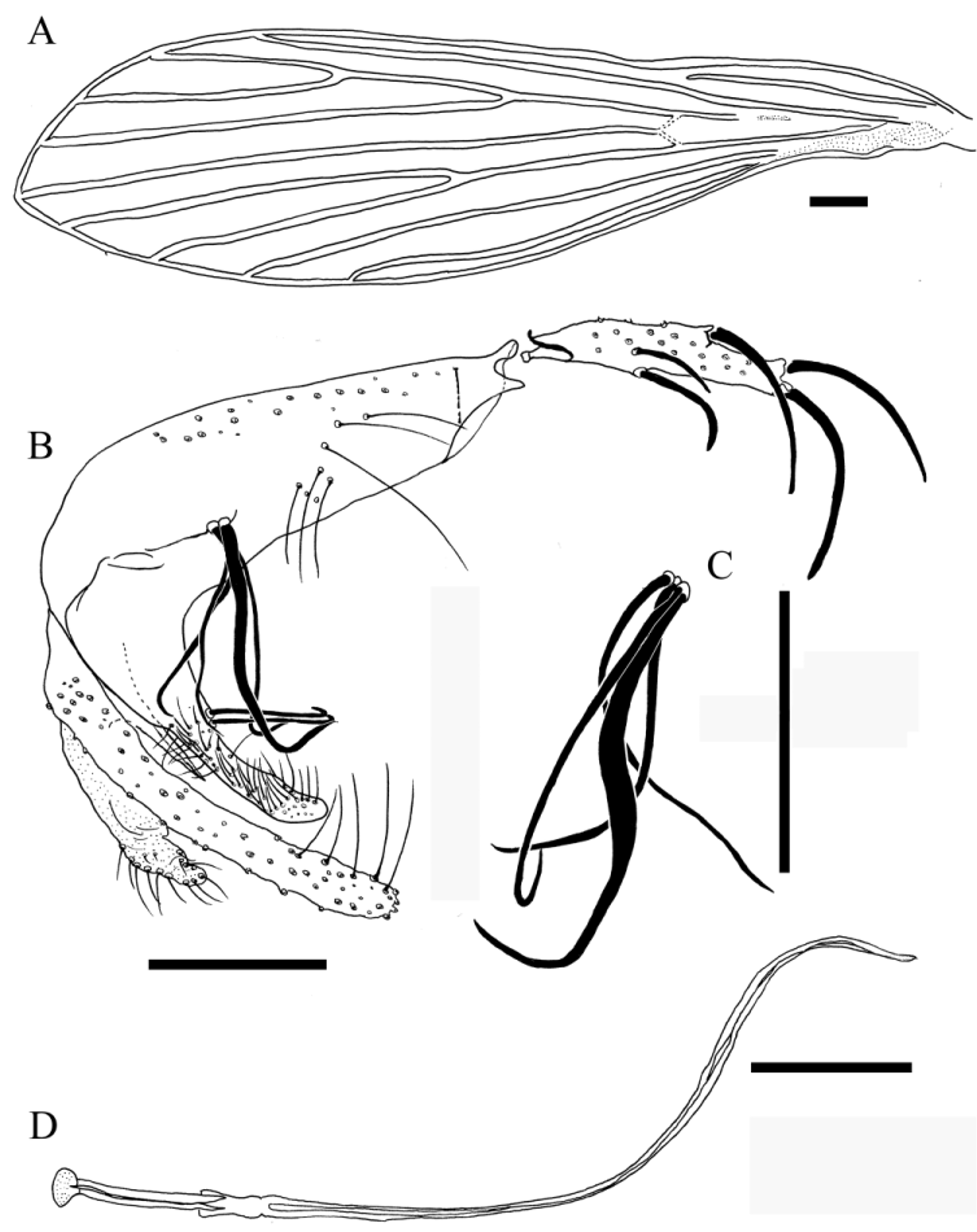
Thorax. Mesonotum 470 (500 and 490) in length. Mesonotum, pronotum, paratergite, anepisternum, metanotum and postnotum brown, pleura off-white. Four proepimeral setae and seven upper anepisternal setae. Setae on the anterior margin of the katepisternum absent. Wing ( Fig. 2 View FIGURE 2 A): 1,782 (1,822 and 1,861) long, 475 (495 and 495) wide; veins: R5 (2,455 and 2,495); alfa (752 and 772); beta (455 and 614); gamma (535 and 594); delta (376 and 297); pi (238 and 337). Legs: anterior, median, posterior: coxa: 310 (300 and 290), 300 (290 and 290), 300 (300 and 300); femur: (673 and 693), 772 (594), (752 and 792); tibia: (772 and 832), 1,247 (1,010), (1,109 and 1,228); tarsomere I: (455 and 495), 732 (594), (634 and 713). Sum of tarsomeres II+III+IV+V: (475 and 594), 713 (673) and (515 and 713).
Abdomen. 1,386 (1,485) long; presence of the tergal papillae on III–VII tergum. Terminalia ( Fig. 2 View FIGURE 2 B): gonocoxite 290 (290 and 290) long, 90 (90 and 100) wide, with basal cluster of four bristles implanted directly on its surface, three of them being fine and one semi-foliaceous ( Fig. 2 View FIGURE 2 B and C). Gonostylus 160 (150 and 150) long, with five spines: two apical, one internal, one upper external and one lower external. The apical and upper external spines are well developed, and the lower external and internal spines are finer. However, the lower external spine is thinner than the internal one. The upper external spine is implanted at a level equidistant between the apical spines and the lower external one; this latter being implanted slightly more basally than the internal spine. Paramere: dorsal margin 159 (161 and 153) long, slightly concave in its middle region, where two bristles with hooked apex are deployed; apical half covered with spiniform setae pointing toward the base of the terminalia; ventral margin 278 (213 and 200) in length, straight with the presence of a few spiniform setae on its middle region. Parameral sheath (aedeagus) conical. Epandrium (lateral lobes) 250 (250 and 260) long, 26 (26 and 26) wide and with rounded apex. Sperm pump (ejaculatory pump) 122 (130 and 127) long; aedeagus (genital filaments) with bevel shaped apex, 570 (540 and 550) long, 4.3 times longer than the ejaculatory apodeme + sperm sac ( Fig. 2 View FIGURE 2 D).
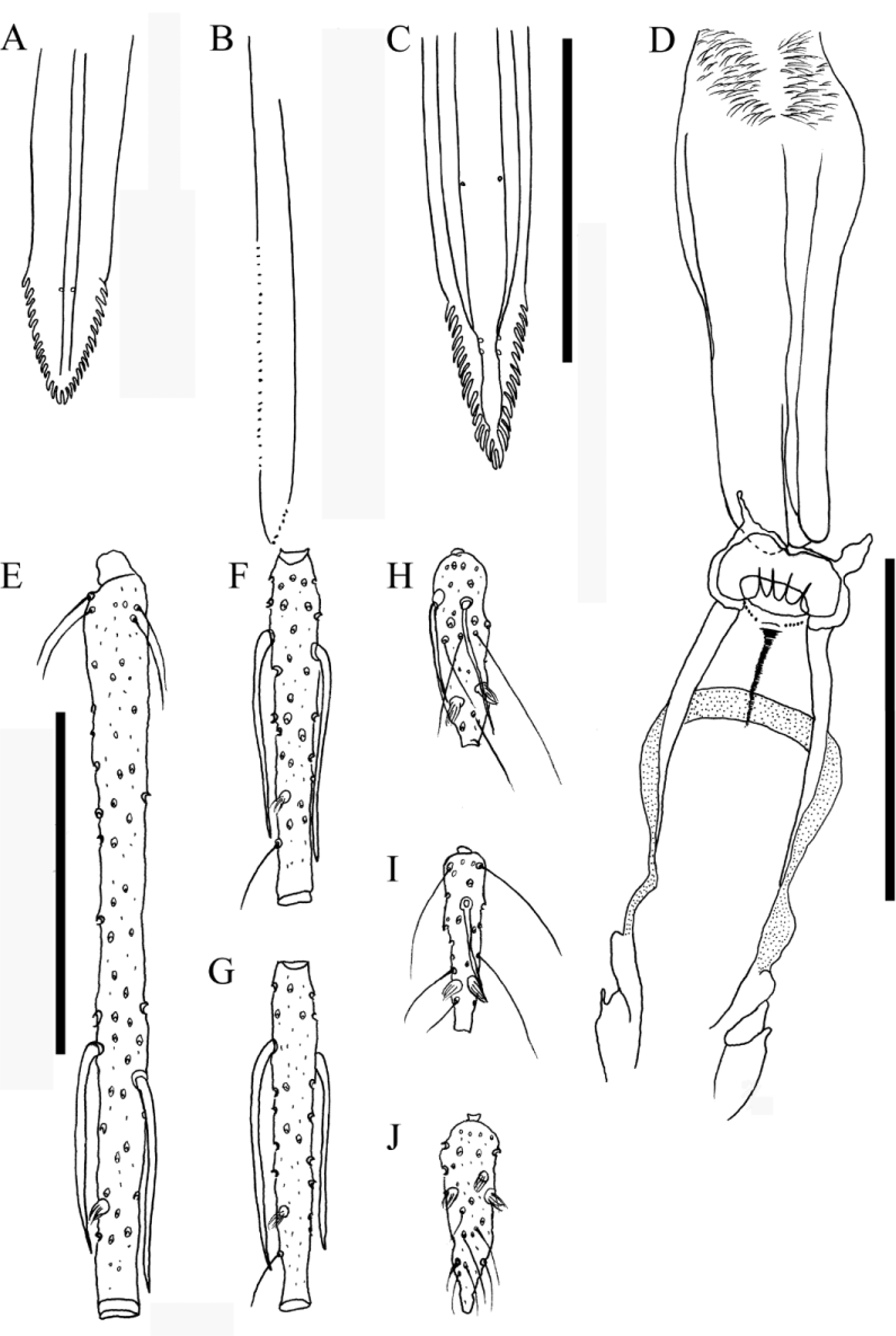
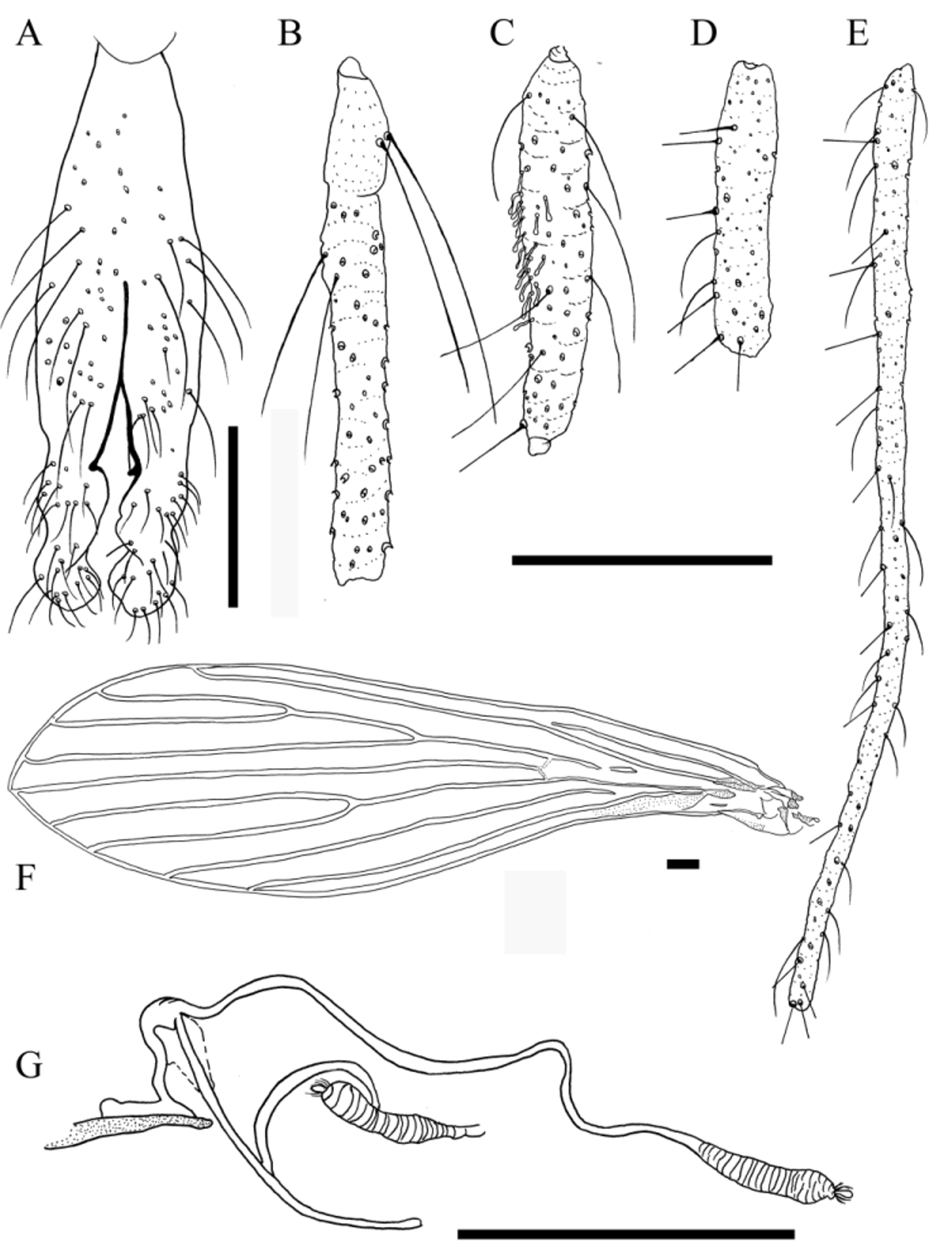
Female. Head 460 long, 440 wide; clypeus 156 long; eyes 221 long; Interocular distance 143 (drawings not presented). Hypopharynx with 28–30 apical teeth ( Fig. 3 View FIGURE 3 A). Lacinia of maxilla with six external teeth and 24 internal teeth ( Fig. 3 View FIGURE 3 B). Cibarium with four posterior teeth well-developed and 10 anterior teeth distributed in one transverse row ( Fig. 3 View FIGURE 3 D); sclerotized area short and triangular; sclerotized arch complete. Labrum-epipharynx 360 and with 30–32 apical teeth ( Fig. 3 View FIGURE 3 C). Antenna ( Figs 3 View FIGURE 3 E–J): flagellomere length: FI 270, FII 120, FIII 120, FXIII 70 and FXIV 70. One antenna was missing but the other was complete (FI–FXIV). Ascoids: absence of long posterior spur; anterior spur long reaching the level of preapical papilla in FI–FXIII; external ascoids implanted more apically than the internal one; antennal formula FI–FXIII 2, FXIV 0; preapical papilla on FI–FIII ( Figs 3 View FIGURE 3 E– G); no papilla on FIV–FXI; papillae on FXII–FXIV. Presence of simple setae on FII–FXIV. Labial suture forming a fork ( Fig. 4 View FIGURE 4 A). Palp ( Fig. 4 View FIGURE 4 B–E): palp length: PI 65, PII 187, PIII 200, PIV 143 and PV 468. Papal formula: 1-4- 2-3-5; Newstead’s sensilla absent on PII; PIII with Newstead’s sensilla dispersed on its middle region ( Fig.4 View FIGURE 4 C).
Cervix. Ventro-cervical sensilla present. Cervical sclerites bearing paired spiniform sensilla.
Thorax. Mesonotum 330 in length. Mesonotum, pronotum, paratergite, anepisternum, metanotum and postnotum brown, pleura off-white. Four proepimeral setae; seven upper anepisternal setae. Setae absent on the anterior region of the katepisternum. Wing ( Fig. 4 View FIGURE 4 F): 2,356 long, 653 wide; veins: R5 1,822; alfa 594; beta 376; gamma 535; delta 297; pi 198. Legs: anterior; median; posterior: coxa: 713; 673; 693; femur: 812; 812; 950; tibia: 950; 1,168; 1,465; tarsomere I: 594; 693; 832. Sum of tarsomeres II+III+IV+V: 733; 752; 871.
Abdomen. 1,782 long; 8th tergum with two to five bristles and 10th sternite with three to five apical bristles. Spermathecae 52 long and 13 wide, ringed with rings of equivalent length ( Fig. 4 View FIGURE 4 G): 52; common spermathecal duct 31 long and 10.4 wide; individual spermathecal ducts 174 long and 5.2 wide. The individual and the common spermathecal ducts are membranous and with smooth walls. Cercus ca. 2.0 times longer than wide.
Material examined. Type series of Lu. renei deposited in the “Coleção de Referência Nacional e Internacional de Flebotomíneos, Centro de Pesquisas René Rachou” ( CRNIF –CPqRR) with the slides numbered as follows: “cotype” n°1 collected on 10-III-1957 (1 ♂), “cotype” n°164 collected on 24-XI-1955 (1 ♂), “cotype” n°165 collected on 24-11-1955 (1 ♂) and n°1,087 (1 ♀). According to the original description, all the “cotypes” were deposited in the collection of “Instituto Nacional de Endemias Rurais”, Belo Horizonte municipality, Minas Gerais state, currently CPqRR-FIOCRUZ. Only three specimens are still deposited in that institution: the specimen collected on 10-III-1955 was designated lectotype, and the two specimens collected on 24-XI-1955 paralectotypes. The remaining three specimens, if found, should also be designated paralectotypes.
Distribution. BRAZIL: Martins et al. (1978, p.25). MINAS GERAIS: Uberlândia, Lemos et al. (2004, p.195); Varzelândia, Andrade et al. (2007, p.981); Parque Nacional Cavernas do Peruaçu, Barata et al. (2008, p.226); Diamantina, Barata & Apolinário (2012 , p.1017); Lassance, Carvalho et al. (2012, p.3). TOCANTINS: Arraias municipality previously belonging to the state of GOIÁS, Martins et al. (1978, p.25). MATO GROSSO DO SUL: Campo Grande, Oliveira et al. (2003, p.936); Silva et al. (2007, p.422).
Medical importance. Coelho & Falcão (1962) demonstrated under laboratory conditions that Lu. renei transmited Leishmania mexicana Garnham, 1962 , cited back then as Leishmania braziliensis by these authors. Later on, Coelho et al. (1967a – b) conducted studies of experimental infections of Leishmania sp., however, there is doubt as to whether the species used was Le. braziliensis or Le. mexicana ( Killick-Kendrick 1986). Gontijo et al. (1987) experimentally infected one male of Lu. renei with Leishmania sp. Currently there is no evidence implicating Lu. renei as a vector of Leishmania spp.
No known copyright restrictions apply. See Agosti, D., Egloff, W., 2009. Taxonomic information exchange and copyright: the Plazi approach. BMC Research Notes 2009, 2:53 for further explanation.
|
Kingdom |
|
|
Phylum |
|
|
Class |
|
|
Order |
|
|
Family |
|
|
Genus |
Lutzomyia (Lutzomyia) renei (Martins, Falcão & Silva)
| Sábio, Priscila B., De Andrade, Andrey J. & Galati, Eunice A. B. 2015 |
Lutzomyia renei
| Alves 2003: 121 |
| Galati 2003: 14 |
| Young 1994: 53 |
| Artemiev 1991: 73 |
| Gontijo 1987: 445 |
| Killick-Kendrick 1986: 135 |
| Galati 1985: 266 |
| Martins 1978: 25 |
| Lewis 1977: 325 |
| Christensen 1972: 55 |
| Forattini 1971: 100 |
| Theodor 1965: 181 |
| Barretto 1962: 92 |
Phlebotomus renei Martins, Falcão & Silva, 1957 : 321
| Sherlock 1964: 332 |
| Coelho 1962: 220 |
| Martins 1957: 321 |
| Sherlock 1957: 547 |