
Macrobiotus noongaris, Coughlan & Stec, 2019
|
publication ID |
https://doi.org/ 10.5852/ejt.2019.573 |
|
publication LSID |
lsid:zoobank.org:pub:7619772F-2300-442E-8950-69559172360E |
|
DOI |
https://doi.org/10.5281/zenodo.4548201 |
|
persistent identifier |
https://treatment.plazi.org/id/A7BF7FF6-451F-4382-A842-CAC1EF51E6B8 |
|
taxon LSID |
lsid:zoobank.org:act:A7BF7FF6-451F-4382-A842-CAC1EF51E6B8 |
|
treatment provided by |
Plazi |
|
scientific name |
Macrobiotus noongaris |
| status |
sp. nov. |
Macrobiotus noongaris View in CoL sp. nov.
urn:lsid:zoobank.org:act:A7BF7FF6-451F-4382-A842-CAC1EF51E6B8
Figs 1–7 View Fig View Fig View Fig View Fig View Fig View Fig View Fig
Etymology
The name refers to the indigenous Australians who live in the region where the new species was found. These are the Noongar peoples, 14 different but related language groups that occupied these lands before western settlement, including the modern city of Perth where the sample was collected. In their languages, the term Noongar means ‘a person of the southwest of Western Australia’.
Material examined
86 animals (including 31 simplex) and 57 eggs. Specimens mounted on microscope slides in Hoyer’s medium (72 animals + 47 eggs), fixed on SEM stubs (10+10) and processed for DNA sequencing (4+0).
Holotype
AUSTRALIA – Western Australia • ♀; Perth, Kings Park ; 31°57′30″ S, 115°350′09″ E; 46 m a.s.l.; moss on soil in an urban park; IZiBB AU.031.12.
Paratypes
AUSTRALIA – Western Australia • 62 paratypes; same collection data as for holotype; IZiBB AU.031.06 to AU.031.14 • 32 eggs; same collection data as for holotype; IZiBB AU.031.02–05 .
Description
Animals (measurements and statistics in Table 2 View Table 2 )
Body transparent in juveniles and white in adults but transparent after fixation in Hoyer’s medium ( Fig. 1A View Fig ). Eyes present in live animals as well as in specimens mounted in Hoyer’s medium. Small round and oval cuticular pores (0.3–0.8 μm in diameter), visible under both PCM and SEM, scattered randomly on entire body ( Fig. 1 View Fig B–C). Granulation present on all legs ( Fig. 2 View Fig A–F). A patch of clearly visible granulation present on external surface of legs I–III ( Fig. 2 View Fig A–B).A cuticular bulge/fold (pulvinus) present on internal surface of legs I–III, with a faint cuticular fold covered with faint granulation and paired muscles attachments just above the claws ( Fig. 2 View Fig C–D). Both structures are visible only if legs are fully extended and properly oriented on slide (particularly in the case of the pulvinus and cuticular fold). Granulation on legs IV always clearly visible and consists of a single large granulation patch on each leg ( Fig. 2 View Fig E–F).
Claws stout, of hufelandi type ( Fig. 3 View Fig A–D). Primary branches with distinct accessory points, a common tract, and with an evident stalk connecting claw to lunula ( Fig. 3 View Fig A–D). Lunulae I–III smooth ( Fig. 3A, C View Fig ), whereas lunulae IV clearly dentate ( Fig. 3B, D View Fig ). Cuticular bars under claws absent. Double muscle attachments faintly marked under PCM but clearly visible under SEM ( Fig. 3A, C View Fig ).
Mouth antero-ventral followed by ten peribuccal lamellae and a circular sensory lobe ( Figs 4A View Fig , 5A View Fig ). Bucco-pharyngeal apparatus of Macrobiotus type ( Fig. 4A View Fig ). Under PCM, oral cavity armature of the patagonicus type, i.e., with only 2 nd and 3 rd bands of teeth visible ( Fig. 4 View Fig B–C). However, in SEM all three bands of teeth visible, with first band being situated at base of peribuccal lamellae and composed of a single row of small fused cone-shaped teeth connected to form a continuous, slightly serrated ring ridge around oral cavity ( Fig. 5 View Fig B–C). Second band of teeth situated between ring fold and third band of teeth and comprises 3–6 rows of small cone-shaped teeth ( Figs 4 View Fig B–C, 5B–C). Teeth of third band located within posterior portion of oral cavity, between second band of teeth and buccal tube opening ( Figs 4 View Fig B–C, 5B–C). Third band of teeth discontinuous and divided into dorsal and ventral portions. Under PCM, dorsal teeth appear as three distinct transverse ridges, whereas ventral teeth appear as two separate lateral transverse ridges and a median tooth ( Fig. 4 View Fig B–C). In SEM, both dorsal and ventral teeth also clearly distinct ( Fig. 5 View Fig B–C). Under SEM, margins of medio-dorsal tooth slightly serrated ( Fig. 5B View Fig ), whereas the medio-ventral tooth slightly anterior to lateral teeth ( Fig. 5C View Fig ). Pharyngeal bulb spherical, with triangular apophyses, two rod-shaped macroplacoids and a small triangular microplacoid ( Fig. 4A View Fig , D–E). Macroplacoid length sequence 2 <1. First macroplacoid exhibits central constriction, whereas second macroplacoid sub-terminally constricted ( Fig. 4A View Fig , D–E).
Eggs (measurements and statistics in Table 3 View Table 3 )
Laid freely, white, spherical or slightly ovoid ( Fig. 6A View Fig ). Surface between processes is of the hufelandi type, i.e., covered with a reticulum ( Figs 6E View Fig , 7 View Fig B–D, F). Meshes of reticulum small (0.1–0.6 µm) and rounded, regular in size and with blurred rims in PCM ( Fig. 6E View Fig ), irregular in size and with thick borders in SEM (meshes in SEM appear as pores; Figs 7 View Fig B–D, F). Interbasal meshes larger than peribasal meshes, but peribasal meshes do not form rings around process bases ( Figs 6E View Fig , 7 View Fig B–D, F). Eggs have 22–30 processes on circumference, 26 on average ( Fig. 6A View Fig ). Processes are of inverted goblet shape, with slightly concave trunks and concave terminal discs ( Figs 6 View Fig C–E, 7B–E). Terminal discs are round and strongly serrated ( Fig. 7 View Fig C–E). Each terminal disc has a distinct concave central area which may contain some scattered granulation within, which is also always present on the margin (visible only under SEM; Fig. 7 View Fig C–E).
Reproduction
The new species is dioecious. No spermathecae filled with sperm have been found in gravid females on the freshly prepared slides. However, the testis in males, filled with spermatozoa, is clearly visible under PCM up to 24 hours after mounting in Hoyer’s medium ( Fig. 6F View Fig ). The new species does not exhibit male secondary sexual dimorphism traits such as lateral gibbosities on legs IV.
DNA sequences
We obtained sequences for all four of the above mentioned DNA markers. All sequenced fragments were represented by single haplotypes except the ITS-2, in which two distinct haplotypes were present:
The 18S rRNA sequence (GenBank: MK737069 View Materials ), 1010 bp long.
The 28S rRNA sequence (GenBank: MK737063 View Materials ), 786 bp long.
The ITS-2 haplotype 1 sequence (GenBank: MK737065 View Materials ), 418 bp long.
The ITS-2 haplotype 2 sequence (GenBank: MK737066 View Materials ), 418 bp long.
The COI sequence (GenBank: MK737919 View Materials ), 658 bp long.
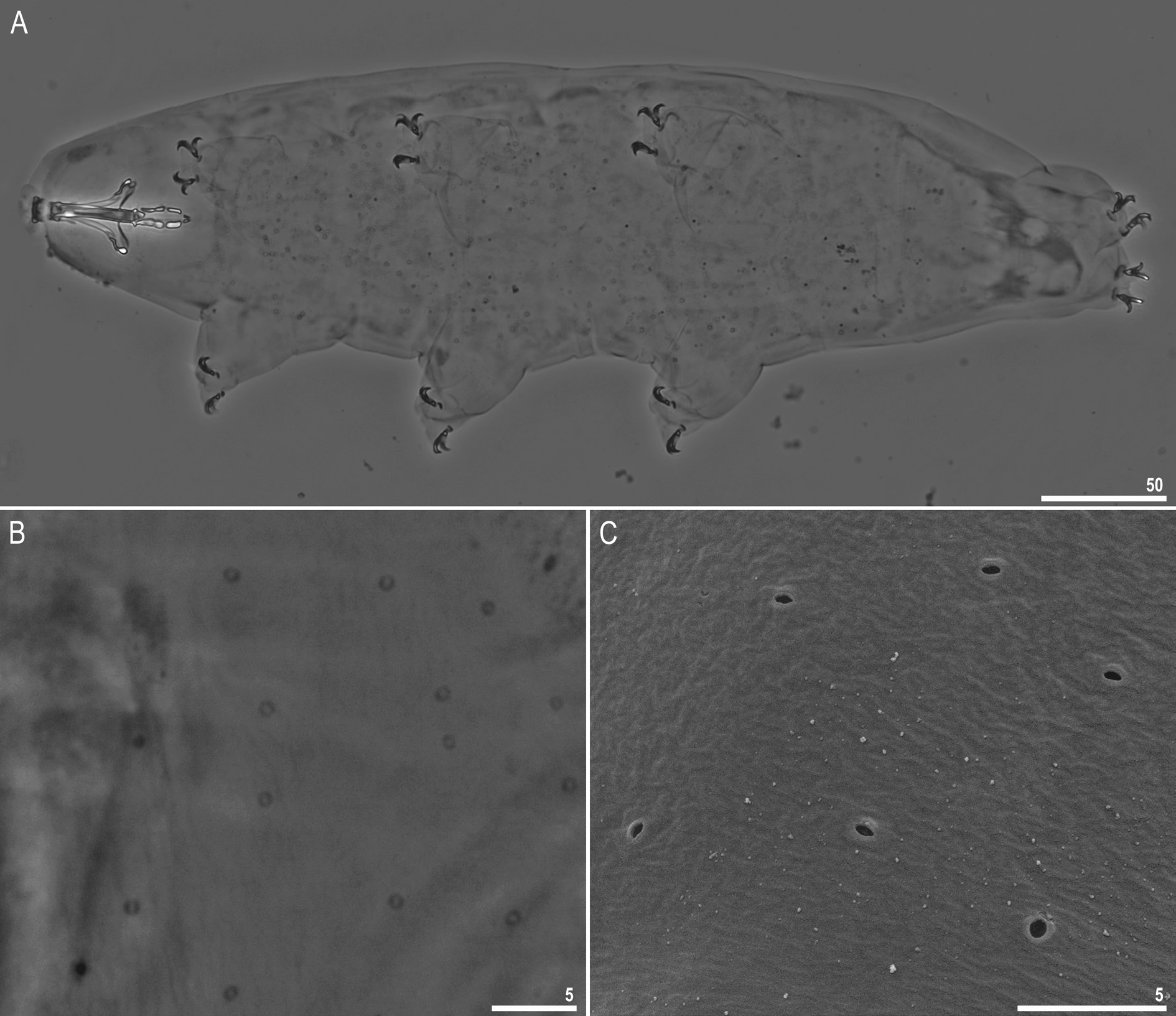
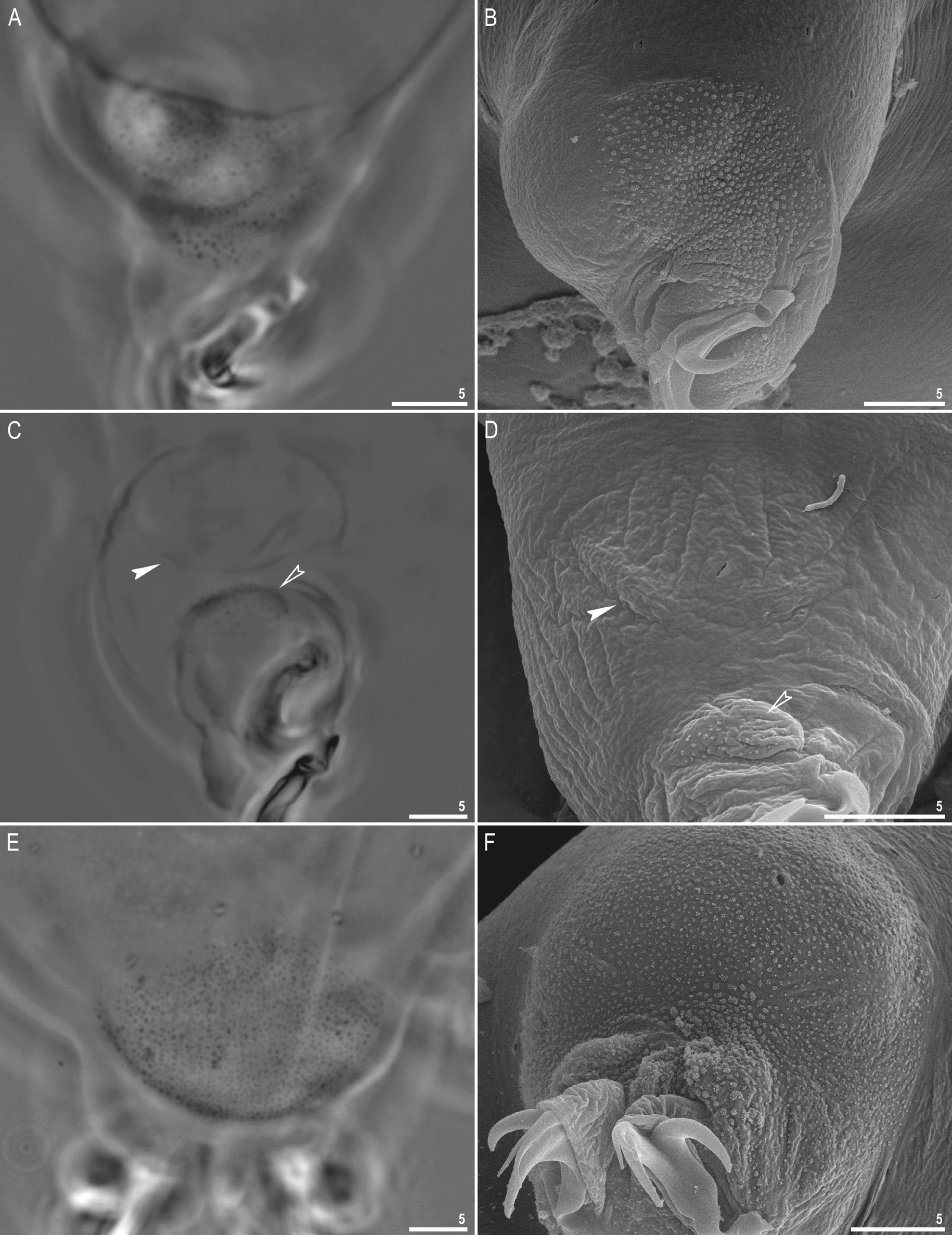
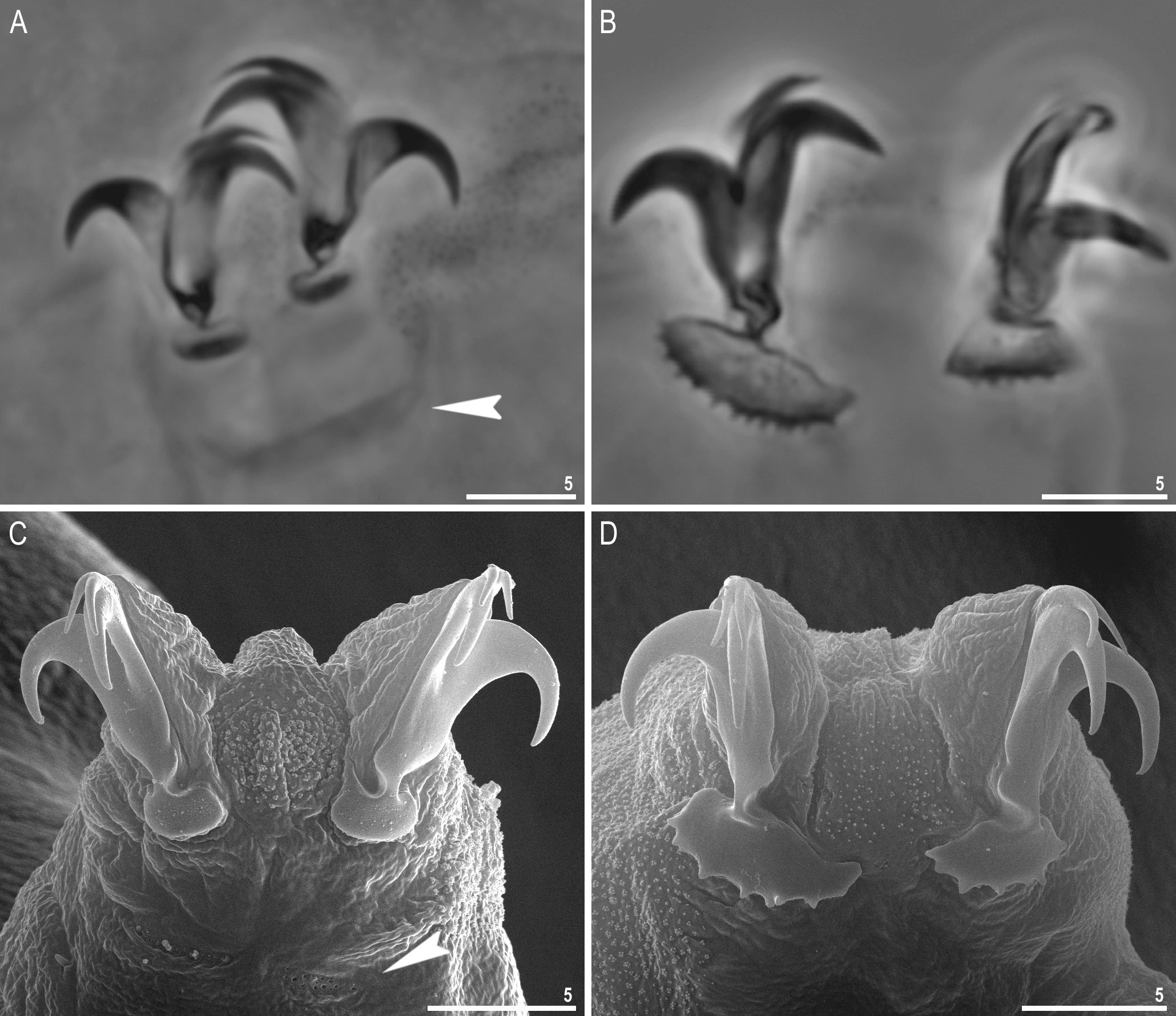
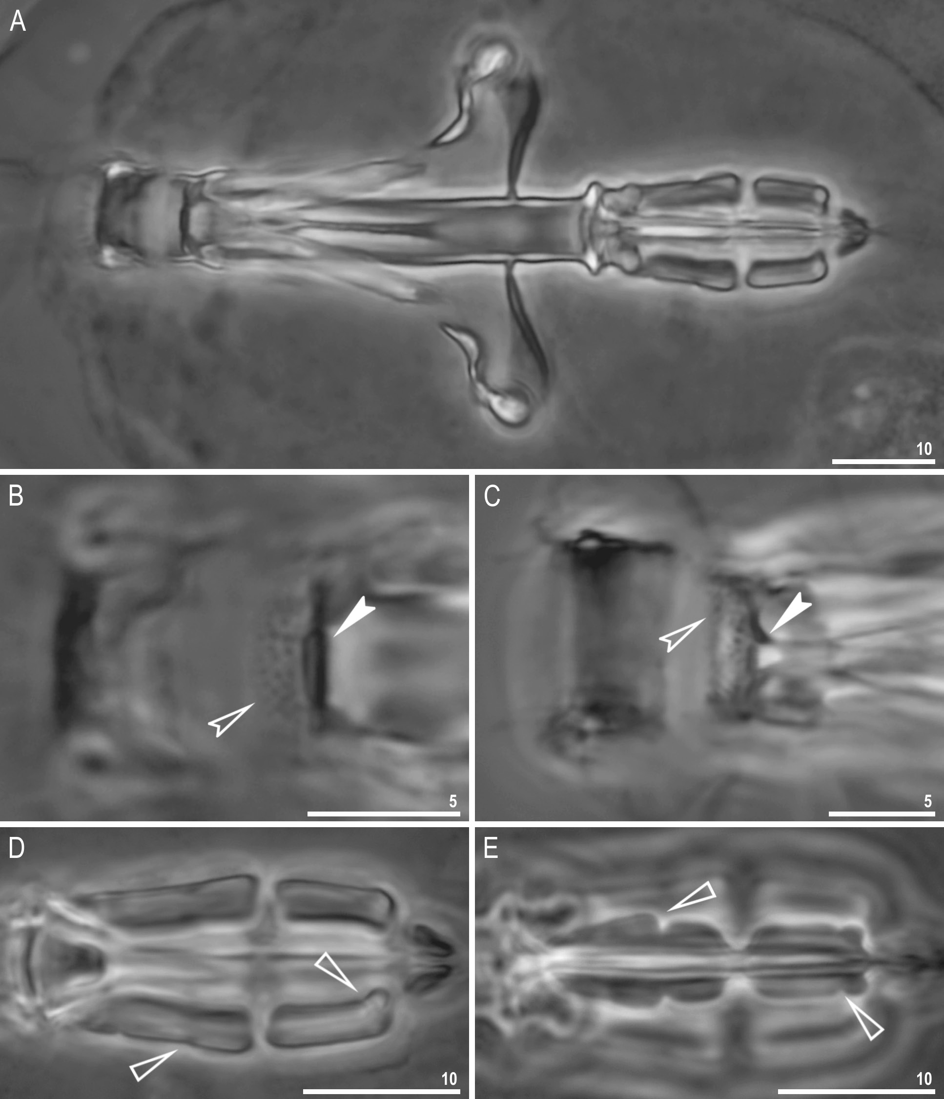

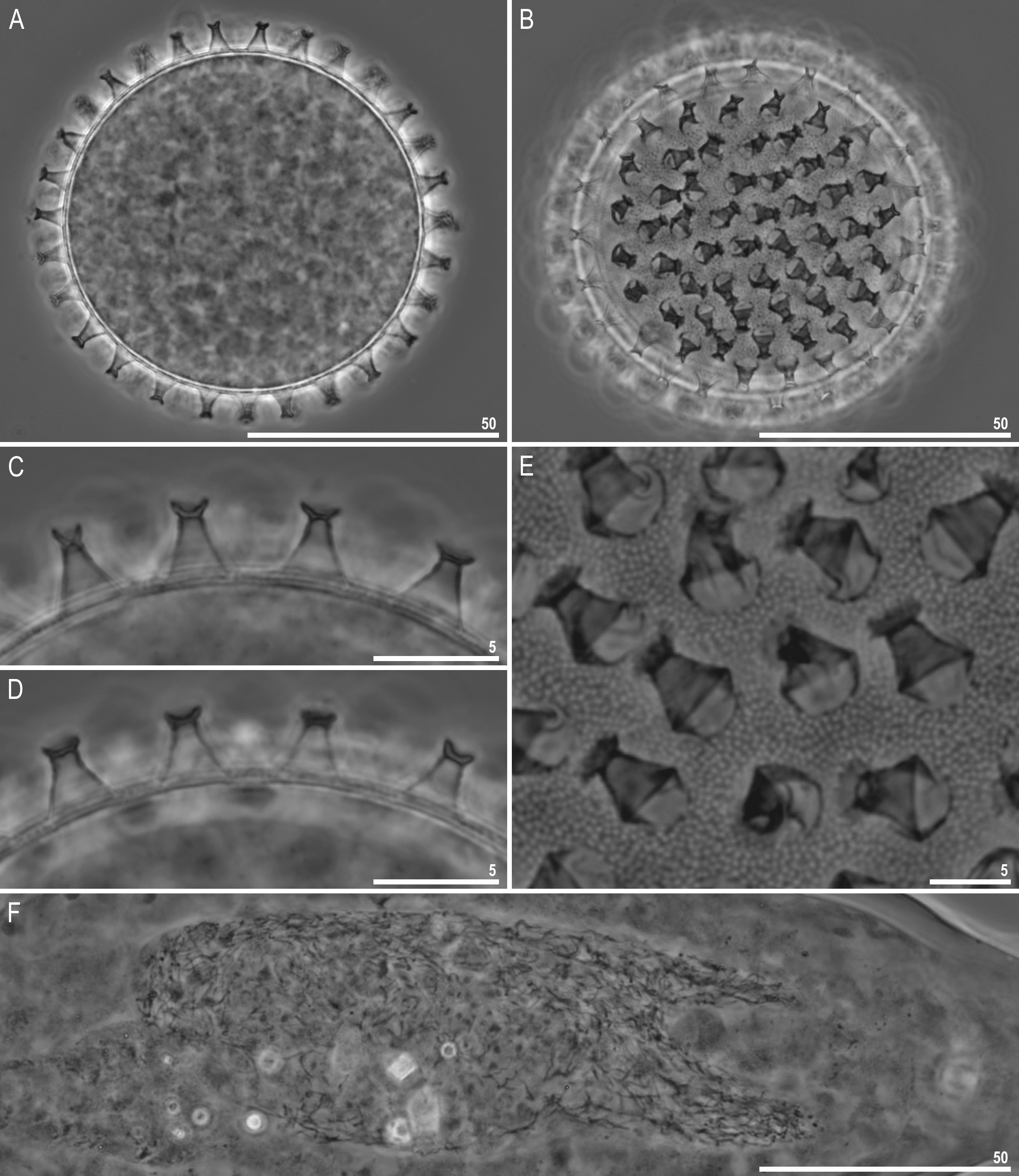
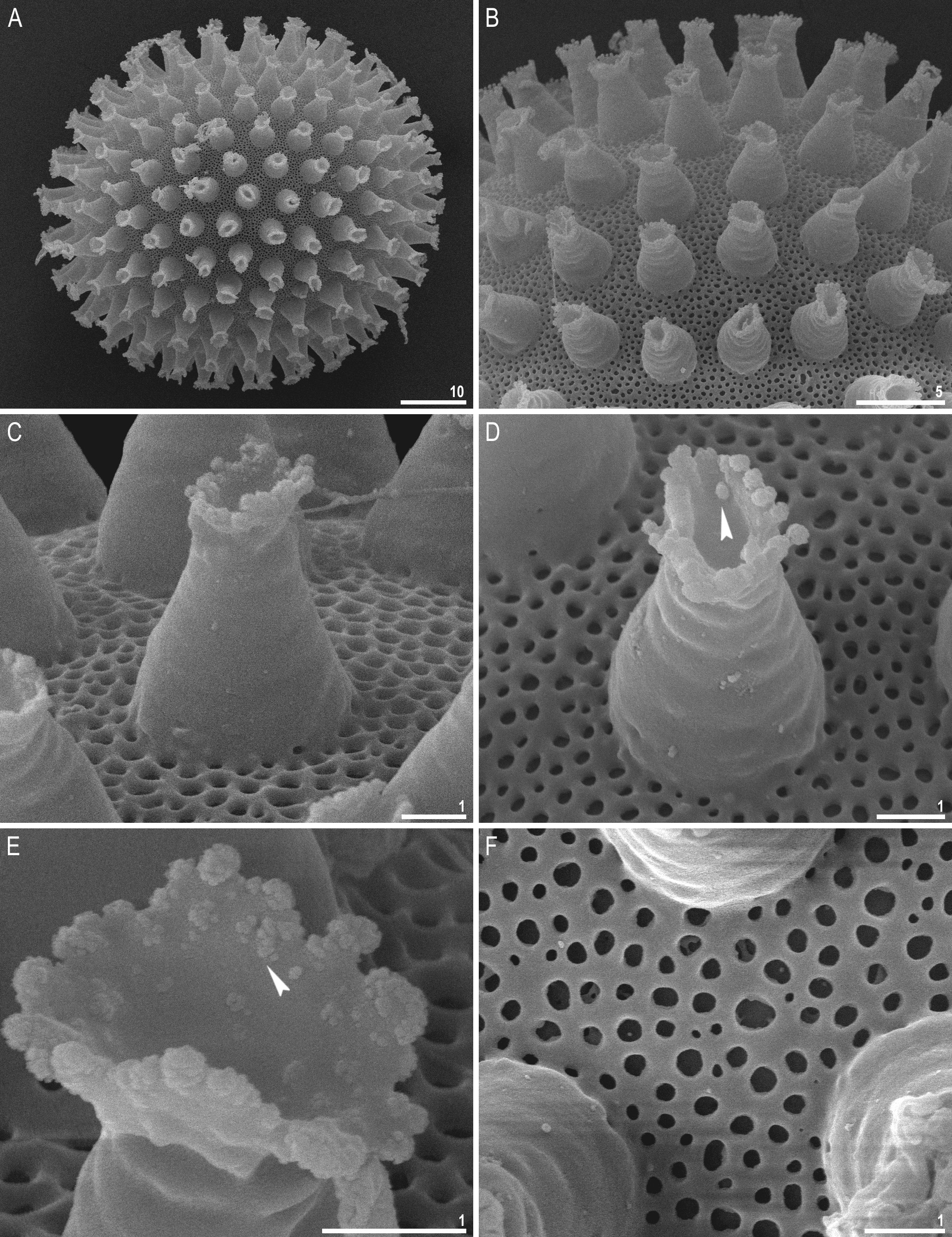
Table 2. Measurements (in μm) and pt values of selected morphological structures of the holotype and paratypes of Macrobiotus noongaris sp. nov. mounted in Hoyer’s medium (N = number of specimens/ structures measured; Range = the smallest and the largest structure among all measured specimens; SD = standard deviation).
| Character | N | Range µm pt | Mean µm pt | SD µm pt | Holotype µm pt | ||||
|---|---|---|---|---|---|---|---|---|---|
| Body length | 30 | 333–557 | 1039 – 1332 | 461 | 1201 | 56 | 71 | 443 | 1205 |
| Buccopharyngeal tube | |||||||||
| Buccal tube length | 30 | 31.8–43.5 | 38.3 | – | 3.4 | – | 36.8 | – | |
| Stylet support insertion point | 30 | 24.7–34.6 | 76.7–81.6 | 30.2 | 78.7 | 2.8 | 1.0 | 28.8 | 78.3 |
| Buccal tube external width | 30 | 4.4–7.4 | 13.8–17.9 | 5.9 | 15.5 | 0.7 | 1.0 | 5.7 | 15.4 |
| Buccal tube internal width | 30 | 2.9–5.0 | 8.0–11.9 | 4.1 | 10.6 | 0.6 | 0.9 | 3.7 | 10.1 |
| Ventral lamina length | 30 | 19.4–27.8 | 55.4–70.2 | 23.4 | 61.1 | 2.1 | 3.2 | 22.0 | 59.7 |
| Placoid lengths | |||||||||
| Macroplacoid 1 | 30 | 8.0–12.7 | 24.5–3.6 | 10.6 | 27.6 | 1.4 | 1.7 | 10.5 | 28.5 |
| Macroplacoid 2 | 30 | 5.2–8.8 | 15.0–21.0 | 7.0 | 18.3 | 1.1 | 1.6 | 6.1 | 16.5 |
| Microplacoid | 30 | 2.0–4.2 | 6.2–9.7 | 3.1 | 8.0 | 0.5 | 1.0 | 2.9 | 7.9 |
| Macroplacoid row | 30 | 13.9–22.7 | 41.9–57.7 | 18.4 | 47.9 | 2.6 | 3.5 | 17.1 | 46.5 |
| Placoid row | 30 | 16.4–26.1 | 51.3–63.7 | 22.1 | 57.5 | 2.8 | 3.3 | 21.2 | 57.6 |
| Claw 1 heights | |||||||||
| External primary branch | 30 | 7.5–10.9 | 20.8–28.7 | 9.6 | 25.2 | 0.9 | 2.1 | 10.4 | 28.3 |
| External secondary branch | 30 | 5.5–9.4 | 14.8–24.3 | 7.5 | 19.6 | 1.1 | 2.5 | 8.4 | 22.9 |
| Internal primary branch | 30 | 6.7–10.2 | 19.2–29.6 | 9.0 | 23.7 | 0.8 | 2.3 | 9.2 | 24.9 |
| Internal secondary branch | 30 | 5.4–8.2 | 16.6–22.1 | 7.3 | 19.1 | 0.7 | 1.7 | 7.8 | 21.1 |
| Claw 2 heights | |||||||||
| External primary branch | 30 | 8.5–12.1 | 22.9–32.9 | 10.5 | 27.6 | 0.8 | 2.5 | 12.1 | 32.9 |
| External secondary branch | 30 | 6.1–9.9 | 16.6–26.5 | 8.4 | 22.0 | 0.9 | 2.3 | 9.1 | 24.7 |
| Internal primary branch | 30 | 7.9–10.3 | 20.9–29.3 | 9.5 | 24.9 | 0.6 | 1.9 | 9.4 | 25.6 |
| Internal secondary branch | 30 | 6.5–8.8 | 16.4–24.9 | 7.7 | 20.3 | 0.7 | 2.0 | 8.8 | 23.9 |
| Claw 3 heights | |||||||||
| External primary branch | 30 | 8.8–11.9 | 23.6–32.7 | 10.5 | 27.4 | 0.7 | 2.1 | 11.3 | 30.6 |
| External secondary branch | 30 | 6.1–9.7 | 17.9–25.8 | 8.3 | 21.8 | 0.8 | 2.1 | 8.9 | 24.2 |
| Internal primary branch | 30 | 7.4–10.7 | 22.4–29.6 | 9.5 | 24.8 | 0.7 | 1.8 | 9.5 | 25.8 |
| Internal secondary branch | 30 | 5.5–9.3 | 16.6–25.2 | 7.5 | 19.6 | 0.9 | 2.0 | 9.3 | 25.2 |
| Claw 4 lengths | |||||||||
| Anterior primary branch | 30 | 8.2–12.3 | 22.7–32.2 | 10.5 | 27.5 | 1.1 | 2.6 | 11.8 | 32.2 |
| Anterior secondary branch | 30 | 5.4–9.7 | 15.7–25.1 | 8.0 | 21.0 | 1.0 | 2.4 | 9.2 | 25.1 |
| Posterior primary branch | 30 | 8.6–12.0 | 23.1–32.7 | 10.5 | 27.5 | 0.8 | 2.6 | 11.1 | 30.3 |
| Posterior secondary branch | 30 | 5.6–9.5 | 16.3–26.5 | 8.0 | 21.0 | 1.1 | 2.6 | 8.9 | 24.1 |
| DNA |
Department of Natural Resources, Environment, The Arts and Sport |
| AU |
Xiamen University |
No known copyright restrictions apply. See Agosti, D., Egloff, W., 2009. Taxonomic information exchange and copyright: the Plazi approach. BMC Research Notes 2009, 2:53 for further explanation.