
Mangalocypria ryukyuensis, Hiruta, Shimpei F. & Kakui, Keiichi, 2016
|
publication ID |
https://doi.org/ 10.11646/zootaxa.4169.3.6 |
|
publication LSID |
lsid:zoobank.org:pub:0794F234-5201-4C55-AEE1-D58ACF678F72 |
|
DOI |
https://doi.org/10.5281/zenodo.6085894 |
|
persistent identifier |
https://treatment.plazi.org/id/FC68836A-E94D-FF8F-B588-F980237CF80F |
|
treatment provided by |
Plazi |
|
scientific name |
Mangalocypria ryukyuensis |
| status |
sp. nov. |
Mangalocypria ryukyuensis View in CoL sp. nov.
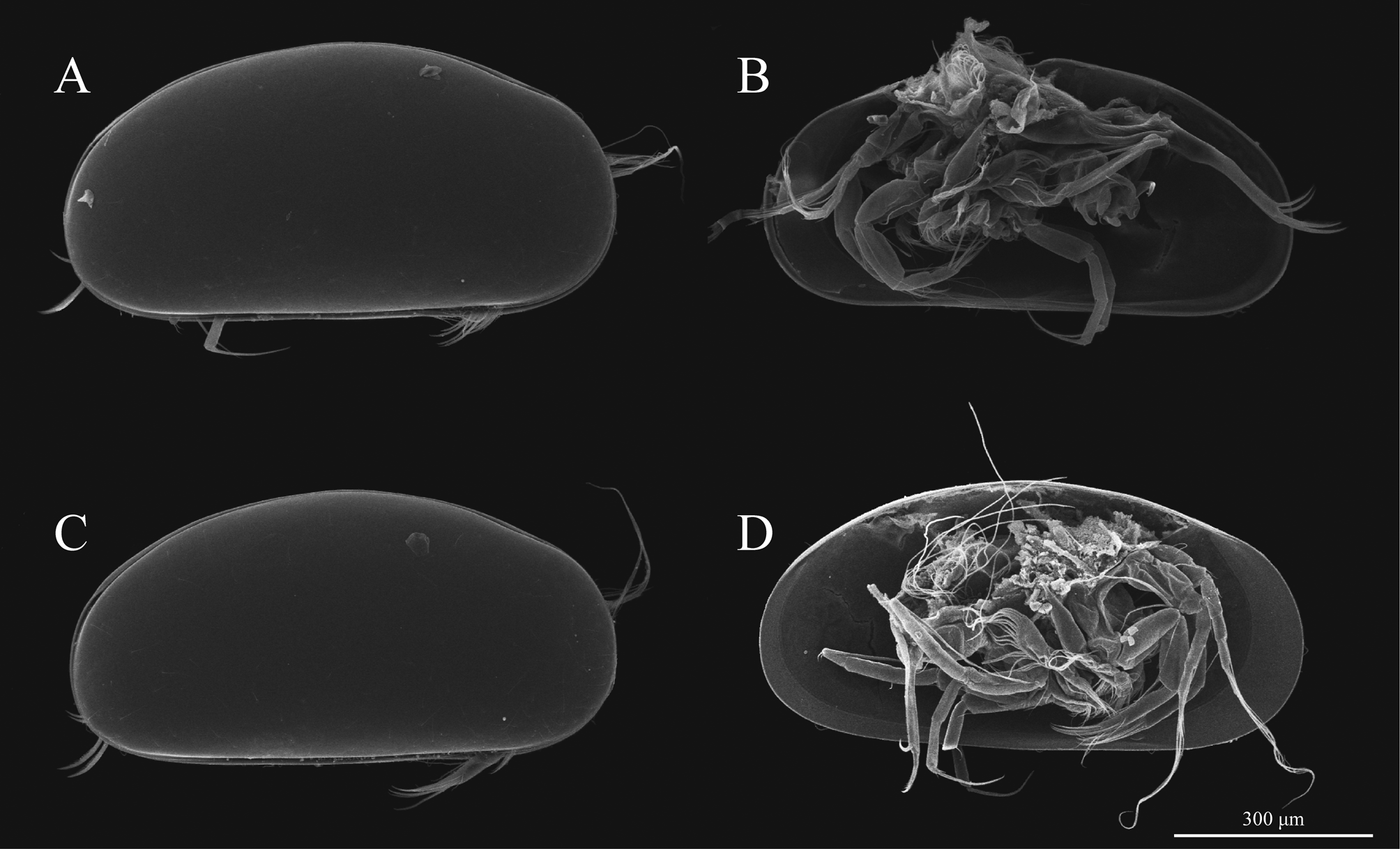
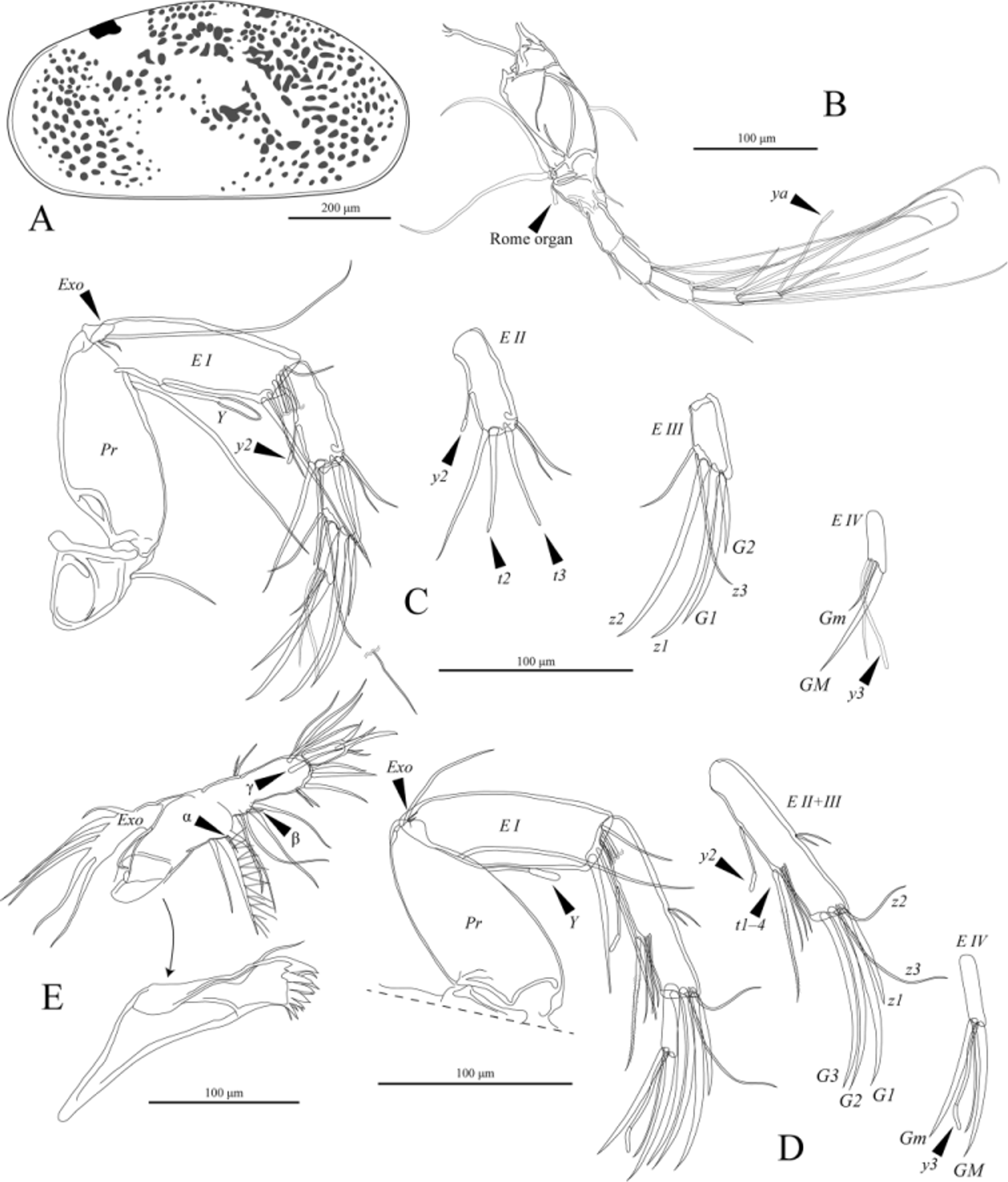
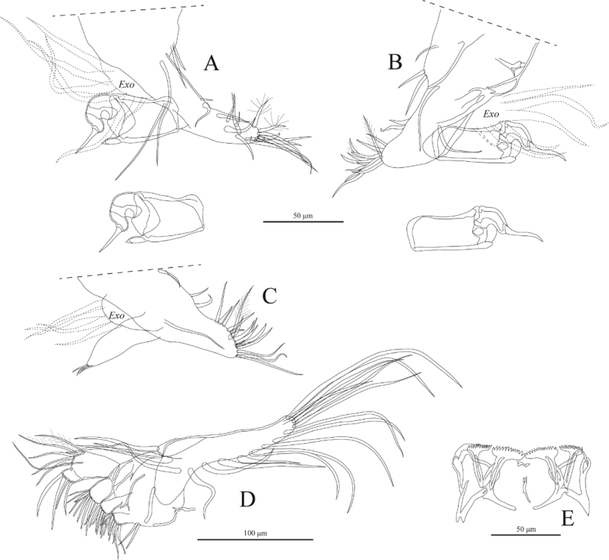
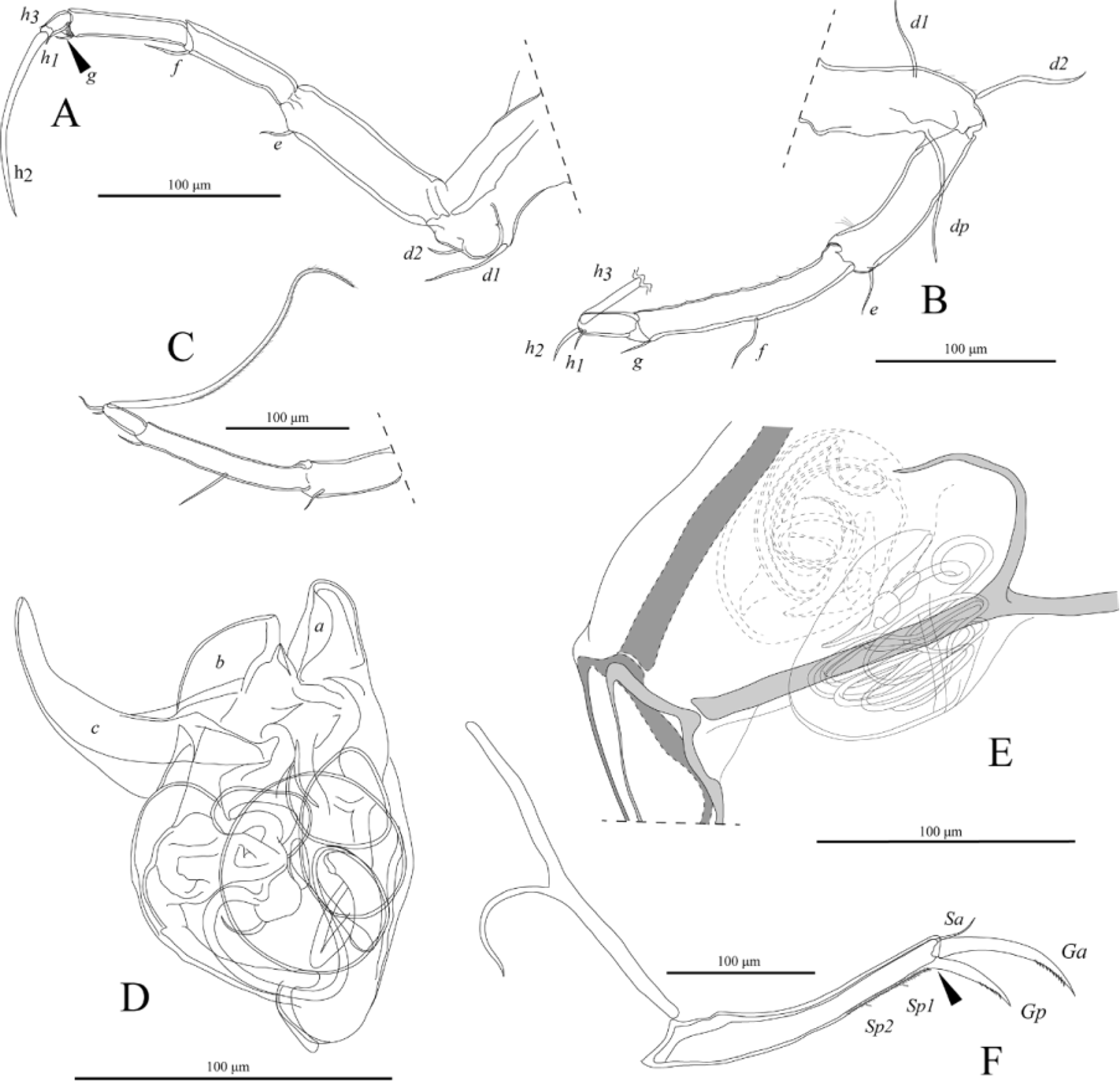
( Figures 2–7 View FIGURE 2 View FIGURE 3 View FIGURE 4 View FIGURE 5 View FIGURE 6 View FIGURE 7 )
Material examined. Type material. Holotype: ICHUM 4895 View Materials (male), 26°33′36″N, 128°2′37″E, Oura River , Okinawa Island, Japan, 26 February 2011, soft parts mounted on 19 slides, carapace mounted on a microfossil slide GoogleMaps . Allotype: ICHUM 4897 (female), collection data same as for holotype, softparts mounted on 17 slides, carapace mounted on a microfossil slide. Paratypes: ICHUM 4896 (male), 4898–4899 (two females), collection data same as for holotype, soft parts mounted on glass slides, carapace mounted on a microfossil slide; ICHUM 4904–4905 (two males), 4906–4907 (two females), collection data same as for holotype, mounted on stubs for SEM observation; ICHUM 4902 (female), collection data same as for holotype, exoskeleton and carapace after DNA extraction mounted on a glass slide.
Additional material. ICHUM 4900 View Materials (male) and 4901 (female), 24°26′32″N, 124°8′21″E, Kabira Bay , Ishigaki Island, Japan, 28 March 2013, softparts mounted on glass slides, carapace mounted on a microfossil slide GoogleMaps ; ICHUM 4903 View Materials (female), 24°26′32″N, 124°8′21″E, Kabira Bay , Ishigaki Island, Japan, 28 March 2013, exoskeleton and carapace after DNA extraction mounted on a glass slide. GoogleMaps
Etymology. The specific epithet is an adjective derived from Ryûkyû, the geographical name for the archipelago extending from Okinawa Island to Yonaguni Island, in combination with the Latin suffix - ensis.
Diagnosis. Medium-sized valves, with evenly convex dorsal margin. Carapace spindle-shaped in dorsal view, widest in the middle. Rome organ of A1 without distal expansion. Natatory setae of A2 reaching slightly beyond tip of claw. Distal setae on third and fourth segments of L6 short. Hook of male clasping apparatus with long apical seta. Dorsal lobe of Hp beaked, median lobe large and curved, ventral lobe strongly developed and widely curved. Uropodal ramus with very short, fine Sp1 and Sp2.
COI barcoding sequences. AB920556 View Materials (658 bp) from ICHUM 4902 View Materials (paratype) collected in the type locality on Okinawa Island ; AB920557 View Materials (658 bp) from ICHUM 4903 View Materials collected on Ishigaki Island .
Description of male. Carapace ( Figures 2 View FIGURE 2 A, C; 3B) 0.76–0.80 mm long, 0.37–0.39 mm high (n = 4); 0.76 mm long, 0.37 mm high in holotype; elongate in lateral view, highest just behind mid-length, broadly rounded at anterior and posterior ends, slightly convex ventrally; conspicuously narrow in dorsal view, pointed at anterior and posterior ends. Valves very thin, transparent, with light-brownish epidermal pigment; surface very smooth, with short, fine setae.
A1 ( Figure 3 View FIGURE 3 B) seven-segmented. First two podomeres fused, with one dorsal seta and two long apico-ventral setae Wouters organ not observed. Third podomere trapezoidal, with apico-dorsal seta and short, flat-end Rome organ; Fourth podomere rectangular, with apico-dorsal seta and apico-ventral seta. Fifth and sixth podomeres both rectangular, each with one apico-dorsal seta and two long apico-ventral setae. Seventh podomere with four long apical setae and one shorter apico-dorsal seta. Eighth podomere elongate and slender, with two long setae, one shorter seta, and long aesthetasc ya.
A2 ( Figure 3 View FIGURE 3 C) five-segmented. First podomere (Pr) with one long apico-ventral seta and one antero-proximal seta. Second podomere (EI) with one normal, one fine apico-ventral setae, aesthetasc Y, and Exo, consisting of one long and two short setae; 4 + 1 natatory setae extending to the tip of A2 terminal claw. Third podomere (EII) with two mid-apical male bristles (t2 and t3), one apico-ventral and two apico-dorsal setae, and mid-ventral aesthetasc y2. Fourth podomere (EIII) with claws G1, short G2, z1, z2, seta z3, and apico-ventral setae. Fifth podomere (EIV) with GM and slender, short Gm, one apical seta, and aesthetasc y3.
Md (figure 3E) consisting of coxal plate and four-segmented palp. Coxal plate with antero-lateral seta and six stout teeth. Palp with smooth alpha, smooth long gamma setae, and plumed very short beta seta. First podomere with exopodal plate (Exo). Terminal podomere of palp with three claws and two setae.
Mx ( Figure 4 View FIGURE 4 D) with elongate vibratory plate, three masticatory processes, and two-segmented palp. First podomere with two long apical plumose setae.
L5 ( Figure 4 View FIGURE 4 A, B) with palp, vibratory plate (Exo), one antero-proximal seta, one antero-apical seta, and one postero-apical seta. Vibratory plate with six filaments. Palp transformed into two-segmented clasping process; first podomere of palp with one apico-ventral peg; terminal segment stout and curved, with apical seta. Masticatory process with numerous setae and two plumed setae.
Lower lips ( Figure 4 View FIGURE 4 E) each with V-process developed as a rake. Arms of both, V-process and T-process with slots. Each rake with around thirteen teeth.
L6 ( Figure 5 View FIGURE 5 A) five-segmented. Second to fourth podomeres elongate. Terminal claw (h2) long.
L7 ( Figure 5 View FIGURE 5 B, C) four-segmented. Penultimate segment fused. First podomere with three setae (d1, d2, dp). Third podomere with one apical (g), one mid-ventral setae (f). Fourth podomere with two short (h1, h2) setae and one long, recurved (h3) seta plumed from the tip to three-fourths of its total length.
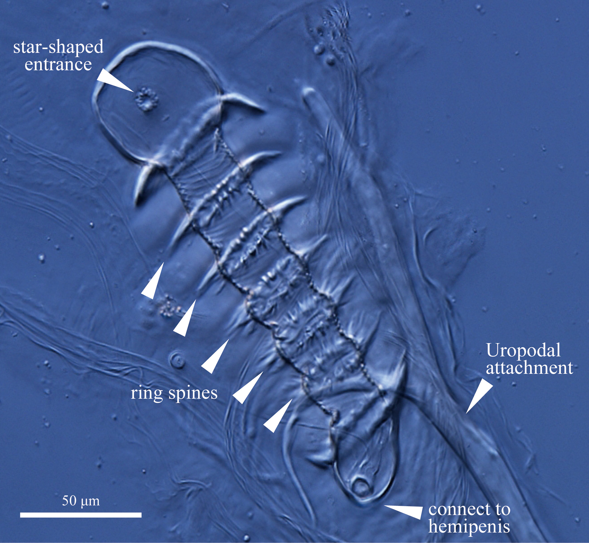
Zenker organ ( Figure 6 View FIGURE 6 ) with 5 + 2 internal rings of spines. Entrance to organ formed as a small star-shaped hole in a spherically shaped proximal end.
Hp ( Figure 5 View FIGURE 5 D) distally with three lobes. Dorsal lobe (a) distally beak-shaped. Median lobe (b) large, outer edge rounded. Ventral lobe (c) conspicuously extruding, hook-like, widely curved.
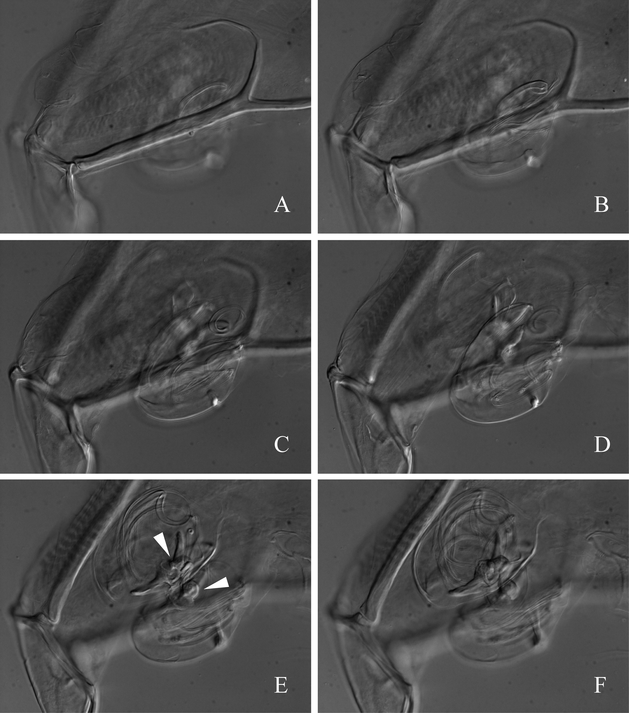
Uropod ( Figure 5 View FIGURE 5 F) with one Sa; with reduced Sp1 and Sp2; Ga and Gp with tiny denticles. Gp without movable joint (arrowhead, Figure 5 View FIGURE 5 F).
Description of female. Carapace 0.75–0.82 mm long, 0.37–0.41 mm high (n = 3); 0.77 mm long, 0.38 mm high in allotype.
A2 ( Figure 3 View FIGURE 3 D) four-segmented. First (Pr) and second (EI) podomeres similar to those of male. Third podomere (EII + EIII) with claws G1, G2, G3 and z1, setae z2 and z3, one mid-ventral aesthetasc y2, one long, plumed mid-ventral seta (t1), and three mid-ventral setae (t2–t4). Fourth podomere (EIV) slender and elongate; Gm longer than that of male.
Palp of L5 ( Figure 4 View FIGURE 4 C) simple, non-segmented, with three short apical setae.
Genital lobe with conspicuous, well-developed spiral canal ( Figures 5 View FIGURE 5 E; 7A–D, Supplementary Data 1).
Vaginal opening rimmed with well-developed sclerotized ring, and forming claw-like process ( Figures 5 View FIGURE 5 E; 7E, F). In lateral view, vaginal opening situated at the center of the spiral canal inside genital lobe. In other characters, female similar to male.
Remarks. This species clearly belongs in genus Mangalocypria due to the presence of a V-process developed as a rake on the lower lips, and a fixed postero-dorsal claw on the uropodal ramus. The genus Mangalocypria Wouters, 1998 contains three species: M. africana (Hartmann, 1974) ; M. appendix Wouters, 1998; and M. eleotridis ( Harding, 1962) . Mangalocypria africana was originally described from Mozambique (Hartmann 1974), and Wouters (1998) also reported it from Papua New Guinea, Indonesia, and the Comoros Islands. Meisch et al. (2007) mention it from Palau, Mecherchar Island (Jellyfish Lake). Mangalocypria appendix was originally described from Papua New Guinea. These two species differ from M. ryukyuensis sp. nov. in the genital organs. Mangalocypria ryukyuensis is generally similar to M. eleotridis , redescribed by Wouters (1998), in having a conspicuous, well-developed, curved ventral lobe on Hp. These two species differ, however, in the shape of the median lobe ( Figure 5 View FIGURE 5 D; b) of Hp (large with the outer side rounded in M. ryukyuensis ; small in M. eleotridis ), the two posterior setae on the uropodal rami (setae very short in M. ryukyuensis ; longer in M. eleotridis ), the terminal segment of the clasping process (stout and elongate in M. ryukyuensis ; shorter in M. eleotridis ), and carapace size (0.76 mm in the holotype of M. ryukyuensis ; 0.98 mm in M. eleotridis ).
The Mangalocypria population on Ishigaki Island was morphologically indistinguishable from the Mangalocypria ryukyuensis population on Okinawa Island, but COI sequences (658 bp) between the two showed a K2P genetic distance of 4.7%. While this seems to exceed typical levels of intraspecific divergence (see Introduction), we sequenced COI for only one individual from each population, and the range of variation and haplotype distributions within and between the populations remain unknown. For this reason, we have included only specimens from the Okinawa population in the type series and regard the identity of the Ishigaki population with Mangalocypria ryukyuensis as requiring confirmation.
No known copyright restrictions apply. See Agosti, D., Egloff, W., 2009. Taxonomic information exchange and copyright: the Plazi approach. BMC Research Notes 2009, 2:53 for further explanation.
|
Kingdom |
|
|
Phylum |
|
|
Class |
|
|
Order |
|
|
Family |
|
|
Genus |