
Maningrida arnhemensis, Golding, Rosemary E., Ponder, Winston F. & Byrne, Maria, 2007
|
publication ID |
https://doi.org/ 10.5281/zenodo.176773 |
|
DOI |
https://doi.org/10.5281/zenodo.6240302 |
|
persistent identifier |
https://treatment.plazi.org/id/03F887BA-9915-5422-118F-C17BFBECFDC8 |
|
treatment provided by |
Plazi |
|
scientific name |
Maningrida arnhemensis |
| status |
sp. nov. |
Maningrida arnhemensis View in CoL sp. nov.
Holotype ( Fig. 2 View FIGURE 2 N): Australia, Northern Territory: Arnhem Land, Maningrida , mudflats off beach at low tide, 27 Mar 1980, P.H. Colman (AMS C.446485).
Paratypes: Same data as holotype (AMS C.446484), 12 specimens.
Other material examined: Australia, Northern Territory: Arnhem Land, Rolling Bay, 25 Aug 1975, under debris at low tide, G. Webb and D. Grace (AMS C.445406); Arnhem Land, east bank Blyth River, 1 Nov 1975, mudflats, unknown collector (AMS C.4d528).
Etymology: From the species distribution in Arnhem Land (Northern Territory, Australia).
Description: Shell ( Fig. 2 View FIGURE 2 N): Very thin, globose, diameter to 6 mm, smooth, slightly reflective, striae faintly visible. Spire short, whorls rounded, lacking shoulder. Umbilicus very narrow, almost closed, lacking umbilical folds. Aperture circular, outer lip thin, with flattened, indented sinus and sinuous axial striae. Exterior pale golden.
Operculum ( Fig. 3 View FIGURE 3 E, 4E, F): Entirely corneous, circular, pale yellow, very thin, transparent. Nucleus marginal on central columellar edge. Striae concentric, elliptical to circular, raised on exterior, external surface of operculum granular. Internal surface of operculum smooth with faint striae. Outer edge of operculum expanded, covering opercular lobe with a corneous sheath.
External morphology ( Fig. 6 View FIGURE 6 D): Tentacles broad, flattened, semilunar, with small lappets on outer edges. Small, black eyes at outer base of tentacles. Posterior upper foot with large, expanded opercular lobe. Head and foot lacking pigmentation in formalin-preserved specimens, bearing large opercular lobe on posterior foot.
Mantle organs ( Fig. 7 View FIGURE 7 F): Opposed ciliary tracts short, restricted to exhalant canal. Hypobranchial gland at right anterior roof of mantel cavity large, yellow in formalin-preserved specimens, raised, composed of mucus-filled chambers. Mantle roof slightly vascularised, with thin layer of white, glandular tissue covering entire internal surface of mantle cavity.
Digestive system: As for A. crenata , with following differences. Oesophagus short, broad, folded at junction with gizzard. Four intestinal coils, two around stomach and gizzard, two around albumen and oviductal glands.
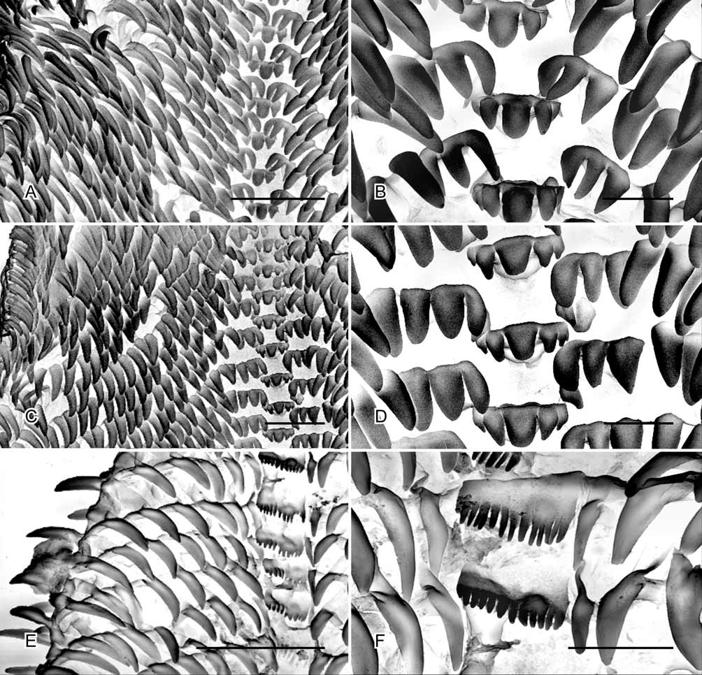
Radula ( Figs. 11 View FIGURE 11 E, F): With comb-shaped central tooth, bearing 11 to 13 equal sized cusps, number of cusps on central tooth variable between individual teeth on a single radula; lateral teeth absent; marginal teeth with approximately 15 on each side of radula, unicuspid, elongate and broad.
Central nervous system ( Fig. 12 View FIGURE 12 G): Pleural ganglia small, adjacent to cerebral ganglia with cerebral-pleural connective not visible. Parietal ganglia small, symmetrical; visceral ganglion with long, thin connectives to parietals. Parietal-visceral connectives crossed near visceral ganglion (slightly streptoneurous), right connective superior. Position of osphradial ganglion at anterior right of mantle cavity confirmed by histological section. Presence of parapedal and subcerebral commissures not confirmed.
Reproductive system ( Figs. 13 View FIGURE 13 D, 16): Ovotestis sac-shaped, occupying dorsal surface of upper whorls, adjacent to but not intertwined with digestive gland. Hermaphrodite duct straight, seminal vesicle occupying upper portion; albumen and oviductal glands pink in formalin-preserved specimens. Seminal receptacle small, bean shaped, brown in formalin-preserved specimens, with moderately long, coiled duct leading to carrefour. Spermoviduct dividing immediately below carrefour, forming morphologically indistinguishable muscular tubes lacking any glandular or other differentiation (these presumably correspond to oviduct and vas deferens), which run parallel to each other through the body wall and reunite near the genital atrium. Reunited spermoviduct continuing via a narrow duct, opening directly into the genital atrium. Two accessory sperm transfer structures (ASTSs) opening to genital atrium; one club-shaped organ with internal duct connected to bilobed gland, exterior surface of structure covered with closely-packed papillae which appear to produce glandular products. Large, spherical prostatic gland positioned on left anterior of reproductive complex; long, muscular duct with thick muscular bulb leading to second ASTS at genital atrium. Second ASTS composed of elongate tube lying within an external tube, lined with dense, cuticular, spinous microsculpture. Both ASTSs have retractor muscles.
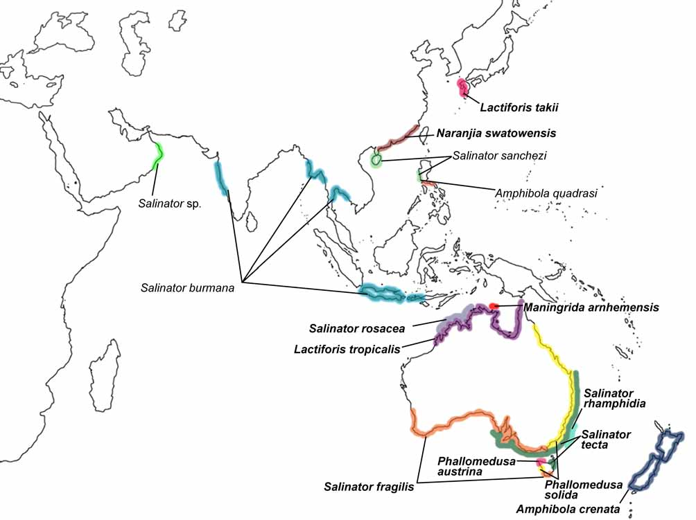
Distribution and habitat ( Fig. 1 View FIGURE 1 ): Known only from three locations in coastal and estuarine mudflats near the deltas of the Liverpool and Blyth River systems in Arnhem Land (Northern Territory, Australia).
Remarks: Anatomical examination of Maningrida arnhemensis was limited by availability of specimens. The only material available was preserved in formalin for a considerable time. Dissection, whole mount and serial histological sectioning were all used to examine the morphology of this species. However, it still remains to examine fresh or recently preserved material to confirm the findings of this study. The anatomical arrangement of the reproductive system suggests that sperm and ova are conducted via separate ducts which reunite at the genital atrium. The method of sperm transfer may involve either of the copulatory structures in the genital atrium, one or both of which presumably function as an intromittent organ.
The known distribution of M. arnhemensis sp. nov. is restricted to a small area in Arnhem Land, northern Australia. It is not yet clear whether this is due to sparse sampling or a narrow distribution. The shell has little resemblance to Salinator and is more similar to those of some Lymnaeidae .


No known copyright restrictions apply. See Agosti, D., Egloff, W., 2009. Taxonomic information exchange and copyright: the Plazi approach. BMC Research Notes 2009, 2:53 for further explanation.