
Megalothorax hipmani, Papáč, Vladimír & Kováč, Ľubomír, 2013
|
publication ID |
https://doi.org/ 10.11646/zootaxa.3737.5.3 |
|
publication LSID |
lsid:zoobank.org:pub:9DF408C6-BC6D-4D4E-BCDE-26E103D4E634 |
|
DOI |
https://doi.org/10.5281/zenodo.5628994 |
|
persistent identifier |
https://treatment.plazi.org/id/03E087E3-FFAC-FFFC-409C-FFD7FE0CFE69 |
|
treatment provided by |
Plazi |
|
scientific name |
Megalothorax hipmani |
| status |
sp. nov. |
Megalothorax hipmani sp. nov.
Figs 32–44 View FIGURES 32 – 35 View FIGURES 36 – 38 View FIGURES 39 – 40 View FIGURES 41 – 44
Megalothorax cf. incertus Mock et al. (2002) Megalothorax sp. 1 Papáč (2009, 2011)
Megalothorax sp. 3 Papáč & Kováč (2012)
Diagnosis. Mucronal lamellae serrate ( incertus -group). Connection of integumentary channels with linea ventralis on head crossed. Unpaired chaeta a0 between the basis of antennae present; posterior part of head with thickened macrochaetae. Basomedian field of labium with 4+4 chaetae. Sensory fields 2–6 with globular sensilla. Macrochaeta p4 on Th. III next to wax rod crypt 2, a6/a5 on Th. III as mesochaeta/macrochaeta. Spherical sensillum s3 on abdomen present. Mesochaeta on subcoxae I (leg I) present. Unguis apparently elongated. Manubrium with 3+3 dorsal chaetae (2+2 proximal, 1+1 distal). Retinaculum with 3+3 teeth.
Type material. Holotype: female on slide (No. 33–02), Slovakia, Kozie chrbty Mts., Važecká Cave, Križovatka (Crossroad), aphotic zone, surface of water pool, 9.x. 2002, leg. Ľ. Kováč. Paratypes: 5 ex. on slidesfemales (No. 33–02), same data as in holotype. Type material (holotype and 3 paratypes) saved in collection of MNHN, Paris. Other paratypes saved in collection of PJSU, Košice, Slovakia.
Other material. Slovakia, Low Tatras Mts., Hipman´s Cave system, Starý Hrad Cave, 1 ex.—subadult female on slide (No. VP 64–08), Studňa radosti (Shaft of Pleasure), bottom of shaft, surface of water pool, 14.vii. 2008, leg. V. Papáč. Demänovská Jaskyňa Mieru (Demänovská Cave of Peace), 1 ex.—female on slide (No. 790–11), near the entrance, water surface, 6.ix.2011, leg. A. Parimuchová.
Spišsko-gemerský Karst, Muránska Plateau, Zlatnica Cave, 1 ex.—female on slide (No. 80–09), hall with „heart“ of cave, 30 m from entrance, aphotic zone, pitfall trap in stony debris, 21.v.–1.x.2009, leg. V. Papáč.
Strážovské Mts., Četníkova Svadba Cave, 1 ex.—juvenile on slide (No. VP 20–12), Vodopádový dóm Hall, surface of water pool, 26.iv.2012, leg. V. Papáč.
Veľká Fatra Mts., Harmanecká Cave, 1 ex.—female on slide (No. 552–12), Nánosová chodba Corridor, surface of water pool, 18.ix.2012, leg. A. Parimuchová.
Description. Body length 0.46–0.57 mm, habitus typical of the genus. Body colour white, with scattered black dots of pigmentation on head, thorax, abdomen and subcoxae. Cuticle finely granulated, integumentary channels observed on ventral side of head and dorsally on posterior parts of head, connection of channels with l. v. ventrally on head of crossed type.
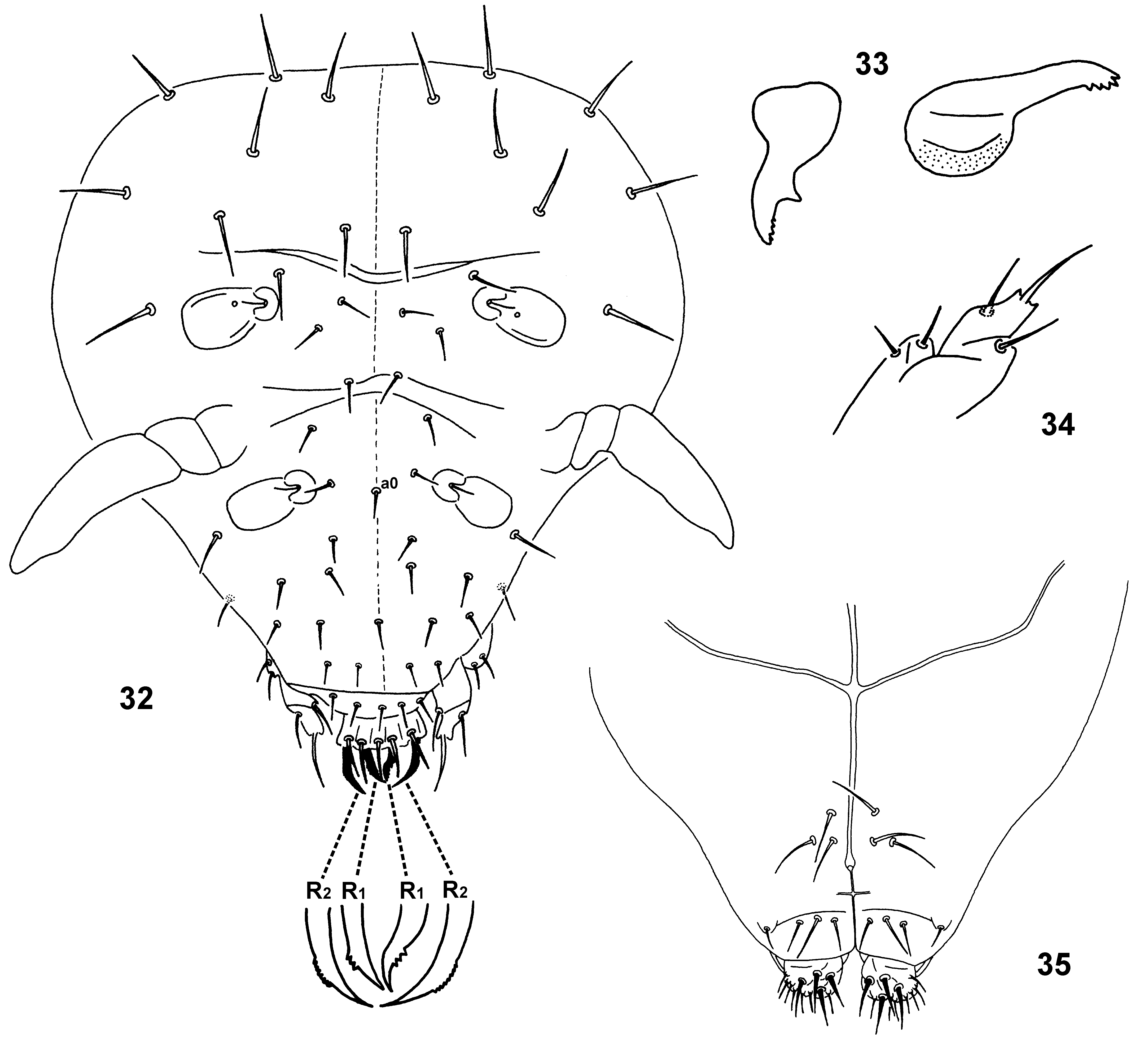
Head. Head length and width as 175 µm and 150 µm, respectively. Dorsal chaetae smooth and pointed ( Fig. 32 View FIGURES 32 – 35 ); unpaired chaeta a0 between the basis of antennae present. Frontal part with ordinary chaetae (8 µm), posterior part behind antennae with thickened macrochaetae (13–18 µm). 1+1 lateral chaetae between labial palp and basis of antenna. Clypeo-labral formula: a0, 2, 4, 5, 4 / 5, 5, 4 ( Fig. 32 View FIGURES 32 – 35 ). Pattern of labral chaetae: a-row with 2R1+2R2, m-row with m+2r1+2r2 and p-row with 5 ordinary chaetae. Anterior chaetae R1 and R2 thick, curved, R2 (14 µm) longer than R1 (9 µm); R1 with external edge roughly serrate; R2 with external edge finely serrate. Medial chaetae (m-row) equal (12 µm), smooth, thicker than p-row. Posterior chaetae (p-row) equal, smooth (10 µm). Integumentary channels present dorsally on headback ( Fig. 60 View FIGURES 58 – 61 ). Basomedian field of labium with 4+4 chaetae, medial ones longer (12 µm) than axial and lateral (10 µm), 1 smaller chaeta laterally near basolateral field ( Fig. 35 View FIGURES 32 – 35 ). Basolateral field with 1 ordinary chaeta, oral fold with 2 chaetae. Ventral side of head with 3+3 smooth postmedian chaetae ( Fig. 35 View FIGURES 32 – 35 ); 2+2 equal anterior macrochaetae thickened and curved (14 µm); posterior 1+1 chaetae as thickened and straight macrochaetae (14 µm). Mandible normal, strong, with 5 apical teeth ( Fig. 33 View FIGURES 32 – 35 ). Maxillary outer lobe with enlarged terminal chaeta, 1 basal chaeta and 1 sublobal hair ( Fig. 34 View FIGURES 32 – 35 ). Maxilla with 1 lamella extending beyond fringed lamellae.
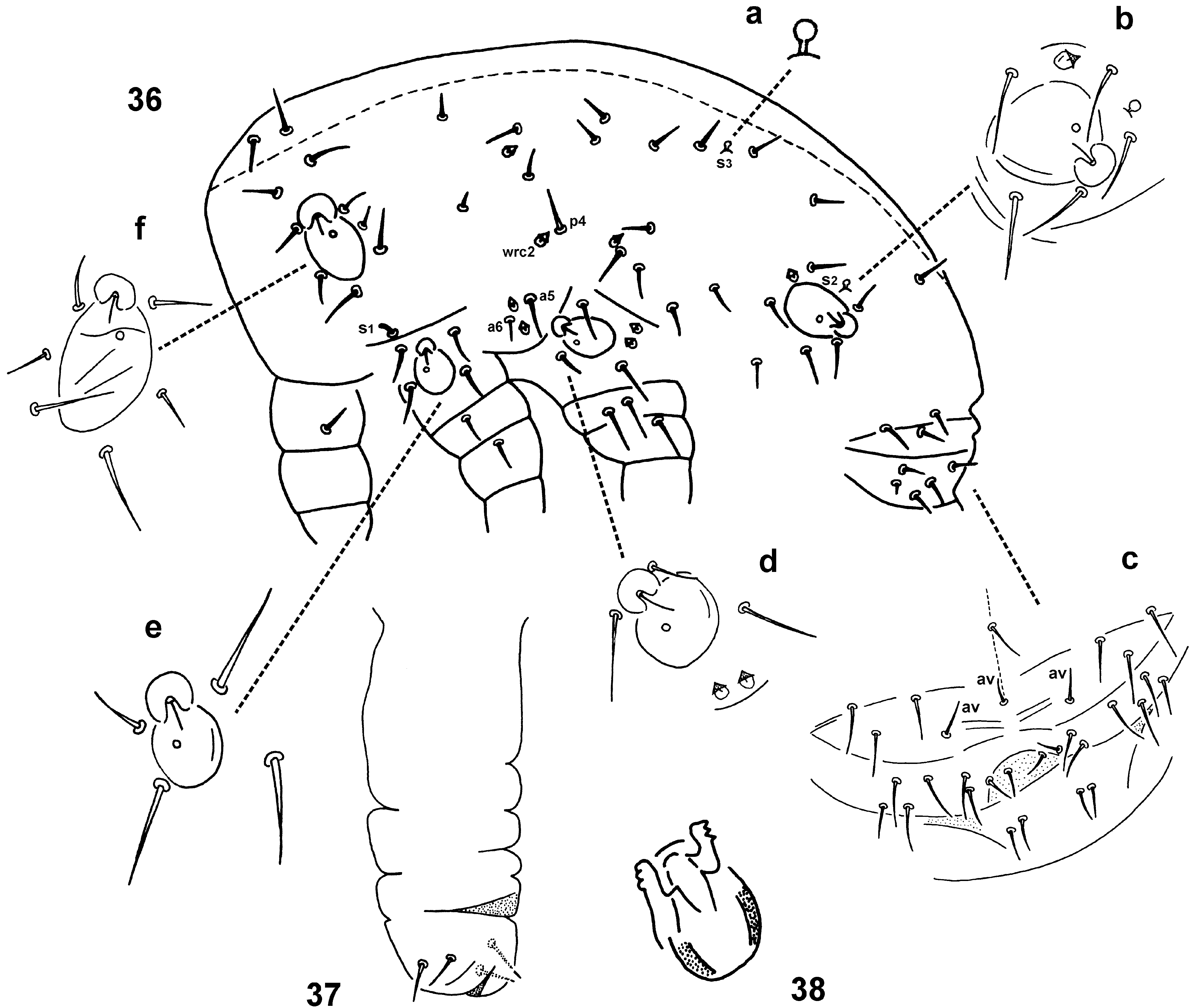
Thorax and abdomen ( Fig. 36 View FIGURES 36 – 38 ). Dorsal side of thorax and abdomen covered with meso- and macrochaetae (10 and 24 µm, respectively) and 8+8 wax rods (wrc1–8) as tiny, straight chaetae (2 µm) placed in small cuticle depressions. Th. II with 8+8 medial chaetae around thoracal sensory fields and 4+4 lateral chaetae around sensory fields at leg II base; 1+1 lateral sensillum s1. Th. III medially with 7+7 chaetae, and 4+4 wrc, laterally at leg III base with 3+3 chaetae and 2+2 wrc around sensory field; macrochaeta p4 next to wrc2; chaeta a5 stouter and longer than a6. Abd. I–V with 18+18 dorsal chaetae, 2+2 wrc and 2+2 globular sensilla s2 and s3, chaetae β2 and ε4 absent ( Figs. 36 View FIGURES 36 – 38 a, b). Abd. VI with 9 dorsal chaetae ( Fig. 36 View FIGURES 36 – 38 c). Anal field with 3 anal valves, each with 1 chaeta ( Fig. 36 View FIGURES 36 – 38 c). Abd. VI with 8+8 ventral chaetae (2+2 axial shorter). Female genital plate furnished with 2+2 chaetae. Manubrium base (Abd. IV sternum) with 2+2 chaetae and laterally with 2+2 broad neosminthuroid chaetae with bristle at tip (8 µm).
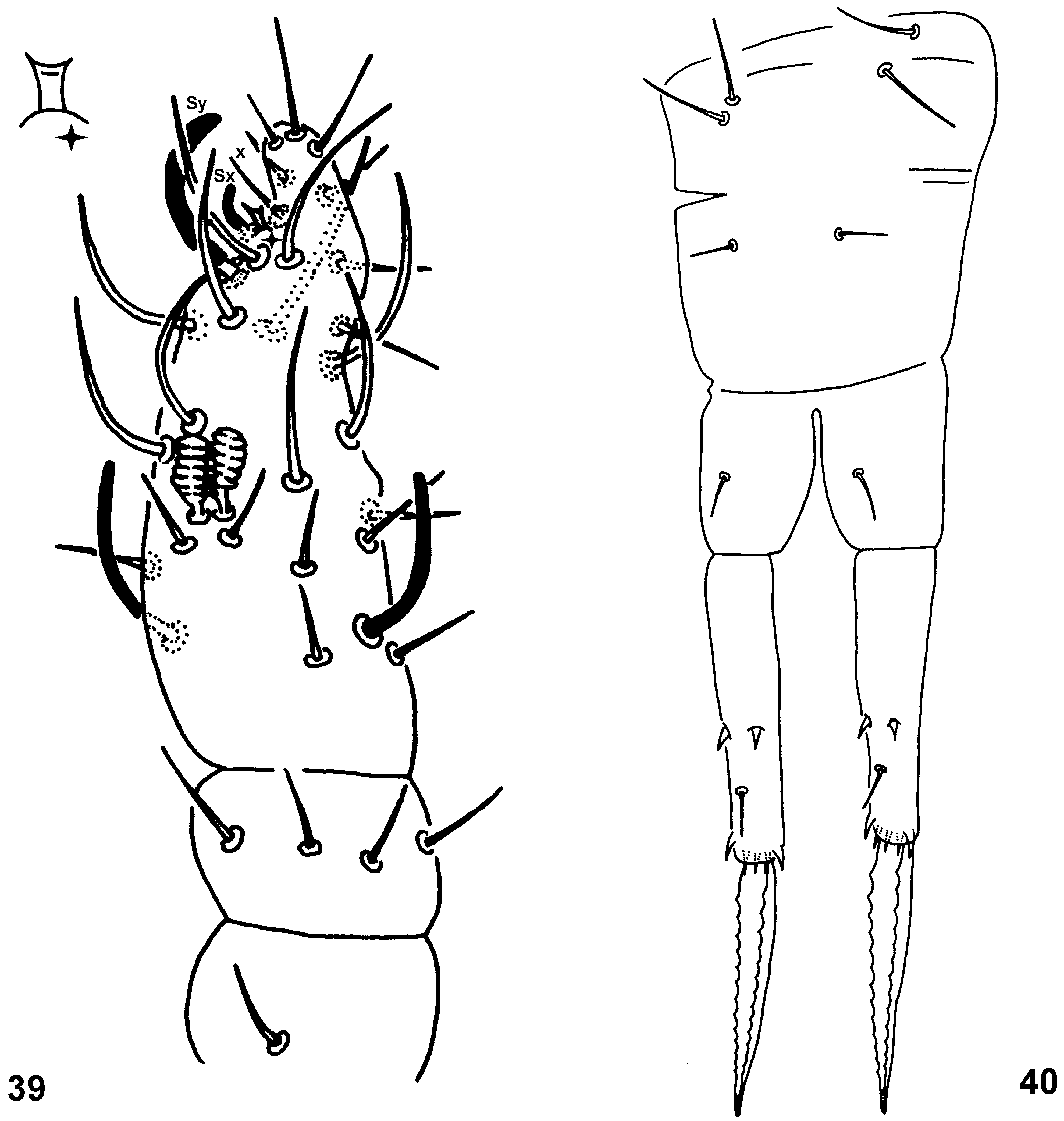
Appendages. Antennal segments III and IV not separated ( Fig. 39 View FIGURES 39 – 40 ). Length of antennae up to 92 µm, ratio antenna/head = 0.50; lengths of antennal segments I, II, III-IV as 11, 13, 58 µm, respectively. Ant. I with 1 mesochaeta (10 µm). Ant. II with 4 chaetae arranged in whorl, 1 dorsal slightly longer than others (12 µm vs. 10 µm). Ant. III with 8 ordinary chaetae, 2 of them in proximal position. Ant. III organ consists of 2 ovoid sensilla with striate rim (10 µm), 2 long, transparent sensilla Sg, external and dorsal (14 µm each). Ant. IV segment with 6 ordinary chaetae arranged mostly on internal side, dorso-external side with 10 thin and curved macrosensilla S finely blunt at tip (20 µm); ventrally with 1 long and thick sensillum Sy (14 µm), 1 thick and shorter sensillum Sx (6 µm) with curved tip and 1 ordinary chaeta x; dorsally with cup-like subapical organite Or (4 µm); subapically with 5 ordinary chaetae (6–8 µm) and apically with 2 rod-like chaetae a and sa (12 µm). Complete chaetotaxy of antenna provided in Table 3. Chaetae numbers of legs I–III ( Figs. 41–43 View FIGURES 41 – 44 , 36 View FIGURES 36 – 38 ), longer chaetae in parenthesis: scx I: 1, 1, 2(2); scx II: 0, 1, 1(1); coxae: 1, 1(1), 1(1); trochantera: 2, 3, 4; femora: 6(2), 7(2), 7(3) and tibiotarsi: 12, 12, 10. Thin meso- or microchaetae in following numbers on leg I: coxa with 1, trochanter with 2, femur with 1; on leg II: trochanter with 2, femur with 2; on leg III: trochanter with 1, femur with 1. For complete chaetotaxy of legs see Table 4. Unguis narrow and elongated, both unguis and unguiculus unequally long in leg I, II and III: unguis 24, 20 and 18 µm, respectively, unguiculus 15, 13 and 12 µm, respectively. Length ratio unguis I (inner margin) / Ti. I width (24 / 15 µm) = 1.60. Unguis furnished with 2 lateral teeth la, lp and 1 tight, long inner tooth Bp in distal 1/ 3 ( Fig. 44 View FIGURES 41 – 44 ). Unguiculus untoothed, apical filament absent, basal tubercle projecting basal lamellae only on leg II and III. Tubus ventralis with 2+2 distal chaetae, posterior lobe absent ( Fig. 37 View FIGURES 36 – 38 ). Retinaculum with 3+3 teeth, chaeta on corpus absent ( Fig. 38 View FIGURES 36 – 38 ). Furca well developed ( Fig. 40 View FIGURES 39 – 40 ), length of manubrium, dens (proximal and distal part) and mucro: 50, 26, 42 and 37 µm, respectively. Manubrium dorsally with 3+3 chaetae, 2+2 proximal ones longer (13 µm) than 1+1 distal (7 µm). Proximal part of dens (dp) with 1+1 dorsal, smooth chaetae (7 µm). Distal part of dens (dd) dorsally with 1 medial, smooth chaeta (8 µm) and 4 broad spines with apical filament (4–5 µm): 2 external and 2 internal; ventrally with 3 tight apical spines in transversal row (5 µm; Fig. 40 View FIGURES 39 – 40 ). Dental spines as leave-shaped integumental structures lacking basal circle. Mucro with both lamellae serrated along whole length (12–14 teeth), gradually constricted in distal part, pointed at the apex; length up to 47 µm, width in the middle part 6 µm.
Sensory fields ( Fig. 32 View FIGURES 32 – 35 , 36 View FIGURES 36 – 38 ). 6+6 s.f placed in small depressions each with secretory rod (8 µm), i.e. blunt, straight chaeta with basal part inserted in cuticle in upper margin of the field. S.f. have following arrangement: (a) anterior and posterior field on head (s.f. 1 and 2, 20 x 15 µm, anterior slightly smaller; Fig. 32 View FIGURES 32 – 35 ), posterior field with 1 internal globular sensillum (3 µm); (b) thoracal field (s.f. 3, 30 x 15 µm; Fig. 36 View FIGURES 36 – 38 f) with 1 internal globular sensillum and 6 external chaetae (2 lateral as macrochaetae); (c) abdominal field (s.f. 6, 30 x 20 µm; Fig. 36 View FIGURES 36 – 38 b) with 1 internal globular sensillum, 5 external macrochaetae, 1 spherical sensillum s2 and wax rod wrc8; (d) field at base of leg II (s.f. 4, 20 x 15 µm; Fig. 36 View FIGURES 36 – 38 e) with 1 internal globular sensillum, 4 external chaetae (1 mesochaeta—12 µm, 3 macrochaetae—18 µm) and 1 sensillum s1 (5 µm) above the field; (e) field at base of leg III (s.f. 5, 20 x 15 µm; Fig. 36 View FIGURES 36 – 38 d) with 1 internal globular sensillum, 3 external chaetae (1 mesochaeta—12 µm, 2 macrochaetae—16–20 µm) and wax rods wrc5,6 on its posterior margin.
Only females known.
Etymology. The new species is named in honour of speleologist Petr Hipman, who considerably contributed to explorations of caves in the Low Tatra Mts. and discovered the deepest cave in Slovakia—Jaskyňa Starý Hrad Cave, part of the Hipman´s Cave system (- 495 m).
Distribution. M. hipmani sp. nov. was discovered in six caves of central Slovakia which are located in five karstic areas: (1) Low Tatras Mts. (Hipman´s Cave, Demänovská Cave of Peace), (2) Kozie chrbty Mts. (Važecká Cave), (3) Spišsko-gemerský Karst—Muráň Plateau (Zlatnica Cave), (4) Veľká Fatra Mts. (Harmanecká Cave) and (5) Strážov Mountains (Četníkova svadba Cave). Specimens of the M. hipmani sp. nov. were sampled mainly from deeper parts of the caves, mostly on surface of water pools.
Discussion. Megalothorax hipmani sp. nov. belongs to „ incertus -group“ by both mucronal lamellae serrated and modified internal chaeta of sensory fields in form of globular sensillum. M. hipmani sp. nov. differs from M. draco sp. nov. by projecting basal tubercle on leg II and III (in M. draco sp. nov. the tubercle hidden behind basal lamellae), 3+3 teeth on retinaculum (4+4 teeth in M. draco sp. nov.), 3+3 manubrial chaetae (4+4 chaetae in M. draco sp. nov.), 12–14 teeth on mucronal lamellae (8–9 teeth in M. draco sp. nov.). M. hipmani sp. nov. differs from M. massoudi by presence of sensilla s3 on abdomen (s3 absent in M. massoudi ). For other differential characters see Tables 3–5.


No known copyright restrictions apply. See Agosti, D., Egloff, W., 2009. Taxonomic information exchange and copyright: the Plazi approach. BMC Research Notes 2009, 2:53 for further explanation.