
Microcosmus pacificus, Monniot & Monniot, 2001
|
publication ID |
https://doi.org/ 10.5281/zenodo.5391440 |
|
DOI |
https://doi.org/10.5281/zenodo.5468118 |
|
persistent identifier |
https://treatment.plazi.org/id/F57D87A3-FF4B-31AD-EA4F-FBB7FDA41080 |
|
treatment provided by |
Marcus |
|
scientific name |
Microcosmus pacificus |
| status |
sp. nov. |
Microcosmus pacificus View in CoL n. sp.
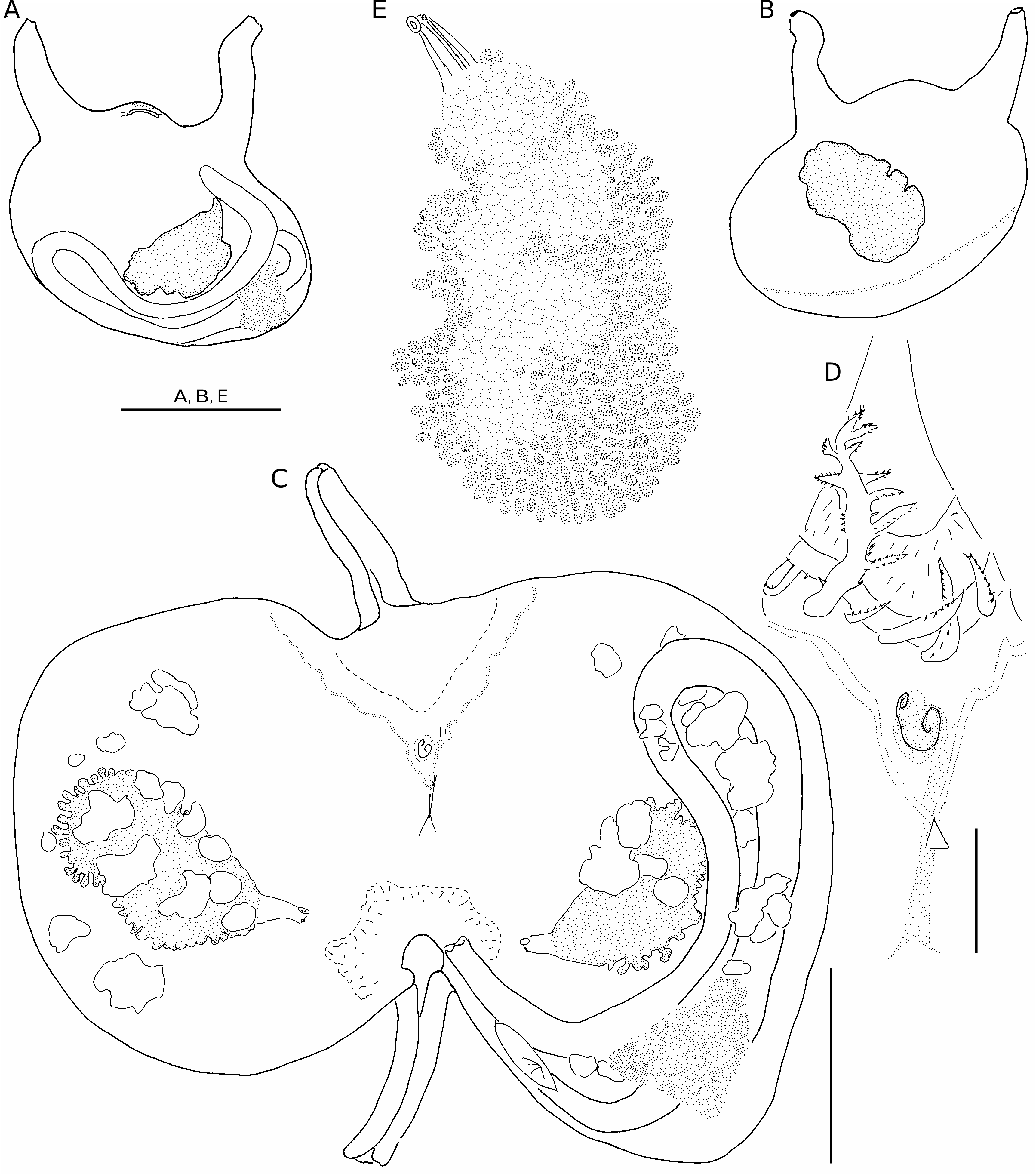
( Figs 108D View FIG ; 109 View FIG )
TYPE MATERIAL. — Papua New Guinea. Milne Bay Province, Samarai Island, 10°36.98’S, 150°39.77’E, 27 m, 10. VI.1998 ( MNHN S2 MIC 159).
ETYMOLOGY. — Named after the Pacific Ocean.
OTHER MATERIAL EXAMINED. — Philippines. Musorstom 3, 11°37’N, 121°43’E, 120-122 m, 5.VI.1985 (MNHN S2 MIC 160).
DESCRIPTION
Both specimens were collected on a sandy bottom. They are very similar, 3 cm in length, covered with coarse sand and broken shells. The siphons are well apart. The body wall is thin and pale in formalin except for the red extremity of the siphons. Internally the siphons are long and narrow. The internal spinules are arranged in chevrons and their section is angular ( Fig. 108D View FIG ). The musculature forms a network of crossed and regularly spaced bundles issuing from both siphons.
The tentacles are inserted at the base of a high velum. There are 12 large, bushy tentacles in two orders of size with three orders of branchings ( Fig. 109D View FIG ); smaller ones are irregularly intercalated. The prepharyngeal band is very close to the branchial tissue. It is deeply indented dorsally. The dorsal tubercle is protruding; its opening is horse-shoe shaped, anteriorly opened with horns inwardly rolled ( Fig. 109D View FIG ). The neural ganglion is particularly long ( Fig. 109D View FIG ).
The dorsal lamina is a high blade of equal height along its whole length.
The branchial sac counts seven folds on each side. The formula on the right side is:
R.E.? 10 2 15 2 17 1 19 1 19 1 19 1 17 2 D.L.
The folds are high, overlapping each other. There are five to six stigmata per mesh between the folds and three to four on the folds. At the top of the folds one or two stigmata are curved. There are parastigmatic vessels.
The gut makes a very long and curved loop with both limbs very close together ( Fig. 109A, C View FIG ). The stomach is only slightly enlarged. It is covered by a massive hepatic gland constituted of lamellae. Many filiform papillae arise from the hepatic lobes ( Fig. 109C View FIG ).
The rectum is cylindrical, its posterior part attached to the oesophagus. The anus is wide open with two lobes. There is one gonad on each side, protruding well above the body wall ( Fig. 109 View FIG A-C). The left gonad lies in the secondary loop of the gut but does not overlap the rectum ( Fig. 109C View FIG ). The general outline of the gonads is oval. The male lobules surround the sinuous ovaries and penetrate them in furrows ( Fig. 109E View FIG ) The gonoducts are joined, opening at the same level.
The endocarps are irregular in size, and lobulated. They lie inside the gut loop, on both sides of the body wall ventrally, and above the gonads ( Fig. 109C View FIG ).
There is a large cloacal velum with an irregular ring of filiform papillae at its base.
REMARKS
By its sediment-covered tunic, the length of its well-separated siphons, its branchial sac, and its long narrow gut loop, this species resembles Microcosmus madagascariensis Michaelsen, 1918 from Madagascar. But the latter has the left gonad overlapping the gut loop. As well, the endocarps are differently distributed in these two species.
Microcosmus agglutinans Hartmeyer, 1919 View in CoL from western Australia is also closely allied. It differs in having its the siphons close together, and it has a more accentuated secondary loop of the gut with the anus level with the top of the primary gut loop. It has no oral velum.
Hartmeyer & Michaelsen (1928) synonymised M. madagascariensis View in CoL and M. agglutinans View in CoL , an opinion that Kott (1985) supported, but we believe that these are different species. Kott’s description corresponds better to M. agglutinans View in CoL . Moreover the description given by Vasseur (1969) for M. madagascariensis View in CoL collected in Madagascar corresponds well to the description of Michaelsen’s type from the same locality. The geographic distance separating them – one in Madagascar, the other in western Australia – is an additional argument for separating the two species.
No known copyright restrictions apply. See Agosti, D., Egloff, W., 2009. Taxonomic information exchange and copyright: the Plazi approach. BMC Research Notes 2009, 2:53 for further explanation.
|
Kingdom |
|
|
Phylum |
|
|
Class |
|
|
Order |
|
|
Family |
|
|
Genus |
Microcosmus pacificus
| Monniot, Françoise & Monniot, Claude 2001 |
Microcosmus agglutinans
| Hartmeyer 1919 |
M. agglutinans
| Hartmeyer 1919 |
M. agglutinans
| Hartmeyer 1919 |
M. madagascariensis
| Michaelsen 1918 |
M. madagascariensis
| Michaelsen 1918 |