
Microphysogobio zhangi, Sun & Zhao, 2022
|
publication ID |
https://doi.org/ 10.6620/ZS.2017.56-08 |
|
DOI |
https://doi.org/10.5281/zenodo.8063702 |
|
persistent identifier |
https://treatment.plazi.org/id/AF7CE82B-FFE0-FFF8-279C-511DFDC6AB37 |
|
treatment provided by |
Valdenar |
|
scientific name |
Microphysogobio zhangi |
| status |
sp. nov. |
Microphysogobio zhangi n. sp.
urn:lsid:zoobank.org:act:C1F8D275-44E3-4E24-BAF4-AE90DE8C3248
Material examined: Holotype: ASIZB 204677 View Materials , 77.1 mm SL, Xiang River , a tributary of Yangtze River, Quanzhou County, Guangxi Province, China (latitude: 25°56'00.0"; longitude: 111°04'49.4"), coll. S.P. Huang and J.C. Huang, 6 November 2015.
Paratypes: ASIZP 0078398, 3 specimens, 65.2-69.1 mm SL, collected with holotype. ASIZP 0078397, 2 specimens, 62.3-63.7 mm SL, Guilin City market, Guangxi Province, China, coll. S.P. Huang and J.C. Huang, 5 November 2015. NTOUP 2010-11-547, 12 specimens, 53.0- 64.1 mm SL, Gongcheng County market, Guangxi Province, China, coll. I-S. Chen, 8 August 2009.
Diagnosis: This new species can be distinguished from other congeners by the following unique combination of features: (1) meristic accounts: anal fin rays 3, 5; pectoral fin rays 11-12; lateral-line scales 35-36; transverse scales 7; predorsal scales 9-10; vertebral counts 4+30-31; (2) lip papillae: barbel length medium, 53.5-69.9% of eye diameter; the medial pad on lower lip divided; and (3) coloration patterns: body with 6-7 indistinct black horizontally-aligned crossbars; interorbital region with a black crossbar; a thin black vertical stripe throughout the cheek; dorsal fin and caudal fin membranes with indistinct vertically-aligned black lines.
Description: The morphometric measurements of this new species are provided in table 2. Body elongated and compressed laterally. Belly flatted. Snout pointed. Eye moderately large, located dorsal half of head. Lateral-line complete and running slightly downward abruptly above the pectoral fin and along the ventral profile into the middle of the caudal fin base. Gill rakers 11-13. Vertebral counts 4+30-31. Dorsal fin rays 3, 7; anal fin rays 3, 5; pectoral fin rays 1, 11-12 (modally 12); pelvic fin rays 1, 7; lateral-line scales 35- 36 (modally 35); transverse scales 7; predorsal scales 9-10 (modally 10) ( Table 3 View Table 3 ). Adult males with pectoral fin reaching anterior margin of anal fin base when compressed. Anterior margin of pelvic fin inserted below second branched ray of dorsal fin. Caudal fin deeply forked, lower lobe slightly longer than upper lobe. Body covered with large cycloid scales. Belly covered with cycloid scales, inter-pectoral fin basal region always naked.
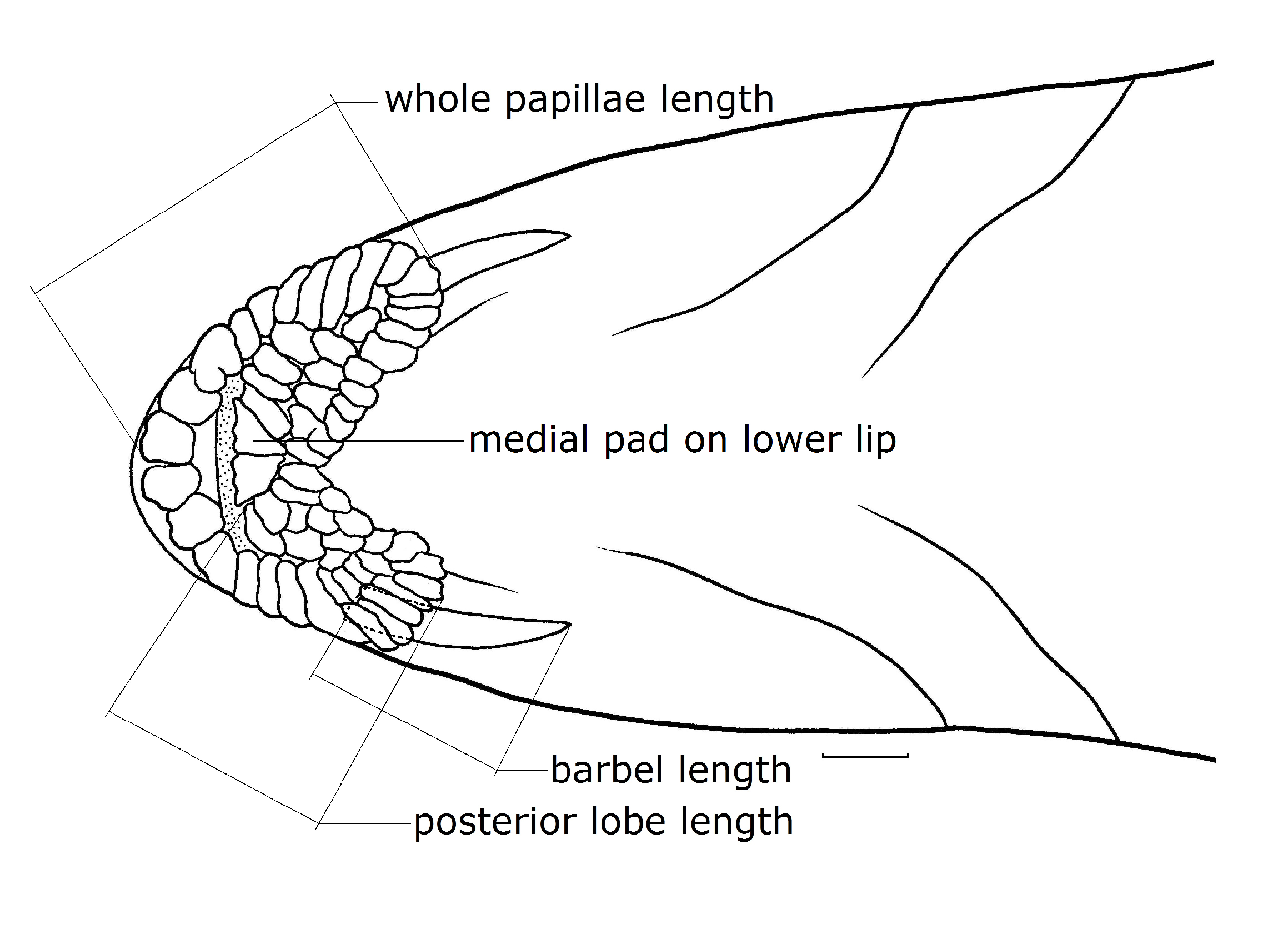
Lip papillae: Mouth horseshoe-shaped. Upper and lower lip thick, covered with pearl papillae. Lip papillae consist of anterior papillae, two posterior lobes and a medial pad on lower lip. Anterior papillae covered with one row of large pearl papillae. Both posterior lobes covered with clusters of well-developed, small pearl-like papillae. The medial pad on lower lip completely divided ( Fig. 1 View Fig ). A pair of barbels located at corners of mouth and rooted at posterior edge of lower jaw, 53.5-69.9% length of eye diameter. Posterior lobes medium length, 60.8-72.5% of eye diameter. Whole papillae long, 89.5-106.4% length of eye diameter.
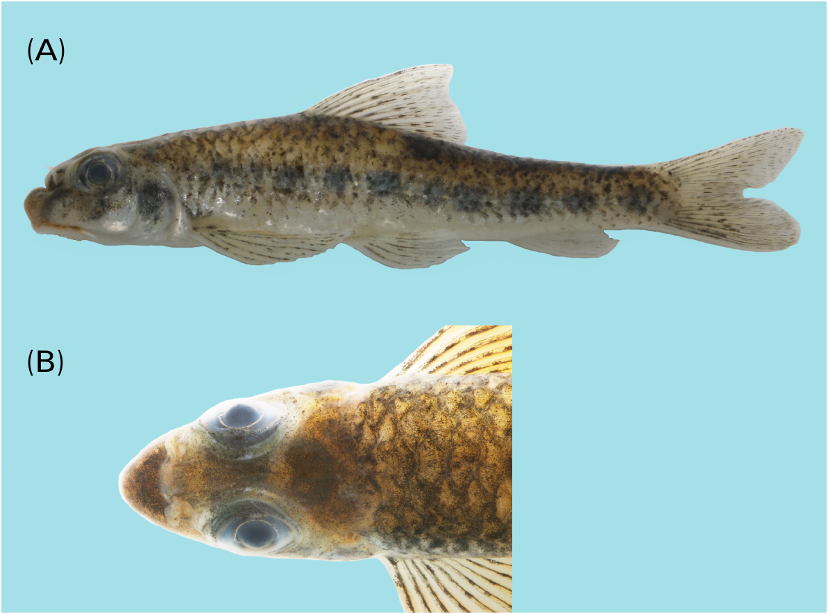
Coloration in fresh specimen: Head and body generally pale yellowish brown ( Fig. 2A View Fig ). Interorbital region with a black cross-band ( Fig. 2B View Fig ). Belly grayish to pale white. Body with 6-7 indistinct, black horizontally-aligned crossbands. Operculum region with a deep brown mark. A thin black stripe running vertically across the cheek. Dorsal fin membrane with one or two rows of indistinct, longitudinally-aligned black lines. Pectoral fin, pelvic fin and anal fin membranes having some tiny black spots. Caudal fin membrane with two or three rows of indistinct vertically-aligned black lines.
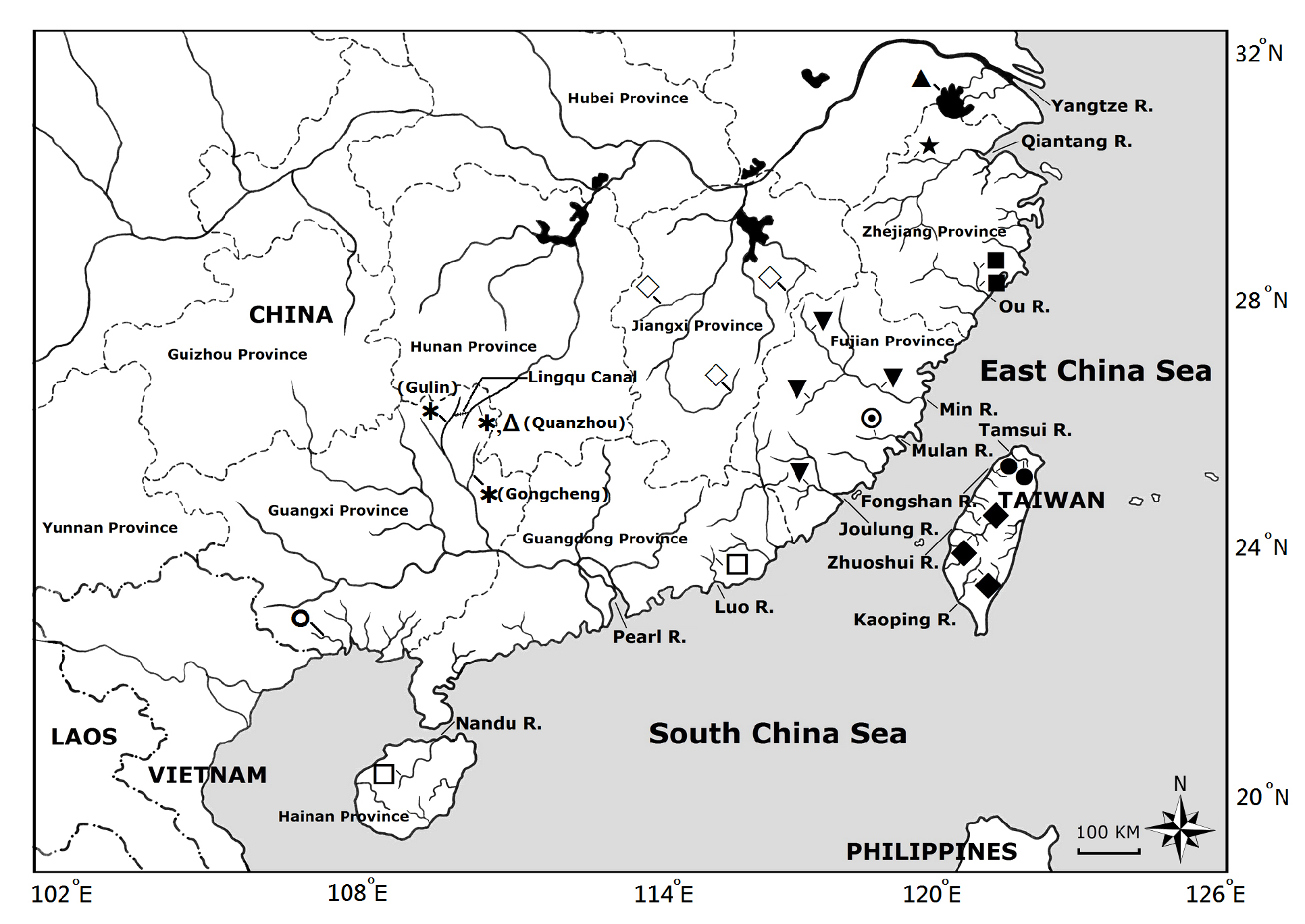
Distribution: Known only from the middle reaches of the Yu River and the Li River, two tributaries of the Pearl River, and the Xiang River, a tributary of the Yangtze River, located in Guangxi and Hunan Provinces of China ( Fig. 3 View Fig ).
Etymology: The Latinized specific name, “ zhangi ” is derived from the name of the Chinese ichthyologist “Professor Chunguang Zhang” in recognition of his great contribution to the fish taxonomic studies in China.
R e m a r k s: T h e m o l e c u l a r p h y l o g e n e t i c evidence of the new species is provided in the next section. Comparing all 19 valid Microphysogobio species from China and two valid species from Taiwan, M. zhangi can be easily distinguished from four valid species ( M. chenhsienensis , M. chinssuensis , M. tafangensis and M. wulonghensis ) by the different types of medial pad on lower lip (centrally divided vs. undivided). As to the remaining 17 species, M. zhangi can be distinguished from M. hsinglungshanensis , M. liaohensis , M. linghensis and M. nudiventris by the different pattern of scale distribution (midventral region covered with scales vs. midventral region naked). Out of the remaining 13 species, M. zhangi can be distinguished from M. amurensis , M. exilicauda , M. tungtingensis and M. yunanensis by having fewer lateral-line scale series (35- 36 vs. 38-39 for M. tungtingensis , 39-42 for M. amurensis , 37-38 for M. exilicauda and 38-40 for M. yunanensis ). Finally, this new species can be distinguished from M. alticorpus , M. brevirostris , M. fukiensis , M. kachekensis , M. kiatingensis , M. microstomus , M. pseudoelongatus and M. xianyouensis by having fewer anal fin rays (3, 5 vs. 3, 6).
Of all the valid species of Microphysogobio from China and Taiwan, M. zhangi appears to be most closely related to M. elongatus based on molecular evidence and some morphological features. Both species share similar pectoral fin rays (modally 1, 12), predorsal scale series (9- 10), spotted dorsal fin and caudal fin. In addition, both species are sympatric ( Fig. 3 View Fig ). However, M. zhangi still can be distinguished from M. elongatus by having (1) fewer anal fin rays (3, 5 vs. 3, 6); (2) fewer lateral-line scale series (modally 35 vs. 37); and (3) different color pattern (presence vs. absence of a black cross-band at interorbital region).
The type specimen of the poorly known species, Microphysogobio chinssuensis multipapillatus Bănărescu and Nalbant, 1973 which was described from Chengtu, China, was also reexamined. Since it shared the same lateral-line scales (37), spotted fins, cheek spot pattern and lip papillae as M. kiatingensis , it should be regarded as a junior synonym of M. kiatingensis .
M. zhangi is further compared with several nominal species of Microphysogobio distributed in Vietnam, Mongolia, and the Yalu River (which forms the border between China and North Korea) and the results are discussed as follows. M. zhangi can be distinguished from M. yaluensis known from the Yalu River ( Mori, 1928) by having fewer anal fin rays (3, 5 vs. 3, 6).
M. zhangi differs from M. anudarini Holcík and Pivnička, 1969 View in CoL , a species known from Mongolia, by having significantly shorter distance between the anus and anal fin origin (15.4- 17.3% of SL, averaged 16.6%, measured from 15 individuals including holotype, 16.3% in the holotype versus 19.0-20.8% of SL, using previous data from the literature reported by Kottelat in 2006). The upper nasal region in M. zhangi can also be easily observed as recessed vs. the region in M. anudarini View in CoL (based on its detailed specimen photograph from the literature reported by Kottelat in 2006).
M. zhangi differs from M. nikolskii ( Dao and Mai, 1959) , one of the two species known from Vietnam, by having fewer lateral-line scales (35- 36 vs. 43). Kottelat (2001b) reported that the taxonomic assignment of M. vietnamica Mai, 1978 View in CoL , the other species from Vietnam, remains unclear. Nevertheless, M. zhangi can be discriminated from M. vietnamica View in CoL by having fewer branched soft rays in the anal fin (5 vs. 6) and smaller dorsal fin (versus a dorsal fin reaching backward almost to the anal fin base).
Molecular phylogenetic analysis
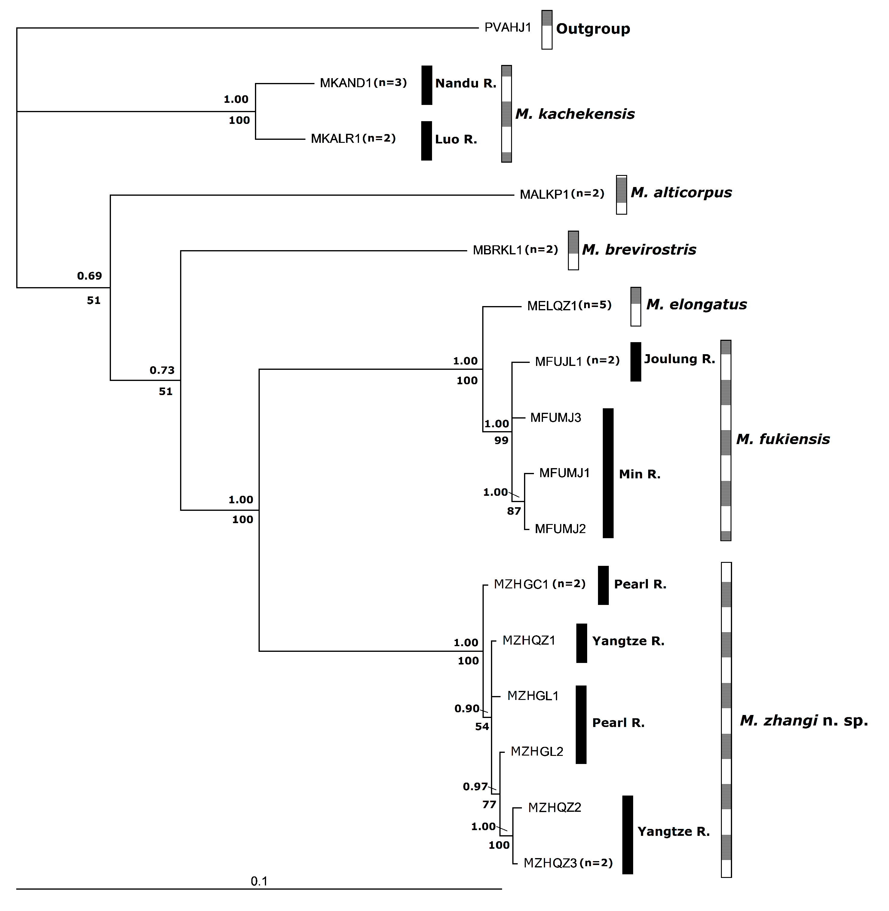
The code of each species and GenBank accession numbers used in this study are given in table 4. Pseudogobio vaillanti ( Sauvage, 1878) was used as outgroup species. The Cyt b sequence from M. zhangi and five closely related species of Microphysogobio . A total of 15 haplotypes from 27 individuals were included in this analysis. The length of Cyt b sequence is 1141 bp in total. This alignment contains 308 total mutations, and 273 polymorphic (segregating) sites. The phylogenetic analyses using both the Bayesian inference (BI) and maximum parsimony (MP) were provided. The phylogenetic tree was reconstructed by the BI analysis based on the HKY+G model. The MP analysis by heuristic search resulted in only one tree, with tree length 507; the consistency index (CI) being 0.7100, retention index (RI) being 0.8088 and homoplasy index (HI) being 0.2840.
The phylogenetic trees reconstructed by the BI and MP methods showed the same tree topology ( Fig. 4 View Fig ) and revealed that M. kachekensis is the earliest offshoot. M. alticorpus and M. brevirostris belong to two independent clades. M. elongatus and M. fukiensis were formed a related sister group, and is sister to M. zhangi . Interspecific nodes between M. zhangi and the closely related species M. elongatus and M. fukiensis sister group with high bootstrap value reach to 100 in MP tree. The posterior probabilities were as high as 1.00 in the BI tree. The interspecific nodes between M. alticorpus and M. brevirostris were supported by lower bootstrap values (51 and 0.69-0.73 in MP and BI, respectively).
The genetic distances of relationships among M. zhangi and five other species were analyzed based on Kimura 2 parameter model (K2P), and ranged from 10.8-15.6%. The genetic distances from 10.8-11.7% and 11.2-11.4% when compared with M. fukiensis and M. elongatus , respectively. The genetic evidence strongly supported M. zhangi to be a distinct species. At the intraspecific level, the genetic distances of M. zhangi from different localities are from 0.1-0.4%. The results are similar to 0.1-0.5% for M. fukiensis which were collected from the Min River and the Joulung River.
| ASIZP |
Academia Sinica Institute of Zoology, Ichthyology Collection |
| R |
Departamento de Geologia, Universidad de Chile |
| T |
Tavera, Department of Geology and Geophysics |
No known copyright restrictions apply. See Agosti, D., Egloff, W., 2009. Taxonomic information exchange and copyright: the Plazi approach. BMC Research Notes 2009, 2:53 for further explanation.
|
Kingdom |
|
|
Phylum |
|
|
Class |
|
|
Order |
|
|
Family |
|
|
Genus |
Microphysogobio zhangi
| Huang, Shih-Pin, Zhao, Yahui, Chen, I-Shiung & Shao, Kwang-Tsao 2022 |
M. zhangi
| Sun & Zhao 2022 |
M. zhangi
| Sun & Zhao 2022 |
M. zhangi
| Sun & Zhao 2022 |
M. zhangi
| Sun & Zhao 2022 |
M. vietnamica
| Mai 1978 |
M. vietnamica
| Mai 1978 |
M. anudarini Holcík and Pivnička, 1969
| Holcik and Pivnicka 1969 |
M. anudarini
| Holcik and Pivnicka 1969 |