
Mnementh brennei, Martens, 2007
|
publication ID |
https://doi.org/ 10.1080/00222930601157183 |
|
persistent identifier |
https://treatment.plazi.org/id/816C6C54-FFE2-FFDC-FE15-56C8FD9A8C04 |
|
treatment provided by |
Felipe |
|
scientific name |
Mnementh brennei |
| status |
sp. nov. |
Mnementh brennei n. sp.
( Figures 2–5 View Figure 2 )
Type locality
A dam near Blinkvlei, Van Rhynsdorp area, Western Cape Province (31 ° 439290 S, 18 ° 559280 E). Approximately 200 males and females were collected on 7 September 2001 by K. Martens and L. Hoenson ( RSA /01/03). Few specimens were collected with a hand net on soft, muddy substrate; the majority of the specimens, however, was handpicked from submerged logs and branches. Accompanying ostracod fauna: Potamocypris humilis ( Sars, 1924) ; P. paludum Gauthier, 1939 ; Isocypris perangusta G. W. Muller, 1908 ; Sarscypridopsis sp. ; Cypridopsinae n. gen. n. sp.; Liocypris grandis Sars, 1924 ; Megalocypris n. sp.
Type material
Holotype: male (OC.2913): soft parts dissected in glycerine in a sealed slide, valves stored dry in a micropalaeontological slide. Allotype: female (OC.2914), dissected and stored as the holotype. Paratypes: two males and one female dissected and stored as the holotype (OC.2915–2917), two males (OC.2920, 2921) and three females (OC.2918, 2919, 2922) stored in toto dry in a micropalaeontological slide after use for SEM; ca 50 males and females stored in toto in 80% alcohol (OC.2923) in one tube, ca 20 males and females stored in toto in 80% alcohol ( SAM.A45530) .
Repository
Holotype, allotype, dissected and dried paratypes, and one tube with wet paratypes are in the Ostracod Collection of the Royal Belgian Institute of Natural Sciences (Brussels, Belgium). One tube with wet paratypes is in the South African Museum (Cape Town , South Africa) .
Other material investigated (all collected by K. Martens and L. Hoenson)
RSA /01/005: one female collected on 7 September 2001 from Blinkvlei, Van Rhynsdorp (31 ° 449210 S, 18 ° 559230 E). Accompanying ostracod fauna: Liocypris grandis Sars, 1924 ; Isocypris priomena G. W. Muller, 1908 ; Megalocypris n. sp.; Homocypris conoidea Sars, 1924 ; Sarscypridopsis ssp.; Potamocypris paludum Gauthier, 1939 .
RSA /01/007: five males and one female collected on 8 September 2001 from a pool near Palmiet Vlei, near Van Rhynsdorp (31 ° 309420 S, 18 ° 519380 E). Accompanying ostracod fauna: Megalocypris n. sp .
RSA /01/008: ca 100 males, females, and juveniles collected on 8 September 2001 from Palmiet Vlei, near Van Rhynsdorp (31 ° 309220 S, 18 ° 519230 E). Palmiet Vlei is a large (diameter ca 500 m), natural, temporary pan, with an estimated maximum depth of 2–3 m. High turbidity, ostracods were collected amongst emerging macrophytes. Accompanying ostracod fauna: Megalocypris n. sp.; Homocypris conoidea Sars, 1924 .
RSA /01/009: 10 females and juveniles, collected on 8 September 2001 from a pool on Rd R 399 Picketburg-Velddrift (32 ° 489320 S, 18 ° 229350 E). Accompanying ostracod fauna: Chrissia ametra (G. W. Muller, 1908) ; Megalocypris hispida Sars, 1924 ; Heterocypris n. sp.; Sarscypridopsis sp. ; Cypridopsinae n. gen. n. sp.; Homocypris conoidea Sars, 1924 .
Derivation of name
The species is named after my friend and colleague, Luc ‘‘brenne’’ Brendonck (University of Leuven, Belgium), in recognition of his major contribution to the knowledge of Anostracan biology, especially in southern Africa, and in fond memory of years of friendship and rewarding collaboration.
Diagnosis
Large species, with globular carapace, very hirsute and pitted. Anterior margin of both valves produced towards the ventral side. Anterior calcified inner lamellae wide. Anterior part in dorsal view pointed like a rostrum, greatest width (. half the length) in the middle. Prehensile palps (male) asymmetrical, with terminal segment in right palp larger than in the left palp. T 1-palp in female short and wide. Hemipenis with dorsal lobe of the lateral shield large, ventral lobe of the lateral shield long and rectangular, distally produced towards the ventral side. T 2 stoutly built. Caudal ramus slender.
Additional description of male
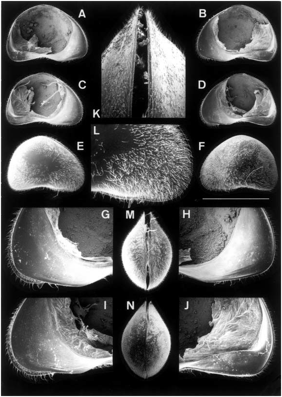
Carapace ( Figure 2N View Figure 2 ) in dorsal view rounded, with greatest width situated in the middle and being more than half of the length; posterior part rounded, anterior part pointed, in both dorsal and ventral view like a rostrum ( Figure 2K View Figure 2 ), RV slightly overlapping LV. Ventrally ( Figure 2M View Figure 2 ), RV overlapping LV in the middle. Valves in external view ( Figure 2E, F View Figure 2 ) highly arched, greatest height situated slightly behind the middle; anterior margin ventrally produced, posterior margin nearly straight; ventral margin strongly sinuous; valve surface very hirsute, except in the centre, and pitted to varying degrees.
RV ( Figure 2D View Figure 2 ) with anterior margin ventrally produced, posterior margin broadly rounded, nearly straight; dorsal margin highly arched; ventral margin strongly sinuous in the middle. Calcified part of the inner lamella very wide anteriorly, narrower along the posterior side. Anterior valve margin with clear, but submarginal, selvage ( Figure 2I View Figure 2 ), antero-ventrally with some traces of inner lists; posterior margin without selvage, but with striking inner list, not running parallel to the valve margin.
LV ( Figure 2C View Figure 2 ) with outline similar to that of RV. Anterior calcified inner lamella wide, with remnants of inner lists, but no selvage; posterior calcified inner lamella narrower, also with large inner list not running parallel to valve margin.
Central muscle scars with mandibular scars large and with scar pattern of adductor muscles conforming to that of the subfamily.
A1 ( Figure 3C) seven-segmented and conform to the subfamily. Terminal segment about as long as wide, all natatory setae long. Aesthetasc Ya less than twice as long as the short seta on this segment. Rome organ minute.
A2 ( Figure 3B) with natatory setae reaching well beyond tip of terminal claws; aesthetasc Y short. Apical chaetotaxy ( Figure 3A) with typical sexual dimorphic characters, but with claw G 1 short; seta of y 3 about twice as long as the aesthetasc and seta z3 as long as claw z1 (seta is longer in Cypris ).
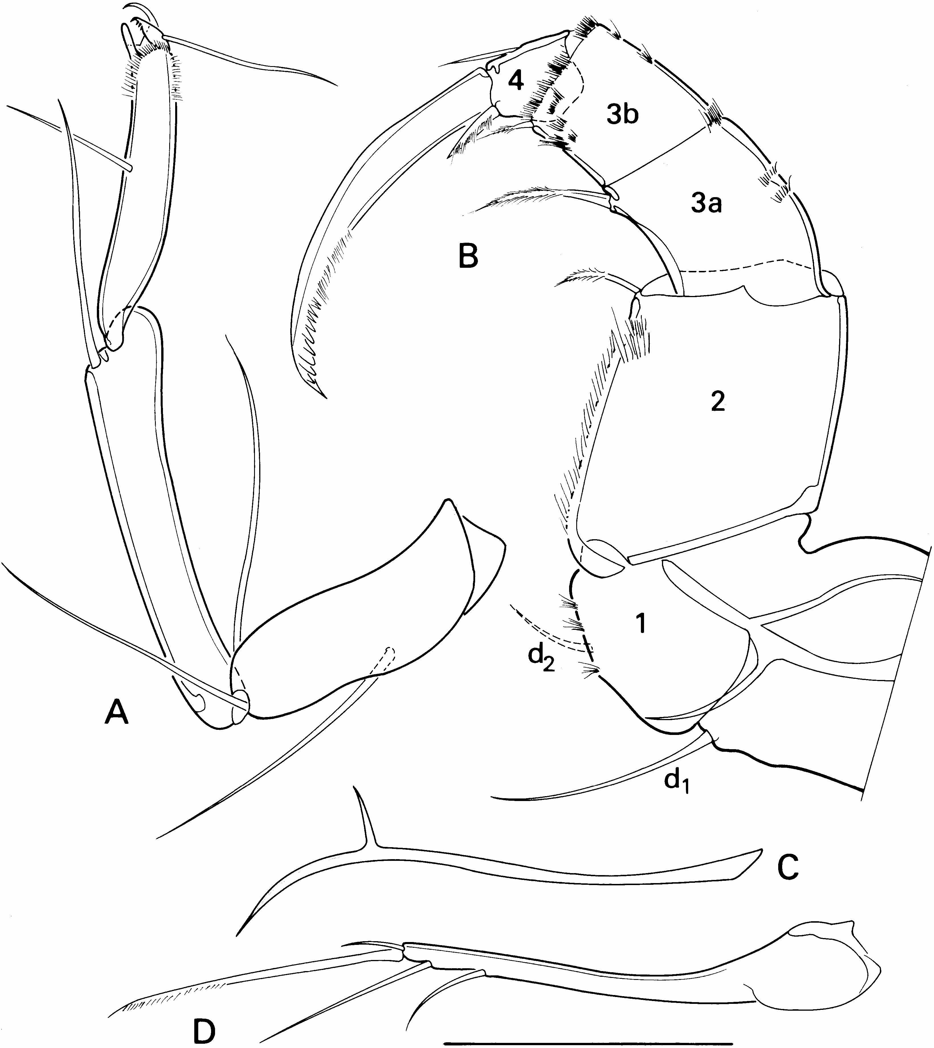
Md with coxa elongated ( Figure 3E). Md-palp (Figure 4A) four-segmented. First segment with large respiratory plate and a group of four apical setae: one long and smooth, one short, narrow and smooth (alpha-seta), the latter flanked by one large ‘‘s’’- seta, set with a double row of setulae, and by one short ‘‘s’’-seta. Second segment with two groups of apical setae: an internal group, consisting of a short and hirsute beta-seta, three long and smooth and one long and barbed setae, external group consisting of two long and one shorter setae. Penultimate segment with four dorsal, subapical setae; apically with one narrow and hirsute gamma-seta of intermediate length, two centrally situated smooth setae and three subequal and subapical setae on the internal side. Terminal segment (not shown) with four claw-like and with two or three short and slender setae, all apically inserted.
Mx1 with three endites, a two-segmented palp ( Figure 3D) and a large respiratory plate. Second palp segment about three times as long as basal width. Third endite with two smooth apical claws and with a long and stout lateral seta, apart from the normal apical setae.
T 1 with asymmetrical prehensile palps, a large endopodite and a respiratory plate (exopodite) with six plumose rays (five long and one short). Endopodite (Figure 4B) elongated, carrying two short ‘‘a’’-setae, one larger, central ‘‘b’’-seta and one lateral ‘‘d’’- seta, ‘‘c’’-seta absent. Apical chaetotaxy consisting of 14 setae of different size and shape.
Right prehensile palp (Figure 4D) with elongated basal segment, carrying one apical sensory seta; terminal segment three-dimensionally curved; when flattened in a slide broad, with a narrow apex and with distal margin showing a blunt angle, apically with one broad sensory organ.
Left prehensile palp (Figure 4E) with basal segment slightly shorter than that of the right palp; terminal segment with broad base, rapidly narrowing towards the tip, the latter bearing a single sensory organ.
T 2 ( Figure 5B View Figure 5 ) stout, with seta d 1 more than three times as long as d 2; all segments short, wide and hairy. Second segment with one short apical seta. Penultimate segment divided; segment 3A with one subapical seta; segment 3B with one large and one minute apical setae. Fourth segment with one lateral seta and, apart from the apical claw, with one other apical clawlike seta; apical claw short and stout and only in its distal half set with a double row of spines.
T 3 ( Figure 5A View Figure 5 ) a cleaning limb with an apical pincer and without further special features.
Caudal ramus ( Figure 5D View Figure 5 ) with ramus narrow and curved, proximally swollen, distally carrying two claws and two setae and with ventral margin serrated with minute setulae. Attachment of the caudal ramus ( Figure 5C View Figure 5 ) distally split into a short ventral and a longer dorsal branch.
Figure 4. Mnementh brennei n. gen. n. sp., Western Cape, South Africa. (A) Male, Md-palp (chaetotaxy of distal segment not shown) (OC.2913); (B) male, left T 1, apical chaetotaxy of endopodite (OC.2915); (C) female, T 1 (OC.2914); (D) male, right prehensile palp (OC.2913); (E) male, left prehensile palp (OC.2915). Scale: 20× (A, D, E); 40× (B, C).
Hemipenis ( Figure 3F, G) with a broadly rounded medial shield, asymmetrically expanded towards the ventral side and with a ventral protuberance; dorsal lobe of the lateral shield symmetrically rounded, ventral lobe of the lateral shield long and distally expanding (but see Figure 3G with pointed vls). Internal anatomy with the normal labyrinth, consisting of the elongated parts ‘‘a’’ and ‘‘c’’ and the rounded hinge-joint ‘‘b’’, followed by the three to five ‘‘8’’-shaped coils of the inner spermiductus, situated distally from the labyrinth, the sclerotized semi-circular loop and the various hollow trabecules, leading to the bursa copulatrix.
Testicle tubes present: 2×4.
Additional description of female
Valves ( Figure 2A, B, G, H, J View Figure 2 ) basically as in the male, but in general somewhat larger and even higher. Carapace in dorsal and ventral view ( Figure 2K, M View Figure 2 ) with greatest width situated in the middle; RV frontally and ventrally overlapping LV.
A2 ( Figure 3A) basically as in the male, apart from the normal sexual dimorphism in the apical chaetotaxy.
T 1 (Figure 4C) with palp undivided, carrying the normal unequal three apical setae and no lateral setae.
T 2 somewhat plumper and heavier than in the male.
Genital lobe undivided and without specific characteristics.
Ovaria on both sides curved upwards.
For measurements see Table I.
| SAM |
South African Museum |
| R |
Departamento de Geologia, Universidad de Chile |
| T |
Tavera, Department of Geology and Geophysics |
| RV |
Collection of Leptospira Strains |
No known copyright restrictions apply. See Agosti, D., Egloff, W., 2009. Taxonomic information exchange and copyright: the Plazi approach. BMC Research Notes 2009, 2:53 for further explanation.
|
Kingdom |
|
|
Phylum |
|
|
Class |
|
|
Order |
|
|
Family |
|
|
Genus |