
Neelus koseli, Kováč, Ľubomír & Papáč, Vladimír, 2010
|
publication ID |
https://doi.org/ 10.5281/zenodo.276298 |
|
DOI |
https://doi.org/10.5281/zenodo.6212256 |
|
persistent identifier |
https://treatment.plazi.org/id/5E348799-1A52-A329-FF0D-5BA4606BEB42 |
|
treatment provided by |
Plazi |
|
scientific name |
Neelus koseli |
| status |
sp. nov. |
Neelus koseli sp. nov.
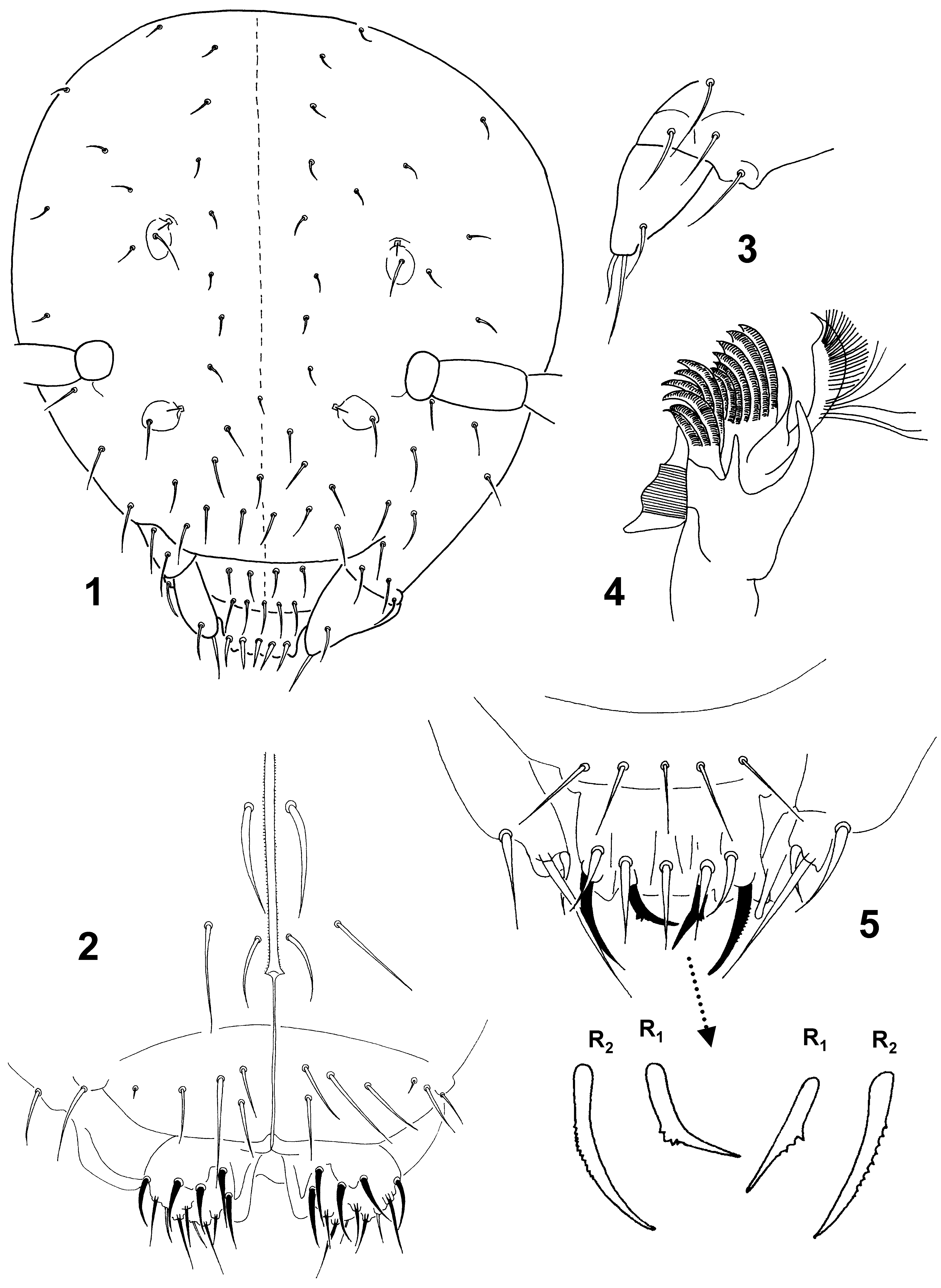
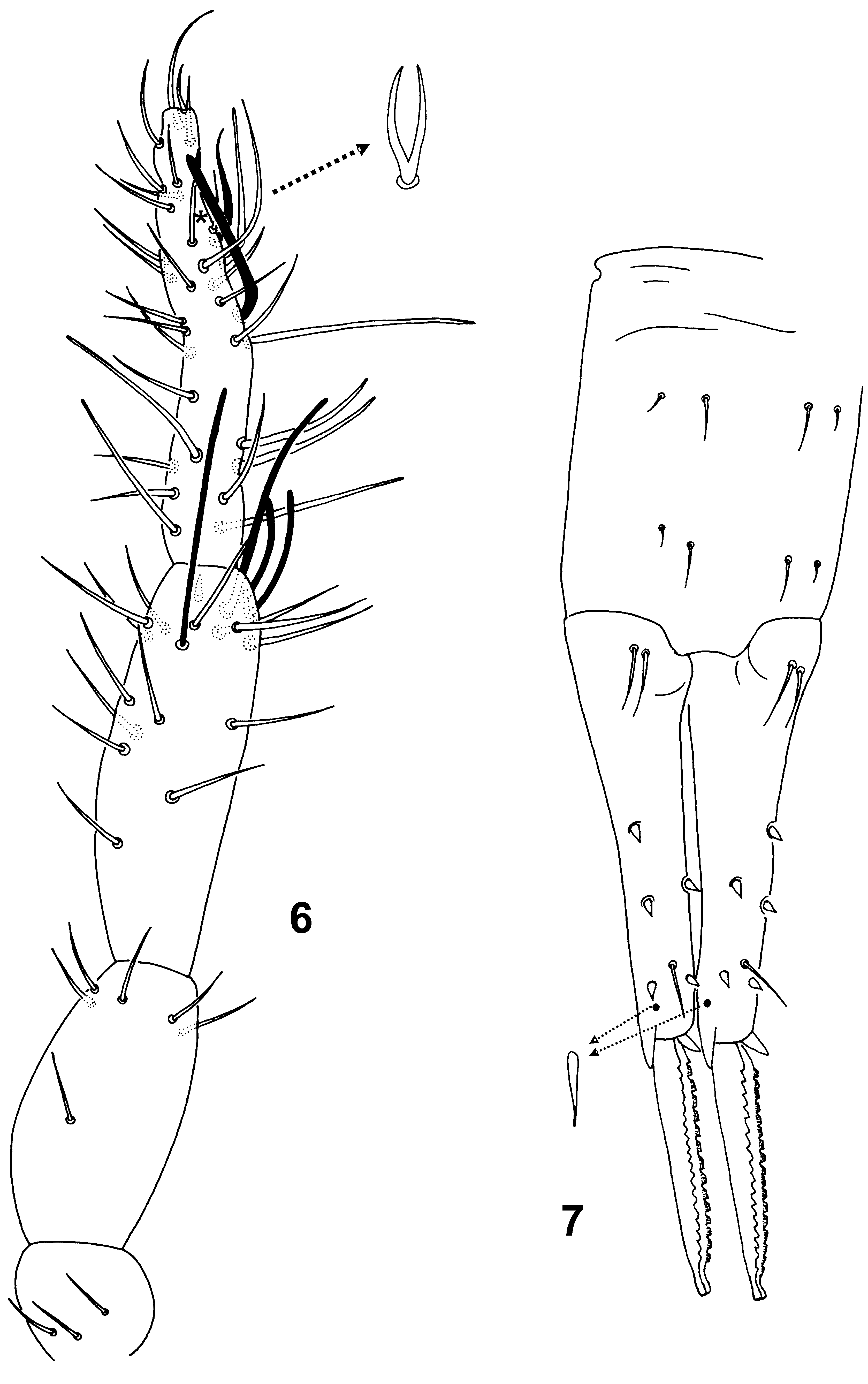
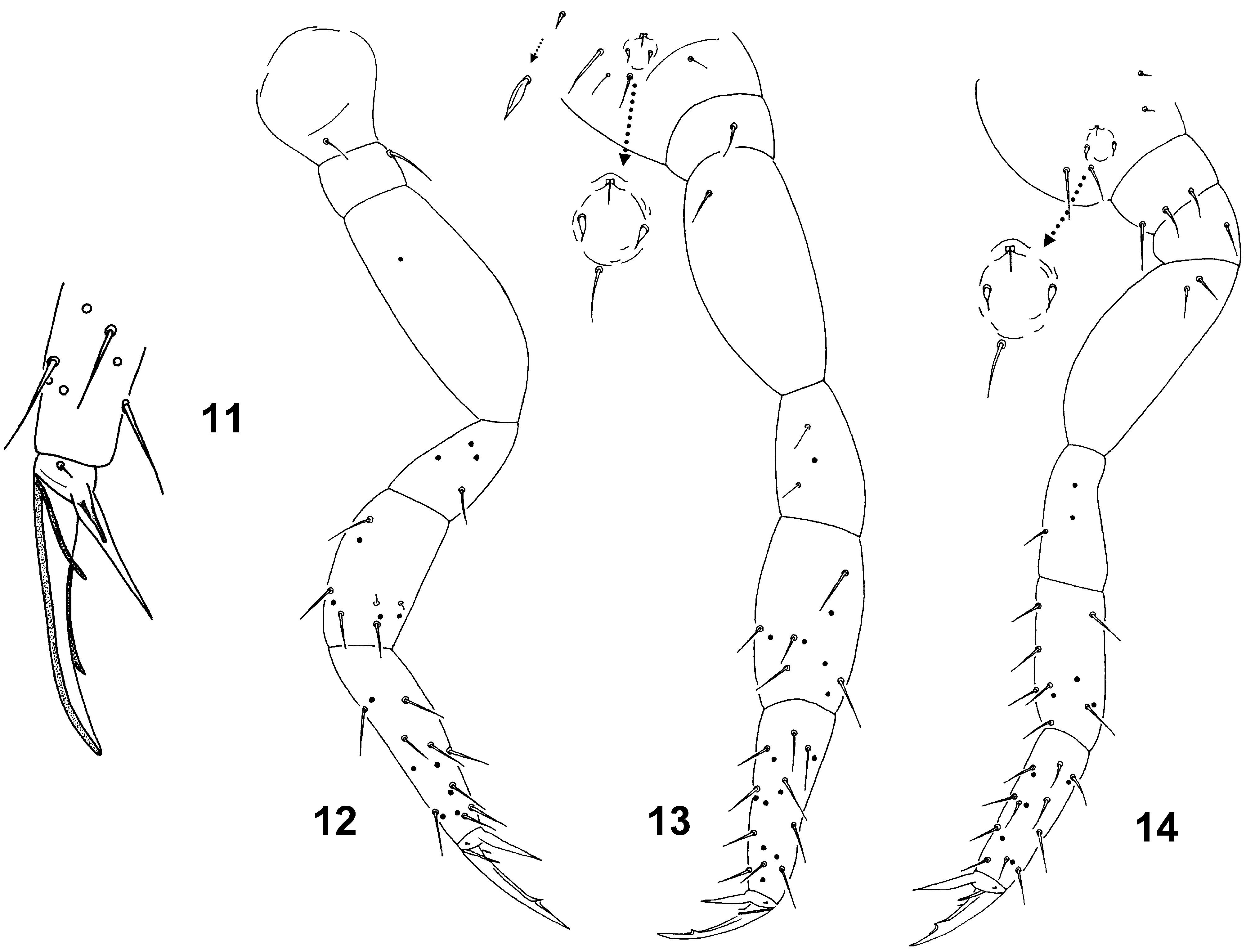
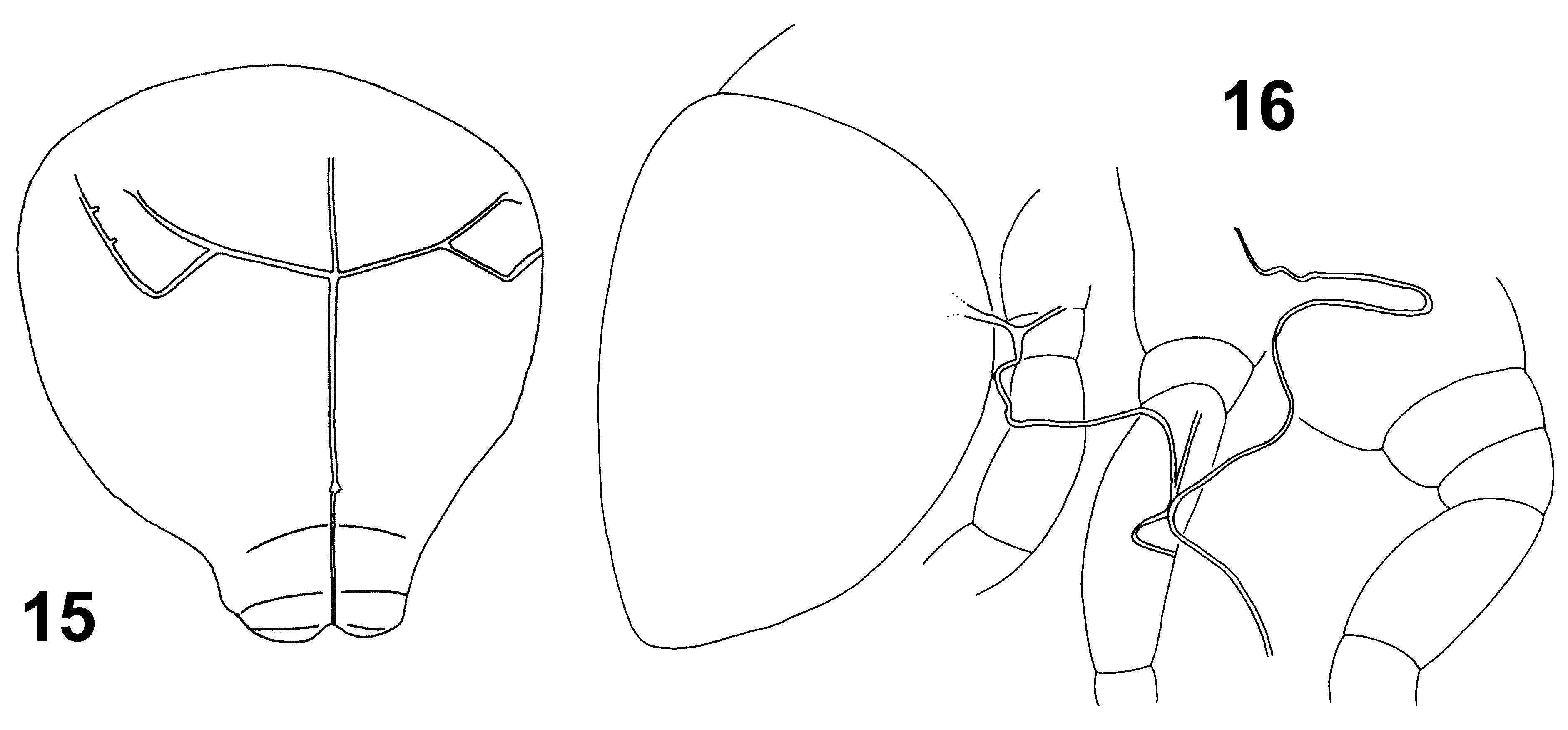
Figs 1–16 View FIGURES 1 – 5 View FIGURES 6 – 7 View FIGURES 11 – 14 View FIGURES 15 – 16
Neelus sp. Kováċ & Krchová (2007)
Diagnosis. Posterior part of head behind antennae with microsetae, dorsal side of hind abdomen with spinelike microsetae situated in 10 rows. Prelabral/labral setae formula (p-, m-, a-row): 4/5, 5, 4. Anterior labral setae R1 and R2 thick, curved; R1 with external edge medially roughly serrate; R2 with external edge finely serrate. Ventral side of head with posterior macrosetae of postmedian area smooth, thickened and curved. Sensory field of thorax with only 2 surrounding (marginal) setae. Ant. I segment with 3 setae, external side of Ant. IV with 8 thin and curved macrosensilla finely blunt at the tip. Unguis apparently elongated furnished with 2 long lateral teeth and 1 inner tooth, weak incision in basal 1/3 present. Manubrium with 4+4 dorsal setae. Mucro with both dorsal lamellae serrated, tip of mucro undivided, rounded.
Type material. Holotype: female on slide (No.162–99), Slovakia, Čierna hora Mts., Humenecká Cave, central part of the cave, aphotic zone, 20 m from the cave entrance, pitfall trap with ethylalcohol, 17.iii.– 27.v.1999, leg. Ľ. Kováč. Paratypes: 2 females and 1 juvenile on slides (No. 162–99), the same data as the holotype; 1 female on slide (No. 163–99), extracted from the rotten wood, 27.v.1999, leg. Ľ. Kováč; 1 female and 1 juvenile on slides (No. 166–99), entrance chimney, dysphotic zone, 5 m from the cave entrance, extracted from the organic material (humus, leaf litter, pieces of decaying wood) deposited on the floor, 27.v.1999, leg. Ľ. Kováč; 2 females on slides (No. 110–00), central part of the cave, hand collecting on rotten wood, 23.vi.2000, leg. P. Ľuptáčik. Type material (holotype and 3 paratypes) saved in collection of the Muséum national d´Histoire naturelle ( MNHN) in Paris; other paratypes kept in the Department of Zoology, Institute of Biology & Ecology, Faculty of Science, P. J. Šafárik University, Košice.
Other material. Slovakia, Čierna Hora Mts., Medvedia Cave, 8 females on slides (No. 255–99, No. 256– 99), entrance passage, dysphotic/aphotic zone, 10 m from the cave entrance, pitfall traps with ethylalcohol, 27.vi.–7.viii.1999, leg. Ľ. Kováč; 1 female and 1 juvenile on slides (No. 12–07), extracted from organic material deposited on the cave floor (humic sediment, rotten wood, scattered bat guano), 16.iii.2007, leg. Ľ. Kováč.
Slovakia, Slovak Karst, Jasovská Cave, 6 females on slides (No.390–03, No. 419–03, No. 564–03, No. 732–03, No. 733–03), Guano Hall, aphotic zone, 50 m from the nearest entrance, thick deposits of old and fresh bat guano, pitfall traps with formaldehyde, 5.iii.–30.x.2003, leg. M. Lukáň, K. Lukáňová and Ľ. Kováč; 1 female on slide (No. 727–03), Komín near Husitská sieň, pitfall trap with beer and glycerol (1:1), 10.ix.– 30.x.2003, leg. M. Lukáň and K. Lukáňová; 1 female on slide (No. 780–03), Melčova chodba, extraction of silty sediment, 1.V.2003, leg. M. Lukáň and K. Lukáňová; 1 female (?) on slide (No. 571–03), Jedáleň, pitfall trap with beer and glycerol (1:1), 8.vii.–10.ix.2003, leg. M. Lukáň and K. Lukáňová.
Description. Body length up to 0.9–1.0 mm, habitus typical of genus. Colour of body mostly pale orange, pigment scattered in small irregular patches over dorsal part of head, thorax, abdomen and subcoxae of legs. In darker specimens dorsal part of abdomen axially with narrow pale longitudinal stripe. Cuticle finely granulated, integumentary channels observed on ventral side of head and lateral parts of thorax ( Figs 15 and 16 View FIGURES 15 – 16 ).
Sensory fields. Sensory fields placed in small depressions each with secretory rod (8 µm), i.e. blunt, straight seta with basal part inserted in the cuticle in the upper margin of field. Fields’ arrangement: (a) on head—anterior and posterior field (20 x 15 µm; Fig. 1 View FIGURES 1 – 5 ) each with secretory rod and 1 internal seta (25 and 15 µm, respectively); (b) thoracal field (30 x 60 µm; Fig. 8a) with secretory rod, 3 curved spines (14 µm) arranged in triangle and 2 marginal external setae (15 µm); (c) abdominal field (30 x 20 µm; Fig. 8c) with secretory rod, 1 curved spine (7 µm), 2 marginal setae (1 internal, 1 external; 10 µm each); (d) fields at base of legs II and III (20 x 15 µm; Fig. 13, 14 View FIGURES 11 – 14 ) each with secretory rod, 2 curved spines (6 µm) and 1 marginal external seta (18 µm). Sharp microsensillum (8 µm) placed above sensory field at the base of leg II ( Fig. 13 View FIGURES 11 – 14 ). Lateral part of abdomen about the base of leg III with small sensory field furnished with short secretory rod (4 µm) and with stronger rod (5 µm) in cup-like depression above it.
FIGURES 8–10. Neelus koseli sp. nov.: 8, hind abdomen, (a) thoracal sensory field (enlarged), (b) spine-like microseta (enlarged), (c) abdominal sensory field (enlarged), (d) neosminthuroid seta (enlarged), e) symetrically swollen microsensillum; 9, lower anal valve and female genital plate; 10, tubus ventralis, posterior side.
Head. Head length and width as 320 and 300 µm, respectively. Eyes absent. Dorsal side with smooth and pointed setae ( Fig. 1 View FIGURES 1 – 5 ); frontal part with ordinary setae (15–20 µm), 2 unpaired axial setae present; posterior part behind antennae with microsetae (6–7 µm). Labrum with 5,5,4 setae, 4 prelabrals ( Fig. 5 View FIGURES 1 – 5 ). Pattern of labral setae: a-row 2R1 + 2R2, m-row m + 2r1 + 2r2, p-row with 5 ordinary setae. Anterior setae R1 and R2 thick, curved, R2 (25 µm) longer than R1 (20 µm); R1 with external edge medially roughly serrate, R2 with external edge finely serrate. Medial setae (m-row) equal (18 µm), smooth, spine-like. Posterior setae (p-row) smooth, lateral ones thicker and longer than those axial (20 and 18 µm, respectively). Maxillary palp simple, with 1 enlarged sublobal seta ( Fig. 3 View FIGURES 1 – 5 ). Ventral side of head with 3+3 smooth postmedian setae ( Fig. 2 View FIGURES 1 – 5 ); 2+2 anterior setae unequal: lateral ones longer, straight (36 µm), axial shorther and curved (20 µm); posterior 1+1 are thickened and curved macrosetae (38 µm). Basomedian field of labium with 4+4 setae ( Fig. 2 View FIGURES 1 – 5 ), medial ones macrosetae (36 µm), others mesosetae (18 µm); basolateral field with 2+2 setae (1+1 axial shorter). Mandible normal, strong, with 5 apical teeth. Maxilla as in Fig. 4 View FIGURES 1 – 5 .
Antennae. Antennal segments III and IV distinctly separated ( Fig. 6 View FIGURES 6 – 7 ). Length of antennae 190 µm, ratio antenna/head = 0.6; length of antennal segments I, II, III and IV as 20, 45, 58 and 67 µm. Ant. I furnished with 3 short setae (10 µm). Ant. II with 1 medial seta and 5 apical setae arranged in a whorl. Ant. III organ consists of 2 rather long sensory rods (20 µm), 2 long guard sensilla (external and dorsal, 38 µm each) and short ventral spine-like seta (4 µm). Ant. IV with ordinary setae mostly on internal side, external side with 8 thin and curved macrosensilla finely blunt at the tip (up to 39 µm); ventrally with 1 long and thick sensillum (23 µm) and 1 apical, basally thick shorter sensillum (12 µm) with abruptly narrowed and curved tip; dorsally with forked subapical seta (8 µm); subapically with 5 short setae (8 µm).
Thorax and abdomen. Dorsal side of thorax and abdomen sparsely covered with microsetae (3 μm), hind part of abdomen with spine-like microsetae (5 μm) situated in about 10 rows (Fig. 8). Mid-abdomen dorsally with swollen symmetrical microsensilla (3 µm; Fig. 8e), their overall number not seen clearly. Upper edge of prefurcal area with 1+1 short, sharply pointed and curved neosminthuroid setae (8 µm) (Fig. 8d). Abdominal segments V and VI cryptic. Genital plate with 4+4 mesosetae (12 µm) and 1+1 axial microsetae (4 µm) (Figs 8 and 9). Anal opening transversal; upper anal valve with 3+3 mesosetae (20–25 µm), 1+1 anterior short setae (12 µm) and 1 unpaired posterior seta (12 µm); lower anal valve with 1+1 macrosetae (33 µm), 4+4 mesosetae (20–23 µm) and 2+2 microsetae (7 µm) (Figs 8 and 9).
Appendages. Setae numbers of leg I–III ( Figs 12–14 View FIGURES 11 – 14 ) (number of longer setae in parenthesis): subcoxae I with 1, 1, 3(1); subcoxae II with 1(1), 1, 1; coxae with 1, 1, 2; trochantera with 4, 3, 3; femora with 10, 10, 10 and tibiotarsi with 15, 18, 16. Some of them as thin meso- or microsetae: leg I—coxa with 1, trochanter with 3, femur with 2; leg II—trochanter with 3; leg III—trochanter with 1, femur with 1. Tibiotarsal tenent seta straight and pointed (24 µm). Unguis narrow and elongated, both unguis and unguiculus unequally long in leg I, II and III: unguis 52, 48 and 42 µm, respectively, unguiculus 23, 25 and 25 µm, respectively. Length ratio unguis I (inner margin)/Ti. I width (40 / 15 µm) = 2.7. Unguis furnished with 2 long lateral teeth and 1 inner tooth, weak incision in basal 1/3 present ( Fig. 11 View FIGURES 11 – 14 ); unguiculus untoothed without apical filament. Tubus ventralis with 2+2 distal setae and posterior lobe (Fig. 10). Retinaculum with 3+3 teeth and no setae. Furca well developed ( Fig. 7 View FIGURES 6 – 7 ), length of manubrium, dens and mucro: 110, 170 and 95 µm, respectively. Manubrium dorsally with 4+4 setae, lateral ones distinctly shorter (10 µm) than those axial (21 µm). Dens in basal part with 2+2 dorsal setae, lateral ones (17 µm) shorter than those axial (21 µm); apically with 1+1 broad, blunt lateral spines and 1 medial sharp spine (12 µm) on ventral side; distal part dorsally with 3 external (E1–E3) and 2 internal (J1–J2) spines (9 µm each), and 1 medial, subapical seta (22 µm). In spines E1 and J1 basal circle apparently absent. Mucro with serrated dorsal lamellae and rounded tip.
Only females known.
Etymology. The new species is named in honour of our friend, zoologist and speleologist, Dr. Vladimír Košel (Comenius University, Bratislava) who added considerably to the knowledge of Oligochaeta and Diptera of the Slovak caves.
Biology. Digestive track with four apparent compartments (diverticula) was filled mainly with clay and organic particles, fungal hyphae were recognized in small extent. In several specimens fungal spores predominated in the gut.
Distribution. Neelus koseli sp. nov. was discovered in three caves of the eastern Slovakia. The caves are located in two adjacent karst areas: (1) Čierna hora Mts. (Humenecká and Medvedia caves), and (2) Slovak Karst (Jasovská Cave). Both areas belong to the same geological unit—“Silicikum“ carbonate platform deposited from the Late Permian until the Late Jurassic and later dissintegrated into smaller structural units during geological history of the territory ( Kováč & Plašienka 2002). That may explain rather disjunctive distribution range of the species. Neelus koseli sp. nov. belongs the most probably to older fauna with Kenozoic origin that inhabited subterranean environments of the ”Silicikum“ platform before its later tectonic fragmentation and separation into smaller units. Discovery of the new species confirms the presence of obligate cave-dwelling fauna in the Western Carpathians, the northern limit of distributional ranges of terrestrial troglobionts in Europe ( Culver et al. 2006). The close affinity of N. koseli sp. nov. to N. klisurensis sp. nov. supports the idea of common historical origin and evolution of obligate cave fauna of the Western Carpathians and the Balkan Peninsula (e.g. Gulička 1975).
Discussion. See Discussion of N. klisuresnsis sp. nov.
| MNHN |
Museum National d'Histoire Naturelle |
No known copyright restrictions apply. See Agosti, D., Egloff, W., 2009. Taxonomic information exchange and copyright: the Plazi approach. BMC Research Notes 2009, 2:53 for further explanation.
|
Kingdom |
|
|
Phylum |
|
|
Class |
|
|
Order |
|
|
Family |
|
|
Genus |