
Neogreenia zizyphi Tang, 1995
|
publication ID |
https://doi.org/ 10.11646/zootaxa.5418.5.1 |
|
publication LSID |
lsid:zoobank.org:pub:0DDD7278-0E9C-4979-ACB9-1915D4209282 |
|
DOI |
https://doi.org/10.5281/zenodo.10787189 |
|
persistent identifier |
https://treatment.plazi.org/id/03C24061-FFC8-D248-FF0F-21E3FE97F9E5 |
|
treatment provided by |
Plazi |
|
scientific name |
Neogreenia zizyphi Tang, 1995 |
| status |
|
Neogreenia zizyphi Tang, 1995 View in CoL
Kuwaniella zizyphi Tang 1984: 123 View in CoL ; Kosztarab et al. 1986: 9; Foldi 2001: 205. Nomen nudum.
Neogreenia zizyphi Tang View in CoL in Tang & Hao 1995: 82. Type data: CHINA: Shanxi Province, Taigu County, on Ziziphus jujuba View in CoL . Syntypes, female and first instar, type depository: Entomological Institute, Shanxi Agricultural University, Taigu, Shanxi, China.
Host plants: Ziziphus jujuba Mill. ( Rhamnaceae ).
Distribution: China (Shanxi).
Remarks. Tang & Hao (1995) described the adult female and briefly described the adult male. Below, we redescribe the adult female and adult male and provide modern descriptions of the second-instar nymph, third-instar female and male nymph and pupa.
Material examined. 3 ♀♀, CHINA: Shanxi Prov., Jinzhong City, Taigu district, Beiguang village , on Ziziphus jujuba , San-an Wu leg., 4.v.2023, mounted singly on 3 slides; same locality and host data, 1 second-instar nymph, 28.vi.2021, mounted singly on 1 slide; 3 second-instar nymphs, 4.v.2023, 3 mounted together on 1 slide; 1 third-instar ♀ nymph, 28.vi.2021, mounted singly on 1 slide; 3 third-instar ♀♀ nymphs, 4.ix.2021, 3 mounted together on 1 slide; 1 third-instar ♂ nymph, 28.iv.2023, mounted singly on 1 slide; 2 third-instar ♂ nymphs, 4.v.2023, 2 mounted together on 1 slide; 2 adult ♂♂, 2 mounted singly on 2 slides and 2 pairs of forewings mounted singly on 2 slides; and 1 ♂ pupal exuviae, 14.v.2023, mounted singly on 1 slide.
Adult female
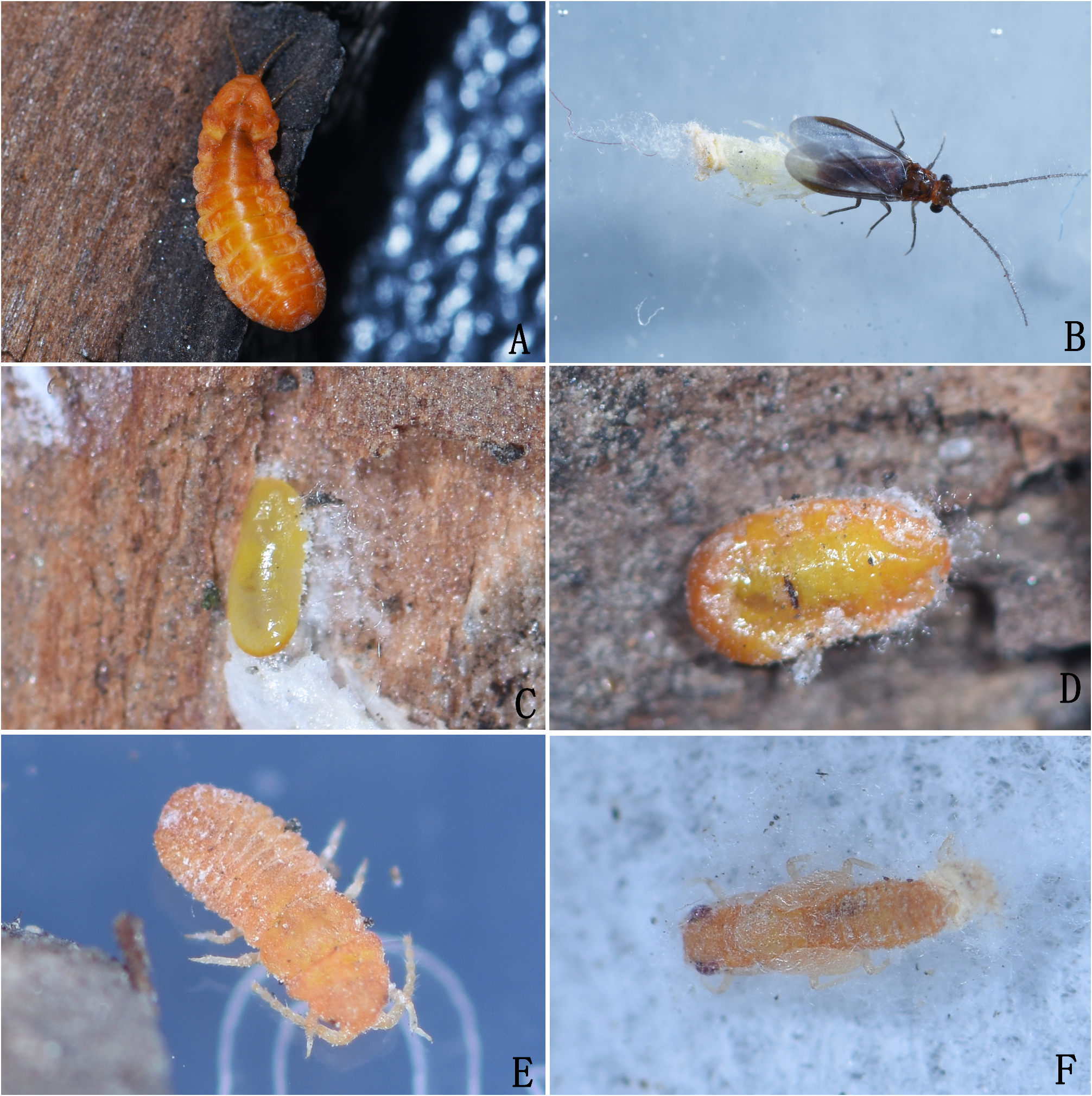
Appearance in life ( Fig. 13A View FIGURE 13 ). Body elongate, with dorsum convex and venter flat; antennae situated on apex of head; legs present; body orange, eyes black.
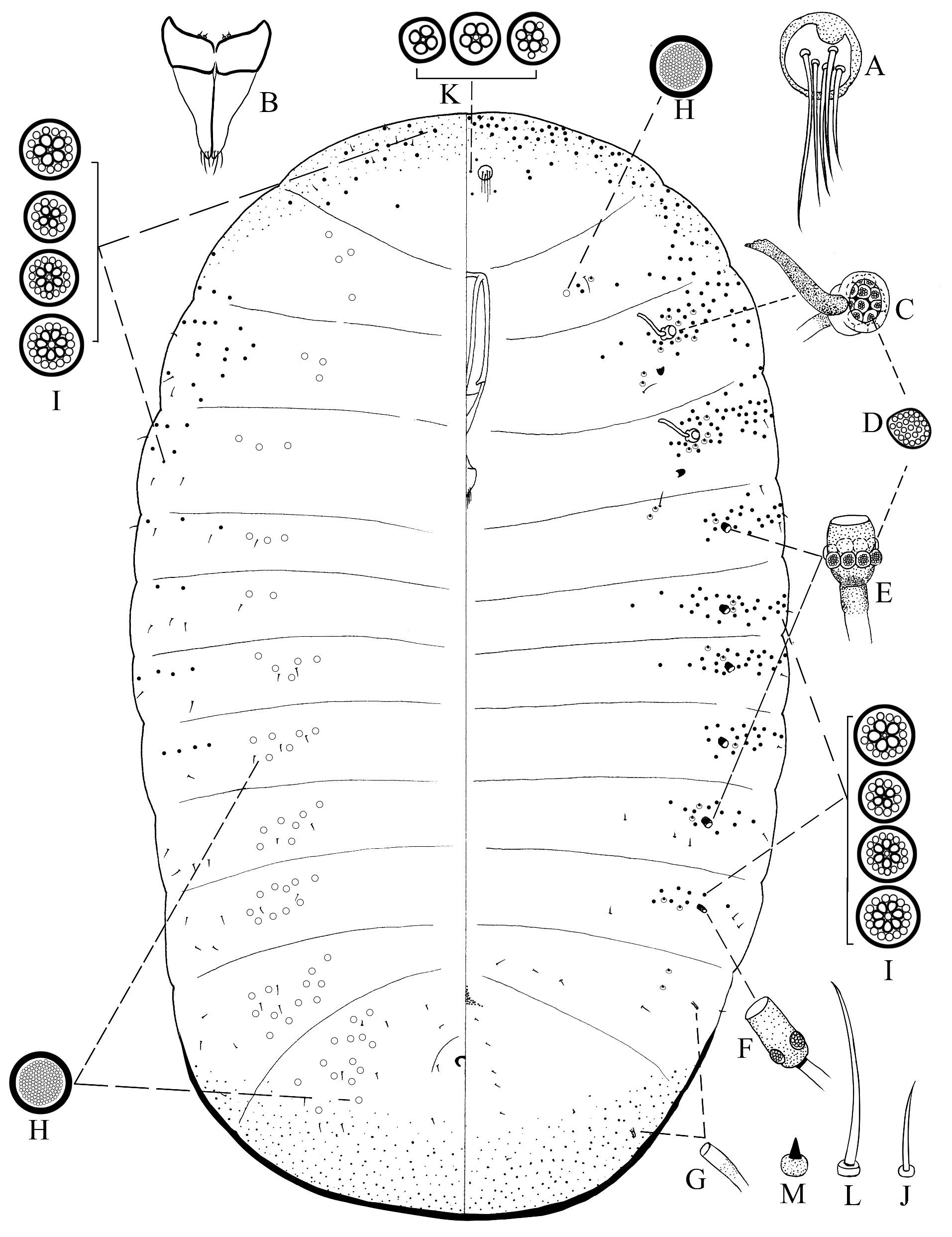
Slide-mounted material (n=3) ( Fig. 14 View FIGURE 14 ). Body 4.1–6.3 mm long and 1.8–2.4 mm wide; derm membranous. Antennae ( Fig. 14A View FIGURE 14 ) 10 segmented, situated close to each other, total length of each 1,100 –1,125 μm long, segment lengths (in µm): I, 130–160; II, 120–140; III, 100–120; IV, 100–120; V, 100–110; VI, 100–110; VII, 90–105; VIII, 90–100; IX, 85–90; and X, 55–60. Each segment with membranous apex and sclerotized base; scape (segment I) largest, 150–198 μm wide; pedicel (segment II) and segment III cylindrical, pedicel 100–125 μm wide, segment III 84–98 μm wide; other segments each more-or-less bowl-like, each 65–80 μm wide. Most setae on antennae hair-like; scape with setae scattered (each 25–43 μm long); pedicel with 3 or 4 circular sensory pores and 1 coeloconic sensillum, and scattered setae (each 30–100 μm long); other segments each with ring of hair-like setae (each 25–108 μm long) except apical segment; segments IV–IX each with 1 pair of sensory setae, each 20–25 μm long; sometimes segments IV, VI and VII each with 1 coeloconic sensillum; apical segment with 6–8 sensory setae and about 10 hair-like setae, also 3 short setae, each 8–11 μm long, and a pair of coeloconic sensilla. Eyes each with sclerotized margin, 60–73 μm wide, situated lateral to each antennal base. Mouthparts ( Fig. 14B View FIGURE 14 ) present: clypeolabral shield longer than labium, 480–530 μm long; without stylets; labium 180–210 μm long, 2 segmented, segment I 63–75 μm long, with margin sclerotized and centre membranous, bearing a pair of short setae (each 8–15 μm long) on each side; segment II 130–146 μm, sclerotized, with 4 apical hair-like setae (each 22–25 μm long) on each side. Thoracic spiracles ( Fig. 14C View FIGURE 14 ) each with opening 30–38 μm in diameter, a sclerotized bar, and a group of 20–27 sieve-like disc-pores at inner end of atrium.Abdominal spiracles numbering 8 pairs, with anterior 6 pairs ( Fig. 14D View FIGURE 14 ) developed, each with opening 18–24 μm in diameter, and a group of 7 or 8 sieve-like disc-pores ( Fig. 14E View FIGURE 14 ) at inner end of atrium; posteriormost 2 pairs ( Fig. 14F View FIGURE 14 ) small and tube-like, each with opening unsclerotized, about 12 μm in diameter, and without pores within atrium. Legs ( Fig. 14G View FIGURE 14 ) developed, lengths (in μm): foreleg: entire length 1,004 –1,178; coxa 140–200; trochanter + femur 340–390; tibia 310–345; tarsus 160–180, and claw 54–63; middle leg: entire length 1,033 –1,281; coxa 160–235; trochanter + femur 350–395; tibia 340–385; tarsus 130–200, and claw 53–66; hind leg: entire length 1,158 –1,343; coxa 200–220; trochanter + femur 360–430; tibia 370–420; tarsus 170–205, and claw 58–68. Hind leg trochanter + femur about 0.7 times as long as tibia + tarsus, tibia 2.0–2.2 times as long as tarsus. Most setae on legs hair-like; setae on coxae each 23–33 μm long; trochanters each bearing 3 or 4 sensory pores on each surface plus 1 long hair-like seta, 185–199 μm long, second-longest seta nearby 53–120 μm long, basal short setae each about 10 μm long, other setae each 28–38 μm long; femora each with inner-side setae long and sturdy, each 23–55 μm long, other setae short and thin, each 15–25 μm long; tibiae each with 10–15 long and sharply-tipped digitules, each 48–68 μm long, remaining setae short, each 18–28 μm long, with inner-side setae sturdy; tarsi each with few setae on distal half and inner-side, each seta 20–28 μm long; claw ( Fig. 14H View FIGURE 14 ) with 3 small denticles and a pair of pointed digitules, each 33–38 μm long, shorter than claw. Anal opening ( Fig. 14I View FIGURE 14 ) 18–30 μm wide, simple, with semicircular sclerotization without pores or setae, located dorsally on medial area of abdominal segment VIII. Vulva ( Fig. 14J View FIGURE 14 ) opening longitudinal and slit-like, 75–90 μm long, situated on venter of abdominal segment VIII.
Dorsum. With disc-pores of 2 types: (i) large simple pores ( Fig. 14K View FIGURE 14 ), each 11–12 μm in diameter, with sclerotized rim, forming small segmental groups on submargins; and (ii) compound multilocular disc-pores ( Fig. 14L View FIGURE 14 ), each 10–11 μm in diameter, with 3–5 subcentral loculi and 10–14 indistinct peripheral loculi, scattered on head and posteriormost abdominal segment, forming transverse bands across other segments. Setae of only 1 type, hair-like setae ( Fig. 14M View FIGURE 14 ), each 24–43 μm long, distribution same as that of compound multilocular disc-pores.
Venter. With disc-pores of 6 main types: (i) large simple pores ( Fig. 14K View FIGURE 14 ), same size and structure as on dorsum but fewer, with a few medially on head and between coxae; (ii) compound multilocular disc-pores ( Fig. 14L View FIGURE 14 ), same structure and size as on dorsum, scattered on head and posteriormost segment, forming transverse bands across other segments; also pores ( Fig. 14N View FIGURE 14 ) each 9–10 μm in diameter, with 4–7 subcentral loculi and an outer ring of 7–14 distinct peripheral loculi, or some pores with irregularly distributed loculi, with 3–8 of these pores around each thoracic spiracle and 0–8 pores around anterior 5 pairs of abdominal spiracles; (iii) bilocular compound multilocular disc-pores ( Fig. 14O View FIGURE 14 ), each slightly oval, about 10 μm wide, with 2 subcentral loculi and a ring of indistinct peripheral loculi, present on 3 posteriormost segments and numerous on apical segment; (iv) thick-rimmed simple pores ( Fig. 14P View FIGURE 14 ), each 5–6 μm in diameter, present on abdominal segment VIII; (v) sieve-like disc-pores ( Fig. 14E View FIGURE 14 ), each about 6 μm in diameter, some slightly polygonal, with many irregularly distributed loculi, forming groups in atria of thoracic spiracles and a few present in anterior 6 pairs of abdominal spiracles; and (vi) thin-rimmed simple pores ( Fig. 14Q View FIGURE 14 ), each about 5 μm in diameter, numbering 1–9 within group of large simple pores on prothorax. Setae of 2 types: (i) hair-like setae ( Fig. 14M View FIGURE 14 ), each 24–120 μm long; short setae present on submargins and margins of each segment and scattered on abdominal segment VIII; longer setae ( Fig. 14R View FIGURE 14 ) present on median areas except on posteriormost segment; and (ii) short conical spine-like setae ( Fig. 14S View FIGURE 14 ) with sclerotized basal sockets, each about 5 μm long; with 7 or 8 around each thoracic spiracle, and 1 or 2 around each of anterior 6 pairs of abdominal spiracles.
Remarks. The adult females of N. zizyphi in this study differ from the original description by Tang & Hao (1995) in having: (i) thoracic spiracles each with a group of 20–27 sieve-like disc-pores at inner end of atrium (Tang & Hao mention 15–16 pores at inner end of atrium); (ii) thick-rimmed simple pores present on abdominal segment VIII (Tang & Hao did not mention these, but it is possible that they might have mistaken these pores for setal sockets whose setae have been lost); (iii) compound multilocular disc-pores each with an outer ring of peripheral loculi (Tang & Hao describe multilocular disc-pores without peripheral loculi, but the structure of peripheral loculi is sometimes difficult to observe); and (iv) vulva situated on venter of abdominal segment VIII (Tang & Hao describe the vulva as being situated between abdominal segments VII and VIII).
Adult male
Appearance in life ( Fig. 13B View FIGURE 13 ). Body elongate, wing-span 4.1–4.2 mm; with head broad, antennae slender and filiform, neck distinct, fore wings broad, and legs slender. Membranous parts of body red brown, sclerotized parts black brown, eyes black, fore wings clear with anterior margin grey to red brown and pterostigma yellow brown; posterior end of abdomen with a dorsal tuft of long waxy filaments.
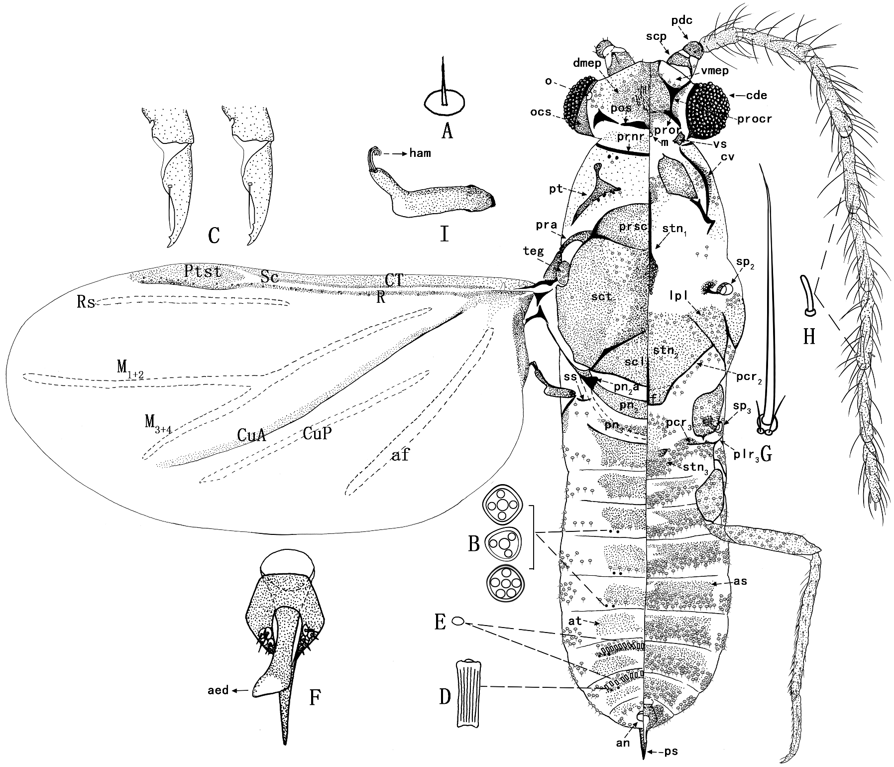
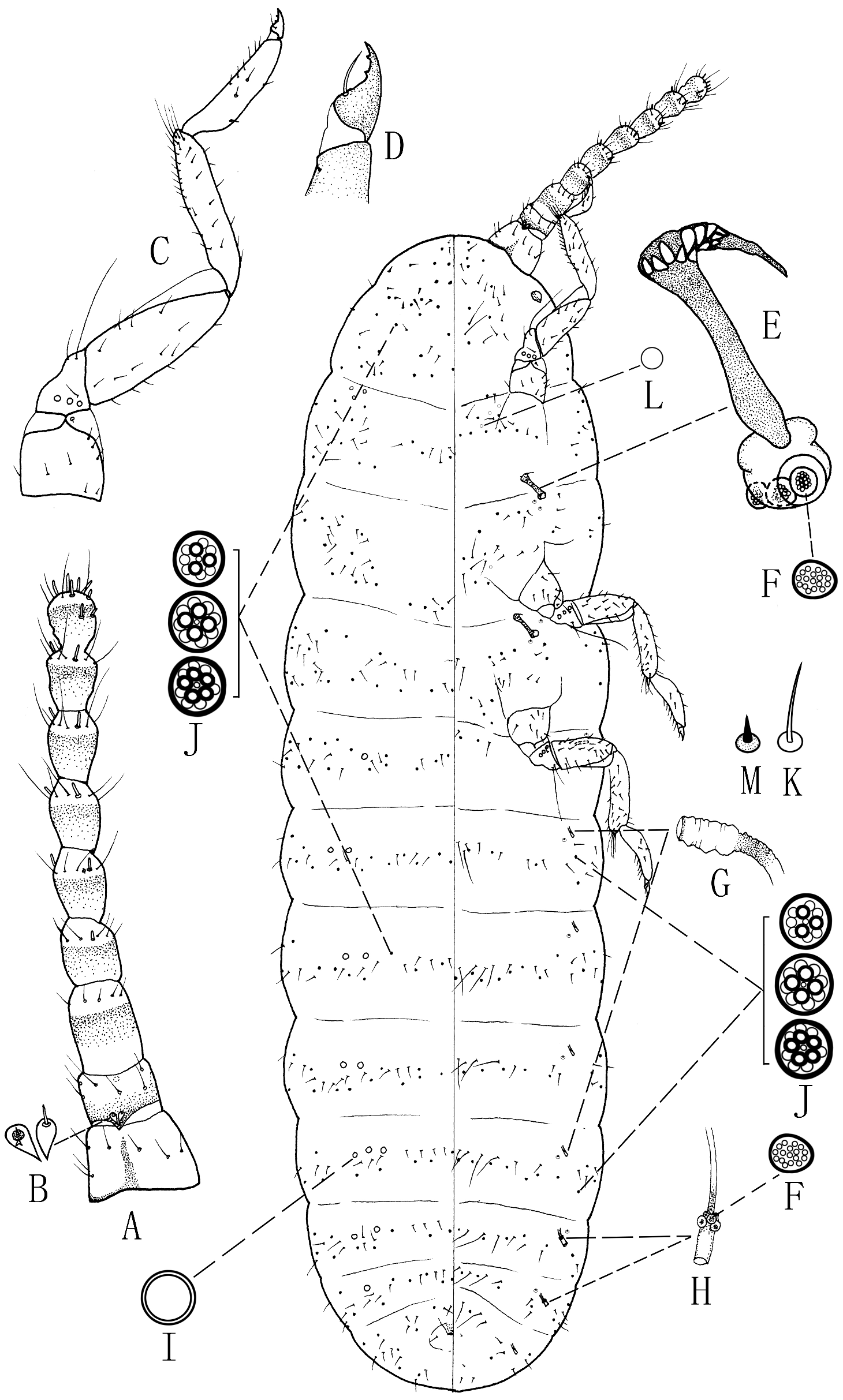
Slide-mounted material (n=2) ( Fig. 15 View FIGURE 15 ). Body 2.2–2.3 mm long, 0.5–0.6 mm wide across prealare. Setae ( Fig. 15A View FIGURE 15 ) spine-like, each 3–14 µm long, with a large convex socket (6–10 µm wide), numerous. Loculate pores ( Fig. 15B View FIGURE 15 ), each 7–8 µm in diameter, with 3–5 loculi, few, present dorsally on pronotal ridge, post-tergite and submedian areas of abdomen. Head with a pair of compound eyes. Antennae 10 segmented, filiform. Post-tergite shaped like inverted “π”. Sclerotized areas without reticular pattern. Legs slender, with some setae bifurcate; each tarsus 2 segmented; claw ( Fig. 15C View FIGURE 15 ) with 1 or 2 denticles and a pair of pointed digitules shorter than claw. Abdomen with tubular ducts ( Fig. 15D View FIGURE 15 ) and thin-rimmed simple pores ( Fig. 15E View FIGURE 15 ) on tergites VI and VII; penial sheath ( Fig. 15F View FIGURE 15 ) situated terminally.
Head: 260–300 µm long, 520–530 µm wide. Dorsum: postoccipital suture (pos) well developed and interrupted, extending across posterior part of epicranium. Midcranial ridge lacking. Dorsomedial part of epicranium (dmep) sclerotized, with 31–40 setae, and a small group of 5–7 setae near compound eyes; membranous area with many sclerotized points. Laterally: compound eyes (cde) each 168–208 µm long, with 195–200 ommatidia, each ommatidium 10–18 µm in diameter. Narrow, sclerotized ocular sclerite (ocs) present along dorsal and posterior margins of each compound eye, each with a single dorsal ocellus (o) situated dorsally, each ocellus 13–18 µm wide. Venter: with strongly sclerotized series of ridges forming medial sub-pentagonal shape, composed of: (i) a pair of antennal sclerites anteriorly and laterally, fusing posteriorly with (ii) a pair of preocular sclerites (procr) and (iii) a pair of preoral ridges (pror) posteriorly. Area surrounded by ridges, posterior to preoral ridges and lateral to preocular sclerites, weakly sclerotized; ventral part of epicranium (vmep) with 60–86 setae. Mouth (m) present. Ventral sclerites (vs) articulating with preoral ridge anteriorly and with ventral projection from ocular sclerite laterally, and with cervical sclerite and pronotal ridge posteriorly.
Antennae each 10 segmented, total length of each 2,000 –2,150 µm, ratio of total body length to antennal length 1: 0.87–0.98. Segment lengths (in µm): I, 70–98; II, 33–40; III, 155–173; IV, 260–305; V, 280–310; VI, 240–265; VII, 240–270; VIII, 245–260; IX, 220–250; and X, 170–180. Scape (scp) (segment I) and pedicel (pdc) (segment II) stout, each with a few spine-like setae; scape with base sclerotized and apex membranous, 95–100 µm wide, with 8–10 setae; pedicel completely sclerotized, 68–78 µm wide, with 10–14 spine-like setae and 1 sensory seta (10–13 µm long), plus 1 circular sensory pore and 1 coeloconic sensillum. Each segment of flagellum parallel-sided, 25–58 µm wide; segment IV or V longest, segment III shortest; flagellum with many long hair-like setae, each 68–118 µm long, and satellite setae ( Fig. 15G View FIGURE 15 ) present; segments IV–IX each with a pair of sensory setae ( Fig. 15H View FIGURE 15 ) near apex, and apical segment with 3 sensory setae, each sensory seta 18–20 µm long.
Thorax. Prothorax: Dorsum with a strong uninterrupted pronotal ridge (prnr), articulating ventrally with cervical sclerite, with 1 or 2 lateral pronotal setae and 0–2 loculate pores on each side. Post-tergite (pt) shaped like an inverted “π”, sclerite with a pair of anterior and posterolateral arms; anterior arms each 70–75 µm long, with anterior sclerotization appearing similar to an apophysis; posterolateral arms each 120–178 µm long; median part broadly or narrowly interrupted, post-tergites 10–110 µm apart; with 3–6 loculate pores and 3–5 post-tergital setae on each side. Laterally with a pair of cervical sclerites (cv) of complicated structure, each articulated anteriorly with both posterior ventral sclerites and pronotal ridge; inner-side margin with a group of 5–7 proepisternal setae, outer side with 2–4 posterior propleural setae. Venter: prosternum (stn 1) with well-sclerotized median ridge, 290–360 µm long; both anterior and posterior parts with sclerotization broad, and with a group of about 44 prosternal setae.
Mesothorax, dorsum: prescutum (prsc) sclerotized and approximately oval, about 195 µm long and 270 µm wide; prescutal ridges strongly sclerotized, prescutal sutures much less sclerotized. Scutum (sct) sclerotized throughout; with 18–20 scutal setae on each side medially. Scutellum (scl) triangular, with a central conical projection on posterior margin, about 180 µm long and 250 µm wide, with about 3 setae. Immediately posterior to scutellum is a membranous area, bordered posteriorly by sclerotized mesopostnotum, which is broadly U-shaped, with each arm very strongly sclerotized, extending anteriorly to articulate with mesopleural ridge. Posteriorly, mesopostnotum (pn 2) extends internally under metathoracic metapostnotum, forming a large mesopostphragma; mesopostnotal apophyses (pn 2 a) developed. Laterally: prealare (pra) elongate; tegula (teg) sclerite slightly sclerotized, with about 9 tegular setae. Mesothoracic spiracles (sp 2) each with outer part of peritreme about 43 µm wide, a group of 2 or 3 antemesospiracular setae and 12–15 lateral mesospiracular setae. Lateropleurite (lpl) without anterior margin, with 13–15 setae, also with 7–9 setae in lateral area of lateropleurite. Venter: basisternum (stn 2) subhexagonal, about 310 µm long and 360 µm wide, median ridge not reaching either anterior or posterior margins; anteriorly, marginal ridge of basisternum absent but laterally, marginal ridge well developed and sometimes bifurcate; basisternum bounded posteriorly by well-developed mesoprecoxal ridges (pcr 2), with 29 mesosternal setae; with 10–12 postmesoprecoxal ridge setae on each side; furca (f) with long arms, each arm about 113 µm long.
Metathorax, dorsum: metapostnotum (pn 3) present and slightly sclerotized, with about 7 metatergal setae. Laterally: suspensorial sclerites (ss) present, each 56–64 µm long. Metapleural ridge (plr 3) well developed; metaprecoxal ridge (pcr 3) well developed and extending medio-ventrally. Metathoracic spiracles (sp 3) similar in structure to anterior spiracles, each with peritreme 27–37 µm wide, with 1–3 metaspiracular setae, 3 or 4 dorsospiracular setae and 31–36 postmetaspiracular setae. Venter: metasternum with area of sclerotization medially; with a group of about 17 anterior metasternal setae and about 47 posterior metasternal setae.
Wings: fore wings quite large and well developed, each 1.70–1.74 mm long and 0.73–0.74 mm wide; ratio of length to width 1: 0.42–0.43; ratio of total body length to wing length 1: 0.75–0.78. Costal thickening (CT) sclerotized; pterostigma (ptst) club shaped, situated near wing apex; subcosta (Sc) present along CT from the wing base toward the apex, rising to pterostigma; radius (R) present posterior to Sc, with a line of 18–29 circular sensoria. Alar lobe fold (alf) sclerotized along proximal posterior margin; cubitus anterior (CuA) originates from Sc at about 1/5 along wing length and runs obliquely to posterior wing margin. Rest of wing membranous, with veins forming clear lines; radial sector (Rs) almost half as long as wing; media (M) branching at middle into M 1+2 and M 3+4; anal fold (af) originates from wing base and runs obliquely across to posterior wing margin; cubitus posterior (CuP) situated between CuA and af. Hind wings (hamulohalteres) ( Fig. 15I View FIGURE 15 ) each about 213 µm long and 53 µm wide, with base and anterior margin strongly sclerotized, remainder weakly sclerotized; apex bilobate, with about 4 hamuli at tip, each hamulus (ham) strongly curved and with apex blunt.
Legs: setose, long and slender, hind leg longest; lengths (in μm): foreleg: entire length 1,123 –1,193; coxa 185–190; trochanter + femur 358–380; tibia 390–410; tarsus 150–170, and claw 40–43; middle leg: entire length 1,129 –1,238; coxa 175–190; trochanter + femur 332–360; tibia 410–460; tarsus 170–180, and claw 42–48; hind leg: entire length 1,198 –1,287; coxa 173–190; trochanter + femur 330–380; tibia 485–505; tarsus 168–182, and claw 45–50. Legs with many hair-like setae, each 7–27 µm long; trochanters each with 2 sensory pores on each surface; femur and tibia with inner-side setae longest, each 29–46 µm; tibia with some inner-side setae conical and a few bifurcated setae distally. Tarsi each 2 segmented, with proximal segment triangular, without setae; segment II with a tarsal campaniform sensillum proximally and some bifurcated setae; claws each broad, with 1 or 2 plantar denticles and a pair of pointed digitules (each 21–24 µm long) shorter than claw.
Abdomen: excluding genital segments, 8 segmented. Segments I–IV each with transverse abdominal tergite (at) complete, segments V –VIII each with tergite discrete. Abdominal sternites (as) present on each segment. Pleural, ventral and dorsal setae present, dorsal setae less frequent than ventral setae. Tergites on segments II–IV, VI and VII each with 1–3 loculate pores present posterolaterally on each side. Segments VI and VII each with a group of large dorsal tubular ducts medially, forming 1 or 2 rows, each duct 19–21 µm long and about 8 µm wide; with 16–20 ducts on segment VI and 17 or 18 on segment VII, also with some thin-rimmed simple pores among ducts, each pore about 2.5 µm in diameter, with about 8–16 pores on each of these 2 segments. Abdominal spiracles not detectable. Anal opening (an) situated dorso-medially near base of penial sheath. Genital segment: penial sheath (ps) sclerotized, 163–175 µm long, extending posteriorly from beneath segment VIII; with anterior area broad and subpentagonal, about 93 µm wide, narrowing posteriorly to an acute apex; with ventral group of 4–6 penial sheath setae on each side. Aedeagus (aed) mostly sclerotized, broadest at base and apex, sides concave, with distal end membranous and truncated, 95–120 µm long, emerging from ventral slit in penial sheath. Eversible endophallus absent.
Remarks. The adult males of only 2 species of Neogreenia , N. zizyphi and N. osmanthus , have been described in detail. The differences between them are (character states for N. osmanthus in parentheses): (i) prescutum with 18– 20 scutal setae on each side (with 9–13 scutal setae on each side); (ii) ventral part of epicranium with subpentagonal sclerotization complete (subpentagonal sclerotization with small circular membranous part anteriorly); and (iii) claw with a pair of pointed digitules, shorter than claw (with a pair of capitate digitules, longer than claw).
Second-instar nymph (cyst) (sexes indistinguishable)
Appearance in life ( Fig. 13C View FIGURE 13 ). Body oval, yellowish, some specimen with posterior part orange; antennae and legs lacking; living under bark, surrounded by flocculent wax.
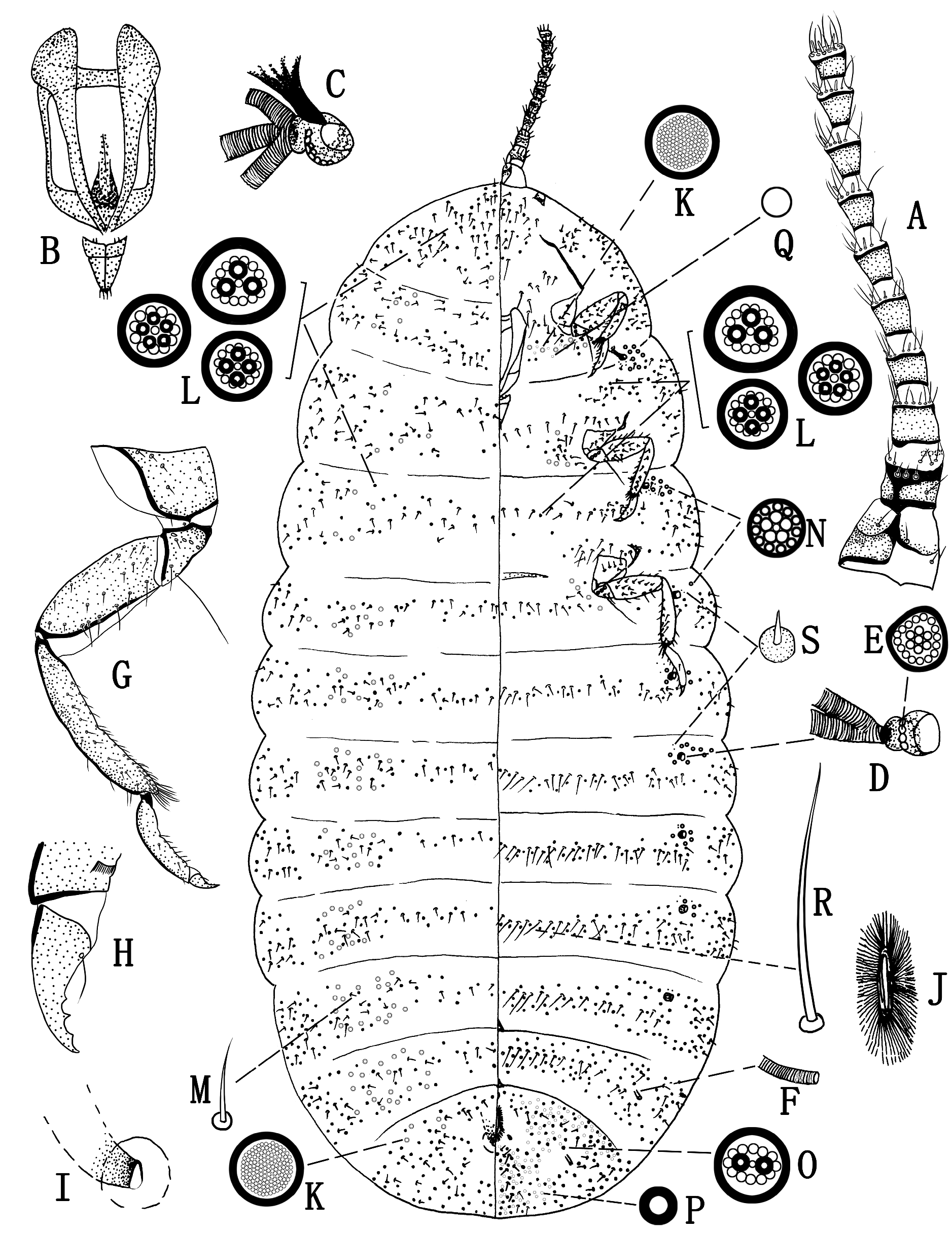
Slide-mounted material (n=4) ( Fig. 16 View FIGURE 16 ). Body 1.1–1.7 mm long and 0.7–1.0 mm wide; derm membranous, but posterior part or body margin becoming sclerotized at late stage of development. Antennae ( Fig. 16A View FIGURE 16 ) reduced to small oval plates, each 15–18 μm in diameter, with margin sclerotized and protruding anteriorly; situated close together medially on head, each bearing 4 or 5 setae, each 20–35 μm long. Eyes absent. Labium ( Fig. 16B View FIGURE 16 ) 2 segmented, 88–112 μm long and 60–85 μm wide; segment I about 38 μm long, with margin sclerotized and centre membranous, and a pair of short setae (each about 7.5 μm long) on each side; segment II 53–63 μm long, sclerotized, with 2 apical hair-like setae (each 12–18 μm long) on each side. Clypeolabral shield longer than labium, 250–280 μm long and 140–150 μm wide; stylets present. Legs absent. Thoracic spiracles ( Fig. 16C View FIGURE 16 ) sclerotized, each opening 10–15 μm in diameter, with a sclerotized bar and a group of 4–6 sieve-like disc-pores ( Fig. 16D View FIGURE 16 ) at inner end of atrium. Abdominal spiracles numbering 8 pairs, with anterior 5 pairs ( Fig. 16E View FIGURE 16 ) sclerotized, each with opening 7–8 μm in diameter and 1 or 2 sieve-like disc-pores at inner end of atrium; posteriormost 3 pairs ( Fig. 16F View FIGURE 16 ) unsclerotized, each small and tube-like, with opening about 2.5 μm in diameter and without pores in atrium. Anal opening with a simple semicircular sclerotized ring 12–15 μm wide, without pores or setae, located medially on posteriormost dorsal segment. Cicatrices absent.
Dorsum. With disc-pores of 2 types: (i) large simple pores ( Fig. 16G View FIGURE 16 ), each 7–10 µm in diameter with sclerotized rim, usually 1 pore present on submargin of each segment, but some segments with 2 pores or none; and (ii) compound multilocular disc-pores ( Fig. 16H View FIGURE 16 ), each 7–8 µm in diameter, with 5–7 subcentral loculi (mostly 5 and 6 loculi) followed by outer ring of 11–16 peripheral loculi, present on margins from head to abdominal segment II or III. Setae of only 1 type: spine-like setae ( Fig. 16I View FIGURE 16 ), each 10–12 μm long, present in submarginal and submedian areas.
Venter. With disc-pores of 3 types: (i) large simple pores ( Fig. 16G View FIGURE 16 ), same size and structure as on dorsum, 1 pore present near each long and hair-like seta on prothorax, sometimes lacking; (ii) compound multilocular disc-pores ( Fig. 16H View FIGURE 16 ), same size and structure as on dorsum, forming a group around each thoracic spiracle and present on margins except for 2 posteriormost segments; also pores ( Fig. 16J View FIGURE 16 ) each about 5 µm in diameter, with 4 or 5 subcentral loculi and without peripheral loculi, numbering 2 pores between antennal bases; and (iii) sieve-like disc-pores ( Fig. 16D View FIGURE 16 ), each 6–7 µm in diameter, some slightly polygonal, with many irregularly distributed loculi, present within atria of thoracic and anterior 5 pairs of abdominal spiracles. Setae of 3 types: (i) hair-like setae ( Fig. 16K View FIGURE 16 ), each 15–25 μm long, situated singly on submedial prothorax and on submargins of mesothorax and abdominal segment I; (ii) spine-like setae ( Fig. 16I View FIGURE 16 ), same size as on dorsum, few on margin; and (iii) short conical spine-like setae ( Fig. 16L View FIGURE 16 ) with sclerotized basal sockets, each about 5 μm long; with 1 or 2 present near each long and hair-like seta and thoracic and abdominal spiracle, except for posteriormost pair of spiracles.
Third-instar female nymph (cyst)
Appearance in life ( Fig. 13D View FIGURE 13 ). Living under bark, surrounded by flocculent wax. Body oval, yellowish, with margin orange; antennae and legs lacking.
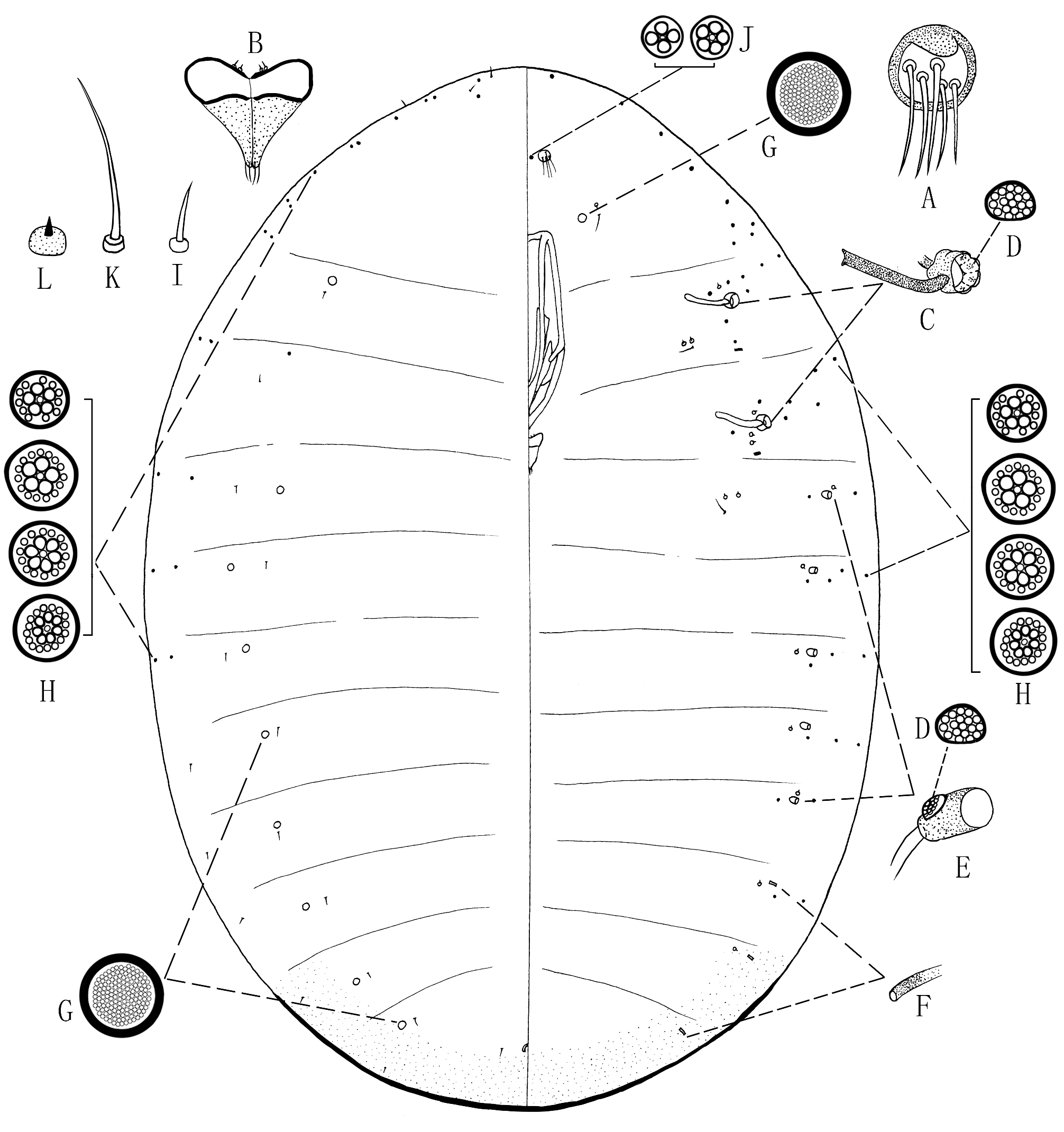
Slide-mounted material (n=4) ( Fig. 17 View FIGURE 17 ). Body 2.2–3.0 mm long and 1.3–1.7 mm wide; derm membranous medially, sclerotized marginally, with posterior end strongly sclerotized. Antennae ( Fig. 17A View FIGURE 17 ) reduced to small oval plates, each 24–29 μm in diameter, with margin sclerotized and protruding anteriorly, and bearing 5–7 setae, each 15–58 μm long; situated close together medially on head. Eyes absent. Labium ( Fig. 17B View FIGURE 17 ) 2 segmented, 148–163 μm long and 95–100 μm wide; segment I 58–60 μm long, with margin sclerotized and centre membranous, and 1 pair of short setae (each about 7.5 μm long) on each side; segment II 90–100 μm long, sclerotized, with 3 apical hair-like setae (each 20–25 μm long) on each side. Clypeolabral shield longer than labium, 320–380 μm long and 205–245 μm wide; stylets present. Legs absent. Thoracic spiracles ( Fig. 17C View FIGURE 17 ) sclerotized, each with opening 22–25 μm in diameter, a sclerotized bar, and a group of 13–18 sieve-like disc-pores ( Fig. 17D View FIGURE 17 ) at inner end of atrium. Abdominal spiracles numbering 8 pairs, with anterior 6 pairs sclerotized; anteriormost 5 pairs ( Fig. 17E View FIGURE 17 ) with each opening 16–19 μm in diameter and 5–8 sieve-like disc-pores at atrium; spiracles ( Fig. 17F View FIGURE 17 ) on abdominal segment VI smaller, each with opening about 7.5 μm in diameter and 3 sieve-like disc-pores at atrium; posteriormost 2 pairs ( Fig. 17G View FIGURE 17 ) unsclerotized, small and tube-like, each with opening about 5 μm in diameter and without pores within atrium. Anal opening with U-shaped sclerotized, 18–34 μm wide, without pores or setae, located medially on posteriormost dorsal segment. Cicatrices absent. With a ventral triangular sclerotization (in position of vulva on adult) situated medially on abdominal segment VIII.
Dorsum. With disc-pores of 2 types: (i) large simple pores ( Fig. 17H View FIGURE 17 ), each 10–12 µm in diameter, with sclerotized rim, forming group on submargins on each side of each segment, with number of pores increasing towards abdominal apex, sometimes head with only 1 pore and posteriormost segment with up to 16 pores; and (ii) compound multilocular disc-pores ( Fig. 17I View FIGURE 17 ), each about 10 µm in diameter, with 4–6 subcentral loculi (mostly 5 and 6 loculi) surrounded by outer ring of 10–15 peripheral loculi, present on margins from head to abdominal segment III. Setae of only 1 type: spine-like setae ( Fig. 17J View FIGURE 17 ), each 12–13 μm long, present in submarginal and submedial areas.
Venter. With disc-pores of 3 main types: (i) large simple pores ( Fig. 17H View FIGURE 17 ), same size and structure as on dorsum, with 0–2 pores near each long hair-like seta on prothorax; (ii) compound multilocular disc-pores ( Fig. 17I View FIGURE 17 ), same size and structure as on dorsum; forming group around each thoracic spiracle and present on margins except for 2 posteriormost segments; also pores ( Fig. 17K View FIGURE 17 ) each 8–9 μm in diameter, with 4 or 5 subcentral loculi, without peripheral loculi or with irregularly distributed loculi, numbering 4–8 pores between antennae; and (iii) sieve-like disc-pores ( Fig. 17D View FIGURE 17 ), each 5–8 µm in diameter, some slightly polygonal, with many irregularly distributed loculi, present within atria of thoracic and anterior 6 pairs of abdominal spiracles. Setae of 3 types: (i) hair-like setae ( Fig. 17L View FIGURE 17 ), each 23–25 μm long, present submedially on prothorax, and submarginally on mesothorax and abdominal segment I; (ii) spine-like setae ( Fig. 17J View FIGURE 17 ), same size as on dorsum, few on margins; and (iii) short conical spine-like setae ( Fig. 17M View FIGURE 17 ) with sclerotized basal sockets, each 5–7 μm long; with 1–3 near each long and hair-like seta, 3–7 around each thoracic spiracle, and 1–4 around each abdominal spiracle except for posteriormost pair.
Third-instar male nymph (prepupa)
Appearance in life ( Fig. 13E View FIGURE 13 ). Body elongate, legs and antennae present, wing buds absent; body orange; medial venter of abdomen, legs and antennae yellowish, eyes black.
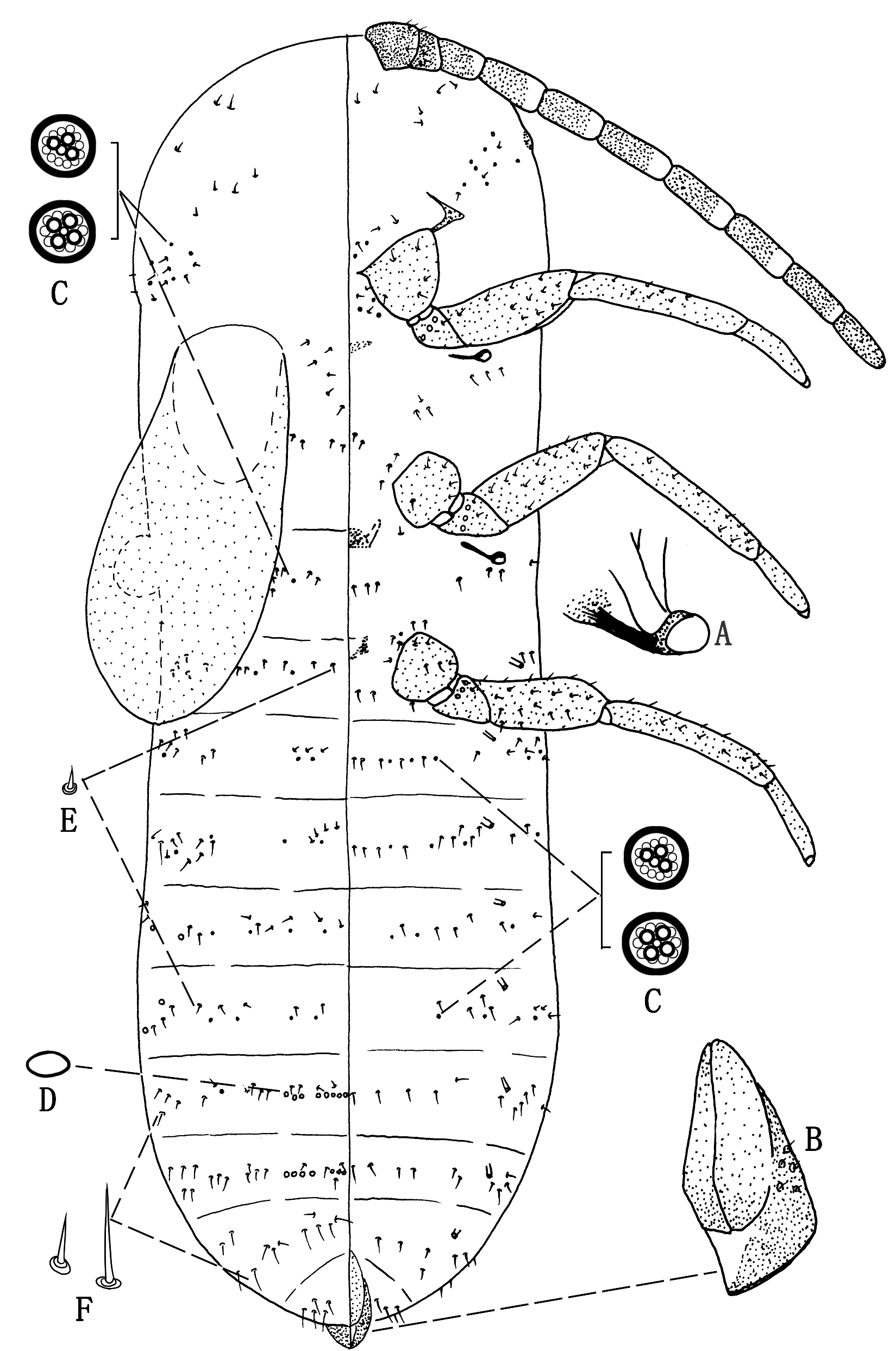
Slide-mounted material (n=3) ( Fig. 18 View FIGURE 18 ). Body 2.3–4.0 mm long and 0.8–1.1 mm wide; derm membranous. Antennae ( Fig. 18A View FIGURE 18 ) 9 segmented, each 700–870 μm long, segment measurements (in μm): I, 93–105 long and 143–150 wide; II, 73–80 long and 105–110 wide; III, 100–127 long and 96–108 wide; IV, 75–110 long and 88–98 wide; segment V, 98–100 long and 80–86 wide; VI, 90–100 long and 78–83 wide; VII, 73–102 long and 68–73 wide; VIII, 88–95 long and 63–78 wide; and IX, 75–84 long and 55–65 wide. Each segment with sclerotized base and membranous apex; scape largest, other segments becoming successively narrower towards apex. Antennae with many hair-like setae, each 27–88 μm long, scattered on scape (segment I) and pedicel (segment II); other segments each with a ring of setae near apex; pedicel with sensory seta and coeloconic sensillum ( Fig. 18B View FIGURE 18 ) near ventral base and circular sensory pore dorsally; sometimes with coeloconic sensillum on segment V; segments IV–VIII each with a pair of sensory setae; apical segment with 5–12 sensory setae, each 13–30 μm long. Eyes 53–59 μm wide, each with sclerotized margin, situated posterolaterally to antennal base. Mouthparts atrophied. Legs ( Fig. 18C View FIGURE 18 ) developed, lengths (in μm): foreleg: entire length 718–840; coxa 90–108; trochanter + femur 250–295; tibia 220– 265; tarsus 125–163, and claw 33–38; middle leg: entire length 720–890; coxa 90–113; trochanter + femur 235– 295; tibia 230–280; tarsus 130–170, and claw 35–43; and hind leg: entire length 720–950; coxa 90–123; trochanter + femur 230–300; tibia 235–310; tarsus 130–183, and claw 35–45. Ratio of length of trochanter + femur to length of tibia + tarsus of hind leg 0.6–0.7: 1; ratio of length of tibia to length of tarsus of hind leg 1.7–1.8: 1. Legs with many hair-like setae, each 14–63 μm long; trochanters each with 3 sensory pores on each surface plus 1 very long hair-like seta 125–150 μm long; tibiae each with sharply-tipped digitules, each 25–58 μm long; tarsi each bent, with setae on inner sides; claws ( Fig. 18D View FIGURE 18 ) each with 2 plantar denticles and a pair of pointed digitules, shorter than claw, each 23–25 μm long. Thoracic spiracles ( Fig. 18E View FIGURE 18 ) each with opening 15–18 µm in diameter, a sclerotized bar and group of 2–4 sieve-like disc-pores ( Fig. 18F View FIGURE 18 ) at inner end of atrium.Abdominal spiracles numbering 8 pairs, situated submarginally on each segment; anterior 5 pairs ( Fig. 18G View FIGURE 18 ) and posteriormost pair unsclerotized and tube-like, without pores in atria, each with opening about 5 μm in diameter; abdominal spiracles ( Fig. 18H View FIGURE 18 ) on segments VI and VII each with opening about 8 μm in diameter and with 2 or 3 sieve-like disc-pores within atrium. Anal opening with sclerotized and semi-circular ring, about 18 µm wide and without pores or setae, situated medially on dorsum of posteriormost segment.
Dorsum. With disc-pores of 2 types: (i) large simple pores ( Fig. 18I View FIGURE 18 ), each 9–10 μm in diameter, numbering 0–3 on each side of each segment; (ii) compound multilocular disc-pores ( Fig. 18J View FIGURE 18 ), each 9–10 μm in diameter, with 3–5 subcentral loculi (mostly 4 loculi) surrounded by 7 or 8 indistinct peripheral loculi, present in transverse rows across each segment except on head and posteriormost segment where scattered. Setae of only 1 type: hair-like setae ( Fig. 18K View FIGURE 18 ), 10–38 μm long, with similar distribution to that of compound multilocular disc-pores.
Venter. With disc-pores of 3 types: (i) thin-rimmed simple pores ( Fig. 18L View FIGURE 18 ), each about 5 μm in diameter, few near coxae, sometimes absent; (ii) compound multilocular disc-pores ( Fig. 18J View FIGURE 18 ), same size, structure and distribution as on dorsum; and (iii) sieve-like disc-pores ( Fig. 18F View FIGURE 18 ), each about 7 µm in diameter, with many irregularly distributed loculi, present within atria of each thoracic spiracle and abdominal spiracles on segments VI and VII. Setae of 2 types: (i) hair-like setae ( Fig. 18K View FIGURE 18 ), each 10–105 μm long, always present in same distribution as multilocular disc-pores, short setae present on submargins and margins, longer setae present medially; and (ii) short conical spine-like setae ( Fig. 18M View FIGURE 18 ) with sclerotized basal sockets, each 7–10 μm long; with 2 or 3 near each thoracic spiracle, and 1 near each abdominal spiracle except for posteriormost pair.
Remarks. The third-instar male nymphs of only two species of Neogreenia are known: N. zizyphi and N. osmanthus . Differences between them are as follows (character states for N. osmanthus in parentheses): (i) posteriormost pair abdominal spiracles lacking sieve-like disc-pores in atrium (posteriormost pair abdominal spiracles with sieve-like disc-pores in atrium); (ii) tibia with sharply-tipped digitules (tibial digitules capitate); and (iii) claw with a pair of pointed digitules, shorter than the claw (claw with a pair of capitate digitules, longer than the claw).
Pupa
Appearance in life ( Fig. 13F View FIGURE 13 ). Body elongate, antennae and legs present but not capable of movement; body orange, eyes red-brown, antennae, legs and wing buds yellow and almost transparent.
Slide-mounted material (n=1) ( Fig. 19 View FIGURE 19 ). Only 1 exuviae available. Body 2.28 mm long and 0.68 mm wide. Antennae each 10 segmented, total length 1,225 µm; segment lengths (in μm): I, 100; II, 85; III, 105; IV, 140; V, 135; VI, 145; VII, 150; VIII, 150; IX, 145; and X, 135. Segments nearly cylindrical, with similar widths, each 35–80 μm long; 2 proximal segments each bearing a few short and spine-like setae, each seta about 13 μm long; remaining segments without setae. Eyes present, represented by a pair of prominences on margin. Mouthparts absent. Legs long and smooth; lengths (in μm): foreleg: entire length 865; coxa 125; trochanter + femur 280; tibia 290, and tarsus 170; middle leg: entire length 915; coxa 140; trochanter + femur 285 tibia 315, and tarsus 175; hind leg: entire length 965; coxa 140; trochanter + femur 295 tibia 340, and tarsus 190. Each coxa, femur and tibia with many short and spine-like setae, each about 7.5 μm long; trochanter with 3 sensory pores on each surface plus 1 short seta; tarsus 1 segmented; claw absent. Fore wing bud broad, 530 μm long and 270 μm wide; hind wing bud small, nearly round, about 80 μm in diameter. Thoracic spiracles ( Fig. 19A View FIGURE 19 ) developed, each without any pores in atria; abdominal spiracles all small and tube-like, numbering 8 pairs. Sub-apex of abdomen with long conical projection ( Fig. 19B View FIGURE 19 ), formed of rudiment of sclerotized penial sheath, about 165 μm long.
Dorsum: with disc-pores of 2 types: (i) compound multilocular disc-pores ( Fig. 19C View FIGURE 19 ) each 7–8 µm in diameter, with 3 or 4 subcentral loculi surrounded by an outer ring of 10–12 indistinct peripheral loculi, few on margins of prothorax, submargins of metathorax and forming sparse transverse rows across abdominal segments I– V; and (ii) thin-rimmed simple pores ( Fig. 18D View FIGURE 18 ), oval, each 4–5 µm in greatest width, forming short transverse rows medially across abdominal segments VI and VII. Setae of only 1 type: spine-like setae ( Fig. 19E View FIGURE 19 ), each about 7.5 µm long, present on margins of head, medial areas of mesothorax and forming transverse segmental rows across abdomen; setae ( Fig. 19F View FIGURE 19 ) on abdominal segments VI –VIII each 10–20 µm long.
Venter: pore and setal types of same sizes and structures as on dorsum. Compound multilocular disc-pores ( Fig. 19C View FIGURE 19 ) present near eyes, coxae of foreleg and hindleg, and forming transverse rows across abdominal segments II– V. Setae present anteriorly on head, on thorax around each coxa, and forming sparse transverse rows across each segment of mesothorax, metathorax and abdomen.
Remarks. The male pupae of only two species of Neogreenia are known: N. zizyphi , which has thin-rimmed simple pores forming short transverse rows medially across abdominal segments VI –VII, and N. osmanthus , which lacks simple pores.
| V |
Royal British Columbia Museum - Herbarium |
| VI |
Mykotektet, National Veterinary Institute |
No known copyright restrictions apply. See Agosti, D., Egloff, W., 2009. Taxonomic information exchange and copyright: the Plazi approach. BMC Research Notes 2009, 2:53 for further explanation.
|
Kingdom |
|
|
Phylum |
|
|
Class |
|
|
Order |
|
|
Family |
|
|
Genus |
Neogreenia zizyphi Tang, 1995
| Zheng, Xinyi, Watson, Gillian W., Zhang, Jiangtao, Tan, Zhixiang & Wu, San’An 2024 |
Neogreenia zizyphi
| Tang, F. D. & Hao, J. J. 1995: 82 |
Kuwaniella zizyphi
| Foldi, I. 2001: 205 |
| Kosztarab, M. P. & Ben-Dov, Y. & Kosztarab, M. 1986: 9 |
| Tang, F. D. 1984: 123 |