
Ophiomonas shinseimaruae, Okanishi & Matsuo & Fujita, 2021
|
publication ID |
https://doi.org/ 10.6620/ZS.2021.60-59 |
|
publication LSID |
lsid:zoobank.org:pub:BC892FF3-4F6A-4C2B-81A3-AEC6ABDD2CA1 |
|
DOI |
https://doi.org/10.5281/zenodo.8055929 |
|
persistent identifier |
https://treatment.plazi.org/id/03884068-1A5B-EE7B-7802-FB70FC00D707 |
|
treatment provided by |
Valdenar |
|
scientific name |
Ophiomonas shinseimaruae |
| status |
sp. nov. |
Ophiomonas shinseimaruae View in CoL sp. n.
( Figs. 2–8 View Fig View Fig View Fig View Fig View Fig View Fig View Fig ) urn:lsid:zoobank.org:act:B45A2DF2-0C3E-450F-B838-9059C21E7DB1
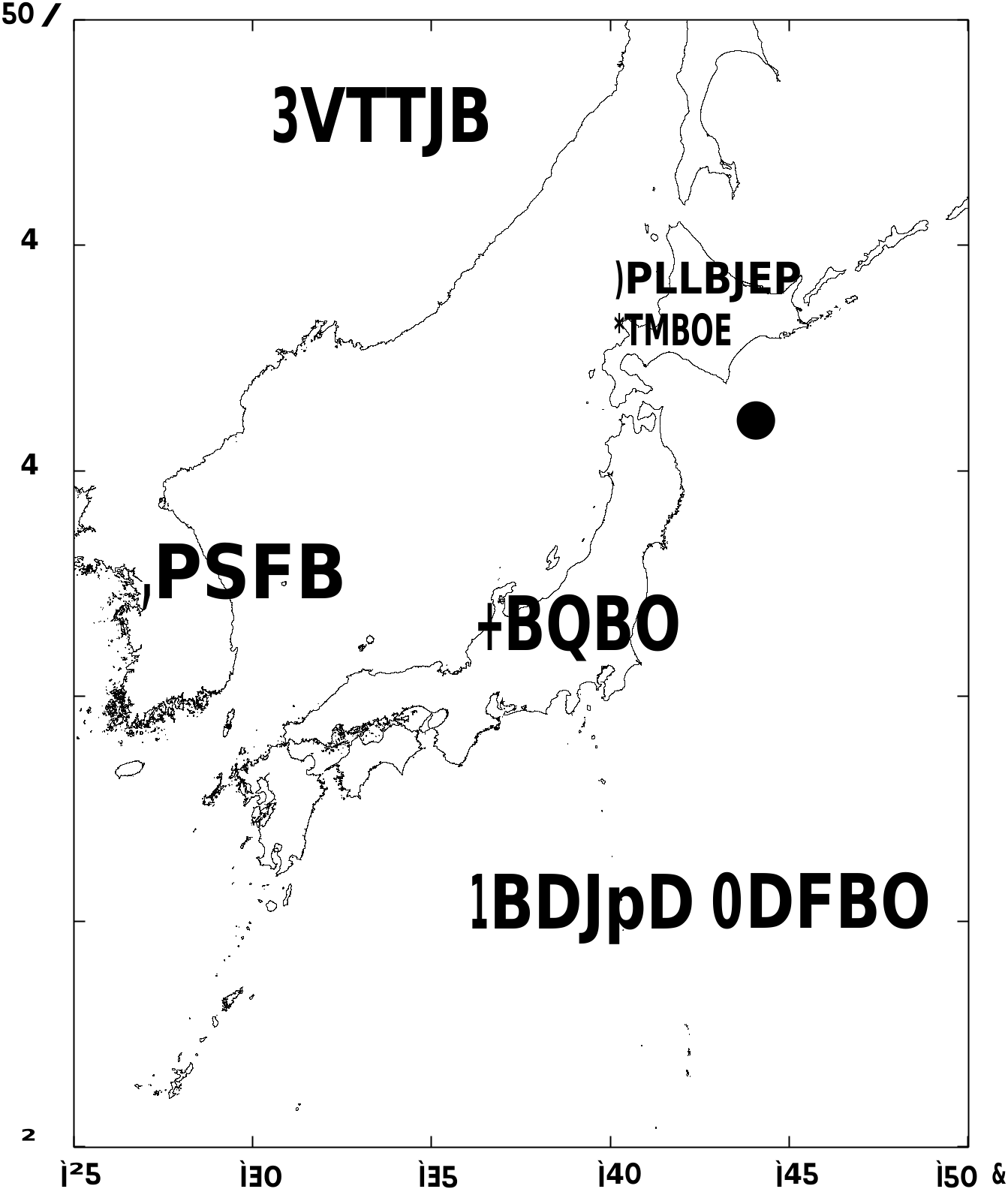
Material examined: Holotype. NSMT E-13546, southeast of Cape Erimo , Hokkaido, Japan (41°17.69'N, 144°08.01'E –41°15.66'N, 144°07.92'E), 3,143–3,176 m depth ( Fig. 1 View Fig ), 19 August 2020, R / V Shinsei-Maru of JAMSTEC, 3 m beam trawl. GoogleMaps
Paratypes: NSMT E-13547, NSMT E-13548, NSMT E-13549, and NSMT E-13550 (each one specimen), the same locality as the holotype.
Etymology: The specific name was derived from R/V Shinsei-Maru of JAMSTEC.
Diagnosis: Radial shields elongate semi-circular and separated; oral shields triangular; tentacle scales at the second tentacle pore flat and broad, two in number; dorsal arm plates octagonal, three times wider than long on proximal portion of arm; arm spines three in number on proximal portion of arm; arm spines on the middle to distal portion of arm acute, spinose.
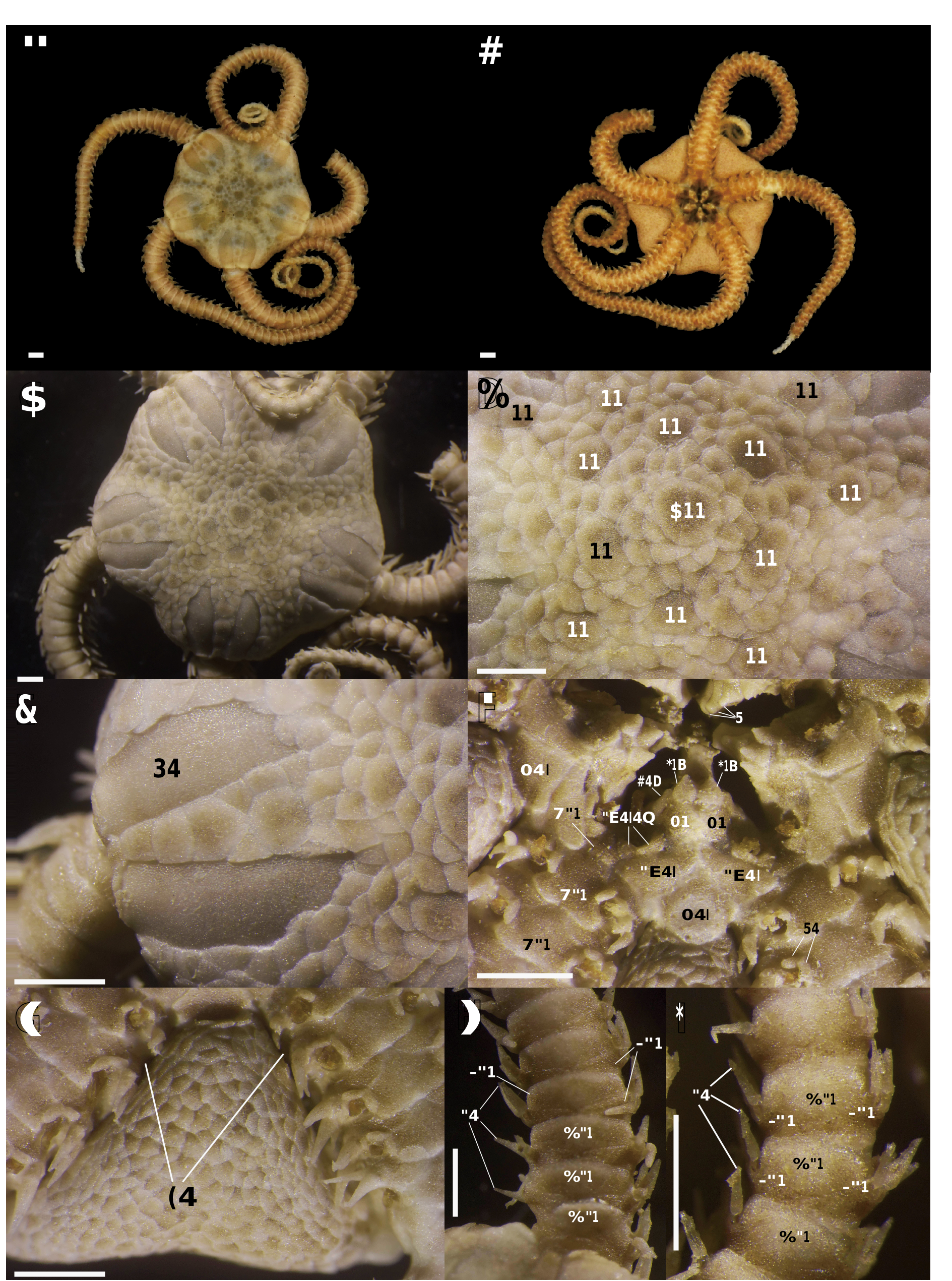
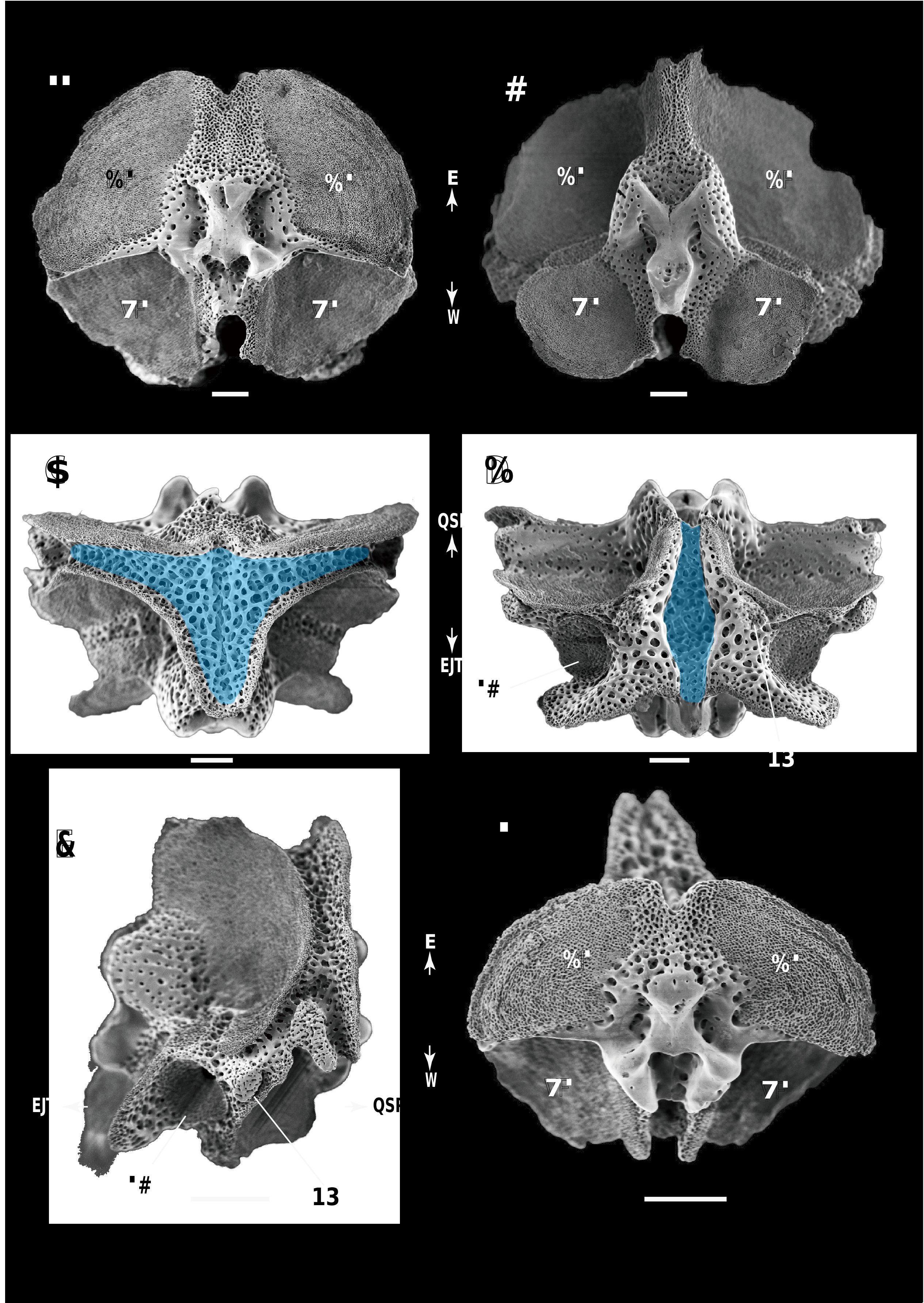
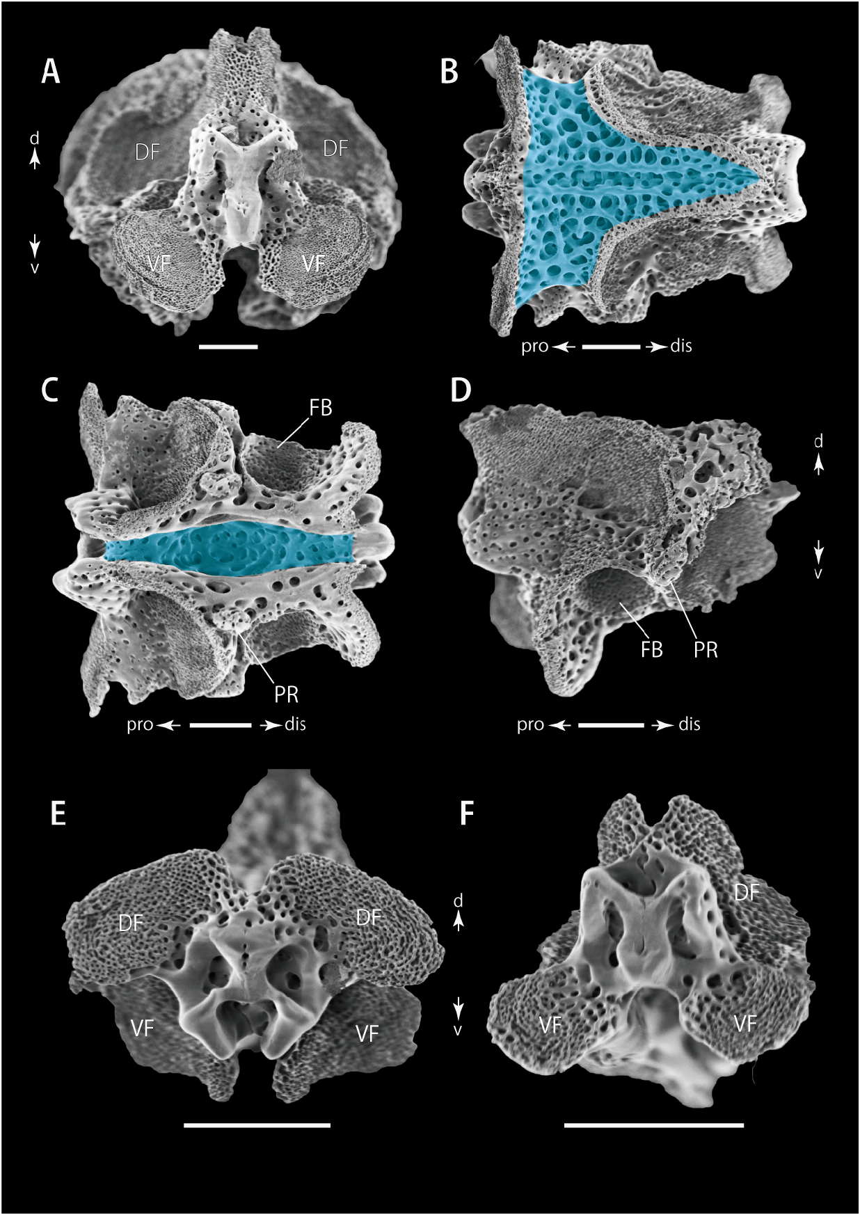
Description of external morphology (holotype, NSMT E-13546): Disc circular, 9.0 mm in diameter ( Fig. 2A, B View Fig ). Dorsal disc completely covered by a circular central primary plate of 1.80 mm in length, other primary plates approximately 1.25–1.80 mm in length, and semi-circular, smooth with imbricating disc scales, approximately 0.60–1.25 mm in length ( Fig. 2C, D View Fig ). Radial shields elongate semi-circular in shape, entirely separated from each other, approximately three times longer than wide; the length approximately half disc radius ( Fig. 2C, E View Fig ). Interradial ventral disc covered by imbricating scales, similar to those on dorsal surface in shape but smaller, approximately 0.20–0.28 mm in length ( Fig. 2F, G View Fig ). Genital slits elongate, from edge of oral shield to lateral edge of disc, approximately 0.15 mm in width and 3.20 mm in length ( Fig. 2G View Fig ). Oral shields triangular, slightly rounded on distal-lateral side, approximately 1 mm in length and width ( Fig. 2F View Fig ). One smaller, hexagonal oral shield with small pores, suggesting it serves as a madreporite ( Fig. 2F View Fig ). Adoral shields triangular, approximately 3 times wider than long ( Fig. 2F View Fig ). Oral plates polygonal, approximately as wide as long, in contact to each other ( Fig. 2F View Fig ). Teeth four and wide, forming a vertical row on dental plate; the ventral top tooth triangular, secondary one trapezoid, and more dorsal ones flat and square ( Fig. 2F View Fig ). A pair of infradental papillae on the ventralmost position of the dental plate ( Fig. 2F View Fig ). One buccal scale situated at ventral edge of each oral plate approximately 2.5 times wider than long ( Fig. 2F View Fig ). Second oral tentacle pores opening on ventral edge of adoral shield with accompanying two flat and broad adoral spines ( Fig. 2F View Fig ).
Arms five, up to 22 mm in length ( Fig. 2A, B View Fig ). Proximally 1.5 mm in width and 1.2 mm in height, rectangular in cross section. Arms tapering gradually toward the tip ( Fig. 2A, B View Fig ). Dorsal arm plates slightly separated from one another along the full distance of the arm. On proximal portion of arm, dorsal arm plates quadrangular, three times wider than long ( Fig. 2H View Fig ). Dorsal arm plates change midway to a more octagonal shape, twice as wide as long ( Fig. 2I View Fig ). Distally, dorsal plates become fan-shaped, as wide as long and gradually decreasing in size towards arm tip ( Fig. 3A View Fig ). Proximally, ventral arm plates are pentagonal, 1.5 times wider than long and in direct contact ( Fig. 3B View Fig ). Midway along the arm length, the ventral plates remain in direct contact but are slightly longer than wide ( Fig. 3C View Fig ). Distally, these plates become approximately twice as long as wide but separate from each other ( Fig. 3D View Fig ).
Arm spines cylindrical, decreasing overall from three proximally to two distally. On the proximal arm region the ventralmost spine longest, with the remaining two spines in series slightly shorter in length than the ventralmost spine ( Fig. 3E View Fig ). Midway along the arm, the three spines all conical, acutely pointed, and approximately the same length as corresponding arm segment ( Figs. 2I View Fig , 3C View Fig ). On the distal portion of the arm, arm spines remain conical and pointed but are half as long as corresponding arm segments as the spine number decreases to two towards arm tip ( Fig. 3A, D, F View Fig ). Tentacle scales two, on proximal and middle portion of the arm, both scales flat and broad at each tentacle pore ( Fig. 3B, C View Fig ). Tentacle scales decreasing in size distally, absent towards arm tip ( Fig. 3F View Fig ).
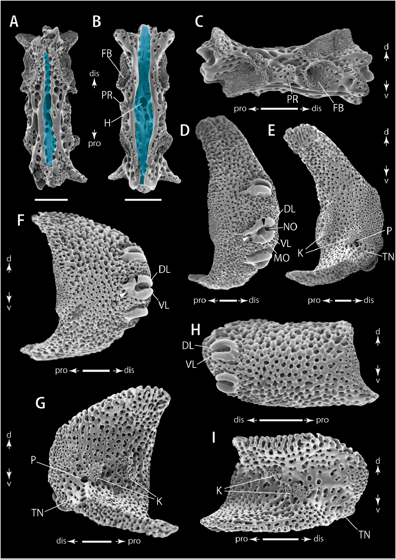
Description of ossicle morphology (holotype, NSMT E-13546 and a paratype, NSMT E-13550): Vertebrae with zygospondylous articulations ( Figs. 4A, B, F View Fig , 5A, E, F View Fig ). On proximal portion of arm, muscle fossae on dorsal sides slightly larger than those on ventral sides on both proximal and distal sides ( Fig. 4A, B View Fig ), becoming clearly wider on dorsal ones than ventral ones on middle to distal portion of arms ( Figs. 4F View Fig , 5A, E, F View Fig ). On proximal and middle portion of arms, groove T-shaped on dorsal side ( Figs. 4C View Fig , 5B View Fig ) and straight furrow, in which the radial water vascular canal and radial nerve are place, on ventral side ( Figs. 4D View Fig , 5C View Fig ). On distal portion of arms, dorsal groove becoming straight on dorsal side ( Fig. 6A View Fig ) similar to ventral side ( Fig. 6B View Fig ). No distinct channels of lateral openings for canals and nerves recognizable from external view of the ventral furrow ( Figs. 4D View Fig , 5C View Fig , 6B View Fig ); foot basins visible laterally on ventral side of the vertebra ( Figs. 4D, E View Fig , 5C, D, 5B, C View Fig ); protrusions present just externally of the middle of ventral furrow ( Figs. 4D, E View Fig , 5C, D View Fig , 6B, C View Fig ). A large hole at centre of dorsal groove on distal portion of arm ( Fig. 6A View Fig ).
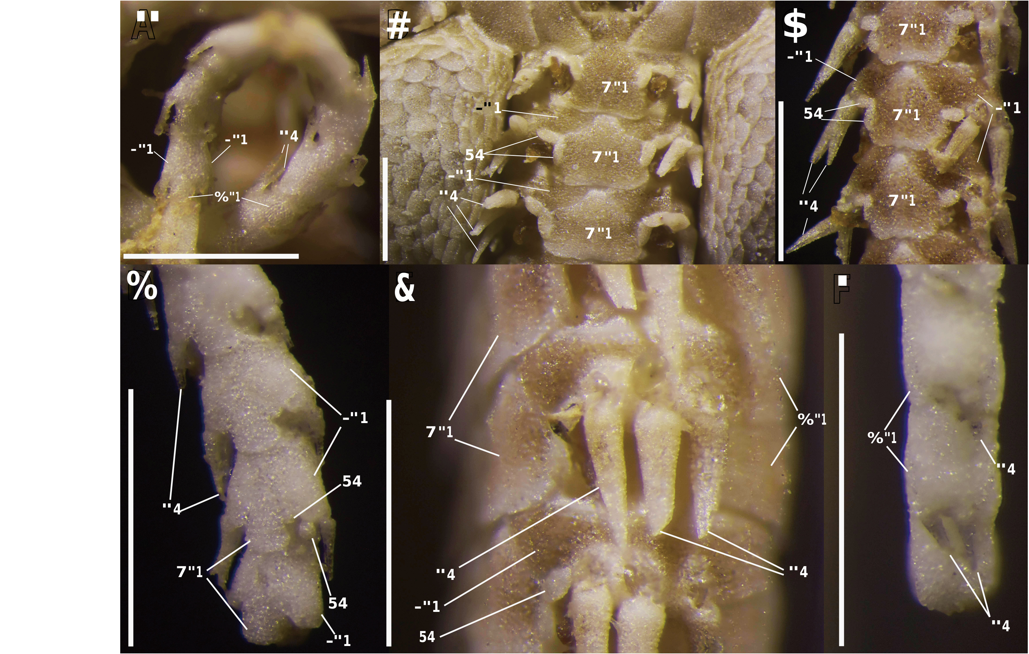
Lateral arm plates much higher than long on proximal portion of arm ( Fig. 6D, E View Fig ); slightly higher than wide on middle portion of arm ( Fig. 6F, G View Fig ); and approximately twice as wide as high on distal portion of arm ( Fig. 6H, I View Fig ). Distal edge convex with dorsal and ventral edges protruding toward proximal side, and proximal edge concave ( Fig. 6D–I View Fig ). External surfaces smooth; stereom on central portion of proximal edges slightly denser than remaining areas ( Fig. 6D, F, H View Fig ). Internal surfaces with two knobs, composed of more densely meshed stereom than remaining area, situated at centre ( Fig. 6E, G, I View Fig ). On proximal to middle portion of arm, a single perforation next to distal-most knob and a tentacle notch extending to distal-ventral edge ( Fig. 6E, G View Fig ). On distal portion of arm, a tentacle notch extends to ventral-distal edge but no perforation recognizable ( Fig. 6I View Fig ). On proximal to middle portion of arm, three spine articulations on distal edge, with nearly horizontal and parallel dorsal and ventral lobes, merging on proximal edge by depressed bridge, with dorsal lobe wider than ventral lobe ( Fig. 6D, F View Fig ). Distally, the number of articulations decreases to two ( Fig. 6H View Fig ), with dorsal and ventral lobes almost equal in width and merged at proximal edge ( Fig. 6H View Fig ). Surrounding structure of the articulations slightly sunken with denser stereom ( Fig. 6D, F, H View Fig ). Muscle and nerve openings clearly recognized and separated by stereom protuberances on proximal to middle portion of arm ( Fig. 6D, F View Fig ) but absent on distal portion of arm ( Fig. 6H View Fig ).
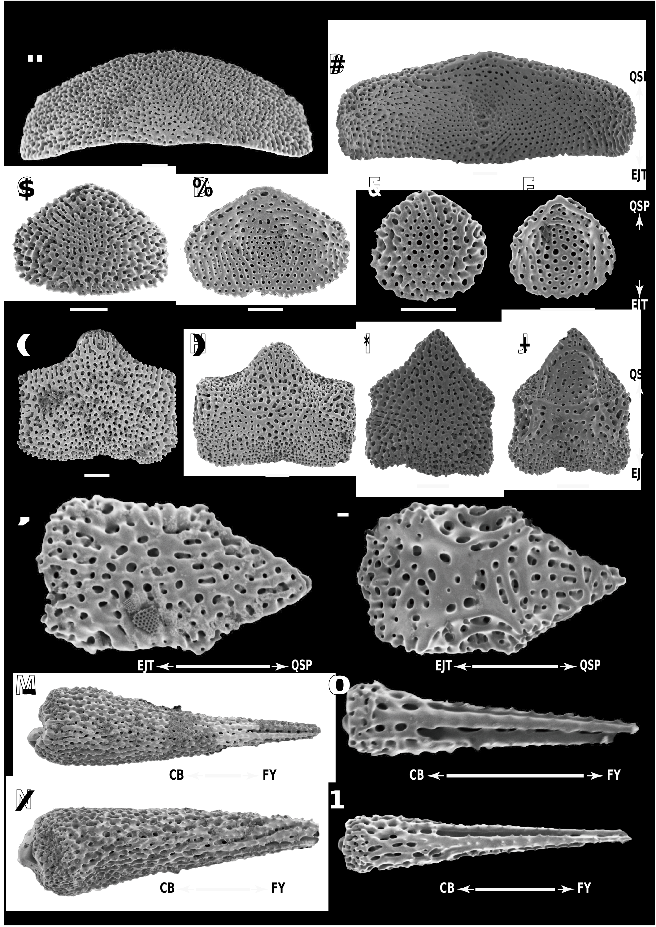
Dorsal arm plates rectangular with straight distal edges and slightly pointing proximal edge, approximately three times as wide as long on proximal portion of arm ( Fig. 7A, B View Fig ); pentagonal with slightly pointed proximal edge, as wide as long on middle portion of arm ( Fig. 7C, D View Fig ); and oval with slightly pointed proximal edge on distal portion of arm ( Fig. 7E, F View Fig ). Surfaces of the dorsal arm plates smooth ( Fig. 7A–F View Fig ).
Ventral arm plates pentagonal with blunt proximal edge on proximal portion of arm ( Fig. 7G, H View Fig ), and with slightly pointed proximal edge on middle to distal portion of arms ( Fig. 7I–L View Fig ); distal edge slightly convex; slightly wider than long on proximal portion of arm, almost as wide as long on middle portion of arm and twice as long as wide on distal portion of arm ( Fig. 7K, L View Fig ). Surfaces of the ventral arm plates smooth ( Fig. 7K, L View Fig ).
Proximally, ventralmost arm spine tapering to acute tip ( Fig. 7M View Fig ) and other two spines tapering to blunt tip ( Fig. 7N View Fig ); distally, arm spine shape similar, tapering gradually to acute tip ( Fig. 7O, P View Fig ).
Dental plates entire without fragmentation, oblong, approximately twice as long as wide, with four sockets for teeth ( Fig. 8 View Fig ). Dorsalmost tooth socket large and circular with two perforating oval holes separated by complete septum ( Fig. 8 View Fig ). Second dorsalmost tooth socket smaller than the dorsalmost one, with small perforating holes separated by complete septum opening as slit-shaped transverse holes on internal side ( Fig. 8 View Fig ). Other two ventral sockets shallow, not penetrating ( Fig. 8 View Fig ).
Variation (paratypes, NSMT E-13547, NSMT E-13548, NSMT E-13549 and NSMT E-13550): Some morphological variation was observed among the four paratypes. Number of tentacle scales on second oral tentacle pores was two for two paratypes (NSMT E-13547, disc diameter [d.d.] = 12.5 mm; NSMT E-13549, d.d. = 11.6 mm), one for one paratype (NSMT E-13548, d.d. = 9.5 mm), and one or two for one paratype (NSMT E-13550, d.d. = 10.9 mm). One paratype (NSMT E-13550) had four arms naturally while other three paratypes have five.
DNA barcoding: We obtained a 774-bp COI gene region for the holotype of Ophiomonas shinseimaruae sp. n. (NSMT E-13546; LC586826 View Materials ). No COI sequence of the genus Ophiomonas was registered in GenBank, and the closest sequence is from Amphiura bidentata H. L. Clark, 1938 ( Amphiuridae : KU895046 View Materials ) with 20.0% K2P genetic distance.
Occurrence: Known only from the type locality, southeast of Cape Erimo, Hokkaido, Japan, 3,143 –3,176 m deep ( Fig. 1 View Fig ).
| NSMT |
National Science Museum (Natural History) |
| R |
Departamento de Geologia, Universidad de Chile |
| V |
Royal British Columbia Museum - Herbarium |
No known copyright restrictions apply. See Agosti, D., Egloff, W., 2009. Taxonomic information exchange and copyright: the Plazi approach. BMC Research Notes 2009, 2:53 for further explanation.
|
Kingdom |
|
|
Phylum |
|
|
Class |
|
|
Order |
|
|
Family |
|
|
Genus |