
Passalus (Pertinax) umbriensis Hincks, 1950, 1044
|
publication ID |
https://doi.org/ 10.1080/00222933.2020.1759721 |
|
DOI |
https://doi.org/10.5281/zenodo.4608954 |
|
persistent identifier |
https://treatment.plazi.org/id/C4008788-691C-9E0C-FEF9-FA2E838F6E06 |
|
treatment provided by |
Carolina |
|
scientific name |
Passalus (Pertinax) umbriensis Hincks, 1950 |
| status |
|
Passalus (Pertinax) umbriensis Hincks, 1950 ( Figures 7 – 8 View Figure 7 View Figure 8 )
Reyes-Castillo and Amat-Garcia 1991: 504; Boucher 2015: 118; Miles 2017: 124, fig. 25; Bevilaqua and Fonseca 2019: 7, fig. 3e
Passalus (Passalus) umbriensis Hincks, 1950: 1044 , fig. 3; Hincks and Dibb 1958: 17; Reyes-Castillo 1970: 204; Amat-García et al. 2004: 177; Fonseca and Reyes-Castillo, 2004: 7 Diagnosis. Small size species; convex body; anterior edge of the front with a notch in the middle whose edges rise reminiscent of secondary mediofrontal tubercles; inconspicuous inner tubercles; mentum with mediobasal area with punctuations and setae; pronotum with protruded and acute anterior angles; abundant pronotal punctuation laterally; almost transverse and deep mesosternal scars; non-delimited metasternal disc.
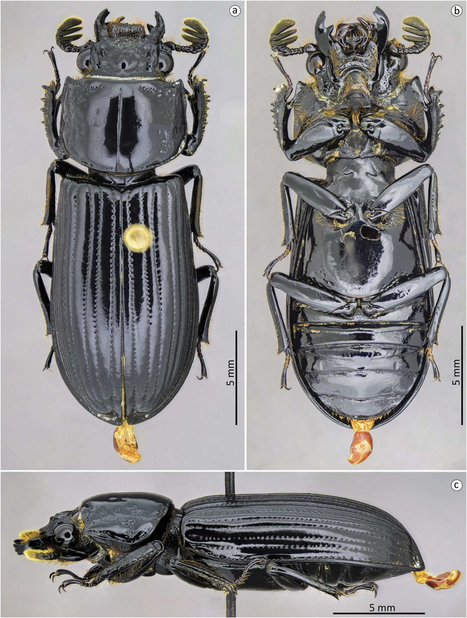
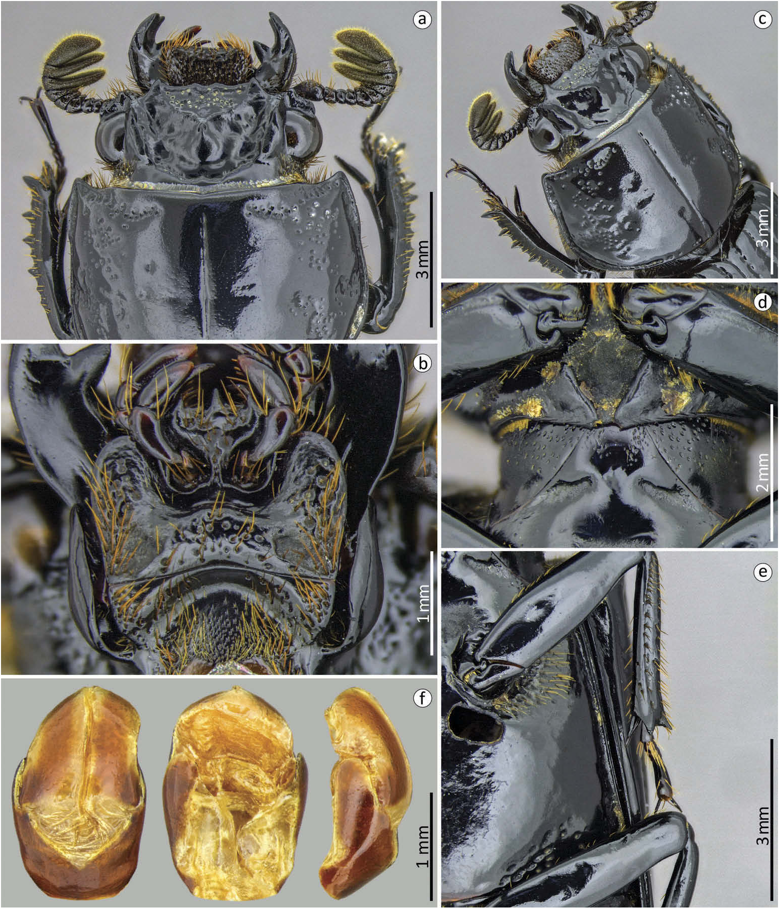
Redescription. Body ( Figure 7 View Figure 7 ): habitus: convex; size: small (> 20 mm in length). Head ( Figure 8 View Figure 8 (a,c)): Labrum: anterior border straight. Clypeus: hidden under the frons; with anterior angles under the lateral + mediofrontal tubercles. Anterior frontal edge: straight, with a notch in the median region whose edges are high resembling secondary mediofrontal tubercles. Secondary mediofrontal tubercles: absent. Laterofrontal + mediofrontal tubercles: conspicuous, acute, and projected forward. Mediofrontal area: caliciform, as long as wide (1.1x), flat and shallow; with punctuations scattered throughout the anterior region. Cephalic nodule: small, not well marked and rounded. Inner tubercles: small, inconspicuous, smaller than the laterofrontal + mediofrontal tubercles, of which they are separated, located midway between them and the apex of the central tubercle. Anterofrontal ridges: straight, smaller, and weaker than the posterofrontal ridges, practically absent. Posterofrontal ridges: low, weak, and slightly sinuous, beginning at the apex of the central tubercle. Laterofrontal areas: flat and smooth surface. Central tubercle: conical, low, with non-free apex. Lateroposterior tubercles: inconspicuous, separated from the central tubercle. Postfrontal area: flat, with smooth surface. Postfrontal groove: well marked, sinuous with a slight notch located medially. Lateropostfrontal areas: shallow, smooth, and glabrous surface. Epicranial sutures: poorly marked. Epicranial pits: shallow but very visible. Anterior angles of the head: well developed, with acute apexes, slightly smaller than the laterofrontal + mediofrontal tubercles. Canthus ocular: apexes rounded not reaching half eye. Antennas: trilamellate, with narrow lamellae, the distal being wider than the other two. Mouthparts ( Figures 7 View Figure 7 (b), 8(b)): Ligula : tridentate with middle tooth larger than the lateral teeth. Hypostomal process: wide, glabrous and separated from mentum. Mentum mediobasal area: with coarse punctuations and bristles scattered throughout the area, with a protruding anterior region with no notch in the middle. Mentum lateral lobes: rounded apexes. Mentum lateral scars: large, shallow, and rounded. Mandibles: incisor lobe with three well-formed teeth at apex; robust suprainternal teeth; inconspicuous infrabasal pits. Maxilla: lacinia bidentate at the apex. Prothorax. Pronotum ( Figures 7 View Figure 7 (a,c), 8(a – c)): Anterior edge: straight. Anterior angles: acute and fairly protracted. Marginal groove: well marked, deep and with coarse punctures throughout its length, without dilation at the apex; reaching four-fifths of pronotal width. Lateral fossae: small, well marked and softly deep; slightly rounded shape. Pronotal punctuations: coarse and fairly dense, diffused throughout pronotum, concentrated mainly above the lateral fossae. Prosternum ( Figures 7 View Figure 7 (b), 8(d)): Prepisternum: not dilated in the anterior region; finely pubescent in lateroposterior region. Prepimerum: glabrous. Prosternelum: rhomboid with narrow apex. Mesosternum ( Figures 7 View Figure 7 (b), 8(d)): smooth, shiny, and glabrous. Mesosternal scars: long, narrow, and deep; practically transversal; without punctuations or pubescence. Metasternum ( Figures 7 View Figure 7 (b), 8(e)): Metasternal disc: poorly delimited, but more dilated than the lateral region of the metasternum. Metasternal punctuations: group of punctures only in the lateroposterior region; setigerous punctuations in the anterior region. Metasternal pubescence: only in the anterior region quite sparsely. Metasternal lateral groove: narrow, thinner than mesotibiae, deep, without punctures or bristles. Elytra ( Figure 7 View Figure 7 (a)): Approximately 2.6x longer and 0.9x wider than pronotum. Striae: narrower than the interstriae, marked with round and fine punctures, well defined in both the dorsal and lateral striae. Epipleura : glabrous. Humeri: with small and sparse bristles. Legs ( Figures 7 View Figure 7 (a,b), 8(e)): Profemur: ventral anterior border with well-marked groove, not reaching the apex of the profemur; ventral posterior border with few setae only near the apex. Protibiae: not dilated. Mesotibiae: one small spine on the outer face. Metatibiae: without spine. Abdomen ( Figure 7 View Figure 7 (b)): sternite VII with full groove and well marked; rough sides. Aedeagus ( Figure 8 View Figure 8 (f)): Median lobe wider than the parameres and the basal piece together and almost as long as these. In the ventral view with two plaques completely sclerotised with a recess in the basal region; basal piece and parameres fused forming a tegmen; parameres projections reaching less than one-third of the length of the median lobe; arched parameres anterior edge. In lateral view, the apex of the parameres is narrow and rounded. In dorsal view with widely separated parameres.
Dimensions. Total length: 21 mm; cephalic length: 2.1 mm; cephalic width: 4.8 mm; mediofrontal area length: 0.7 mm; mediofrontal area width: 1.9 mm; canthus ocular length: 0.6 mm; canthus ocular width: 0.3 mm; area of the mediofrontal area: 1 mm 2; mandibles external angle: 139º; antennal club length: 0.7 mm; antennal club width: 1.4 mm; distal lamella length: 0.4 mm; medial lamella length: 0.1 mm; width of the mentum at the lateral scars: 2.2 mm; mentum mediobasal area width: 1 mm; diameter of the mentum lateral scars: 0.4 mm; pronotal length: 4.7 mm; pronotal width: 6.4 mm; length of the pronotal anterior groove: 2 mm; elytral length: 12.4 mm; elytral width: 7.4 mm; humeral width: 6.1 mm; profemur length: 3.1 mm; length of the anterior ventral groove of the profemur: 2.4 mm; protibiae width: 0.8 mm; protarsal length: 2.4 mm; length of the last protarsomer: 0.9 mm; mesotibiae width: 0.5 mm; metasternal lateral groove width: 0.3 mm; metasternal disc length: 4.8 mm; abdomen length: 5.7 mm; aedeagus length: 2 mm; aedeagus width: 1.2 mm; median lobe length: 1.7 mm; paremeres projection lenght: 0.4 mm.
Material examined. Holotype (♂) F2439.49. COLOMBIA: Umbria/Guines Fluss/ Columbien//Ex Staudinger &/Bang Haas, Dresden.// Passalus Type/( Passalus )/ umbriensis Hincks /det. W.D. Hincks//Manchester Museum/ HOLOTYPE // ♂ dissect/S. Boucher det. 2012. F2439.49 ( MMUE).
Remarks. Described in P. ( Passalus ) in the session ‘ Phoroneus ’, this species had its combination reviewed by Bevilaqua and Fonseca (2019) who allocated it within the subgenus P. ( Pertinax ).
Distribution. Colombia (Umbria) (locality type) and Brazil (Acre) Bevilaqua and Fonseca 2019.
| MMUE |
Museum of Manchester University |
No known copyright restrictions apply. See Agosti, D., Egloff, W., 2009. Taxonomic information exchange and copyright: the Plazi approach. BMC Research Notes 2009, 2:53 for further explanation.
|
Kingdom |
|
|
Phylum |
|
|
Class |
|
|
Order |
|
|
Family |
|
|
Genus |
Passalus (Pertinax) umbriensis Hincks, 1950
| Bevilaqua, Marcus & Fonseca, Claudio Ruy Vasconcelos da 2020 |
Passalus (Passalus) umbriensis
| Amat-Garcia G & Vargas EB & Reyes-Castillo P 2004: 177 |
| Reyes-Castillo P 1970: 204 |
| Hincks WD & Dibb JR 1958: 17 |
| Hincks WD 1950: 1044 |