
Phalloptychus januarius (Hensel)
|
publication ID |
https://doi.org/ 10.1590/S1679-62252005000300004 |
|
publication LSID |
lsid:zoobank.org:pub:9B60A10B-F68C-4462-9A6C-39B139514394 |
|
persistent identifier |
https://treatment.plazi.org/id/0714F61A-5A00-FFD8-1DCB-C524FA61BCA6 |
|
treatment provided by |
Carolina |
|
scientific name |
Phalloptychus januarius (Hensel) |
| status |
|
Phalloptychus januarius (Hensel) View in CoL
Fig. 7; Tables 1-2
Girardinus januarius Hensel, 1868: 360 View in CoL . Type locality: “aus den Pfützen und Gräben um Rio de Janeiro ” [= from the puddles and ditches around Rio de Janeiro].
Type material. Lectotype [present designation]: ZMB 7422 View Materials . Paralectotypes: ZMB 33219 [originally ZMB 7422 View Materials ] , ZMB 7424 View Materials , and ZMB 31497 .
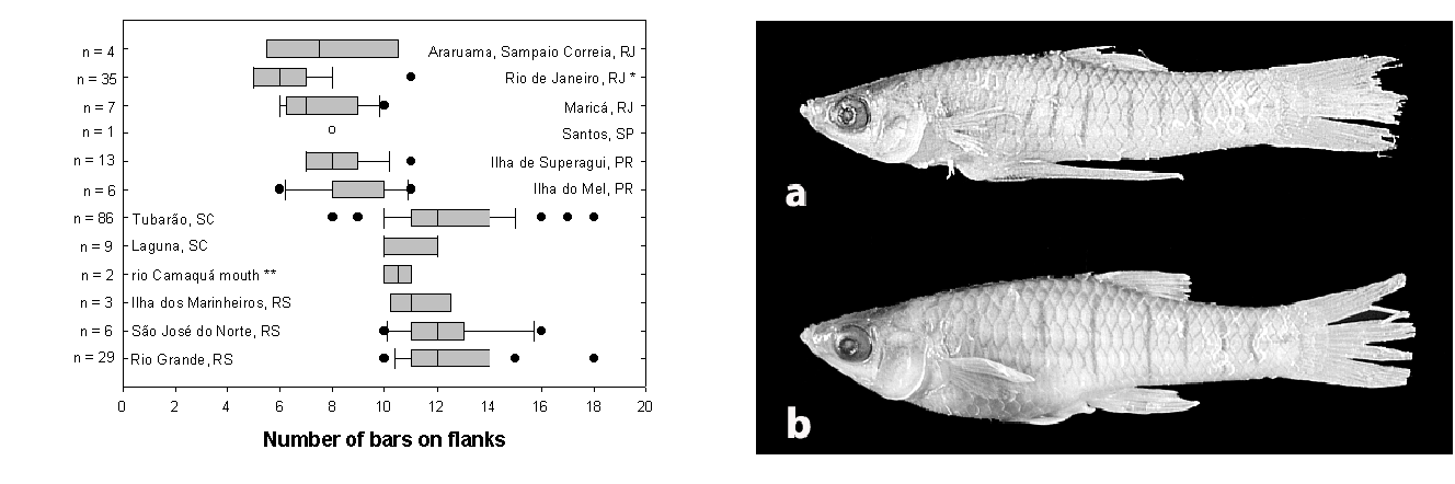
Diagnosis. Phalloptychus januarius can be autapomorphically diagnosed by the possession of nine anal-fin rays in males [86-4]. Furthermore, Phalloptychus januarius can be distinguished from P. eigenmanni by the number of pelvicfin rays in females (5 vs. 6, respectively), by the number of predorsal scales (10-12 vs. 13, respectively), and the shorter predorsal distance in females (55.4-62.2 vs. 64.0-68.6 % SL, respectively). Phalloptychus januarius is distinguished from P. iheringii by the number of epipleural ribs (12-13 [one specimen had 11 in one side and 12 in the other] vs. 10-11, respectively), by the number of gonopodial rays (9 vs. 8, respectively), and by a significantly lower number of vertical bars along body sides of females (range = 5-12, median = 7, vs. range = 8-18, median = 12, respectively).Although some overlap occurs, Mann-Whitney rank sum tests ( Fig. 6 View Fig ) indicate significant differences (P <0.001).
Description. Morphometric data in Tables 1-2. Range of SL: 18.4 to 32.6 mm (females), 13.6 to 19.8 mm (males). Body compressed; width in predorsal region uniform, and about half body depth. Postdorsal region compressed towards caudal peduncle. Dorsal profile of head slightly concave. Predorsal profile convex. Dorsal-fin base oblique. Postdorsal profile concave. Pre-anal profile convex.Anal-fin base oblique; postanal profile slightly concave. Dorsal fin with semi-elliptical border, located posterior to mid-body. Origin of dorsal fin in females at vertical passing approximately through base of second anal-fin ray; in males, origin of dorsal fin posterior to vertical passing through origin of anal fin. Pectoral fin with high insertion. Base of dorsalmost pectoral-fin ray closer to dorsal than to ventral profile. Longest pectoral-fin ray of females reaching about fifth or sixth scale in longitudinal series. In adult males, origin of pectoral fin aligned with origin of pelvic fin. Pelvic fin small, surpassing origin of gonopodium in male; not reaching origin of anal fin in females. Origin of anal fin of male closer to snout tip than to caudal peduncle. Mouth superior, almost aligned with base of dorsalmost pectoral-fin ray.
Dorsal-fin rays: 9[52], 10[1]. Pectoral-fin rays: 10[10], 11[28], 12[9]. Pelvic-fin rays: 4[32] (males), 5[27] (females). Anal-fin rays (females): 10[25]. Gonopodial rays: 9[2]. Caudal-fin rays: 23[3], 24[19], 25[26], 26[1]. Predorsal scales: 11[14], 12[36]. Longitudinal series scales: 26[3], 27[27], 28[21], 29[4]. Scales around caudal peduncle: 16[60]. Scales in transverse row: 8[38]. Epipleural ribs: 12[9], 13[2], (plus one specimen with 11 in one side and 12 in other). Pleural ribs: 13[2], 14[7]. Vertebrae: 31[6]: (11/5/15)[1], (12/4/15)[1], (12/5/14)[4]; 32[4]: (11/5/16)[1], (12/4/16)[1], (12/5/15)[2]; 33 (12/6/15)[1].Expanded neural processes: 4[6], 5[5]. Premaxillary teeth: 7[1], 8[1], 9[1], 10[3], 11[11], 12[2], 14[1], 15[1]. Dentary teeth: 8[4], 9[1],10[2], 11[5], 12[6], 13[4]. Branchiostegal rays: 5[10], 6[1]. Caudal-fin rays attached to hypural plate: 9[11]. Upper accessory cartilages: 2[3]. Lower accessory cartilages: 2[3].
Preorbital ramus of cephalic sensory system represented by one to three grooved neuromasts. Preorbital canal absent. Anterior portion of supraorbital ramus (pores 1 and 2a) parallel to upper lip with three inconspicuous neuromasts on each side. Posterior portion of supraorbital ramus (pores 2b, 3, 4a) composed of two or three grooved neuromasts. Posterior remnants of infra-orbital ramus represented by three neuromasts (pores 4b, 5, 6a) and by one canal opened in both ends (pores 6b and 7). About 15 inconspicuous neuromasts on ventral infra-orbital line. Preopercular ramus represented by large canal (sometimes completely open, forming groove) along preopercular posterolateral border and by prolonged canal along preopercle ventral border opened by four pores. Opercular canal absent. Mandibular ramus composed of two or three superficial neuromasts (pores Z, Ya, and Yb) on anterior border of ventral surface of mandible and by one superficial neuromast near maxillary distal end (pore W).
Gonopodial complex composed of three functional gonapophyses and nine gonactinosts. Gonactinosts 2, 3, 4 fused. Gonactinost 4 with wing-like expansions. Ligastyle present. Gonopodium sinistrally asymmetrical. Eight gonopodial rays. R1 and R2 unbranched and short, with 8 segments. R3 with dorsal convexity located near base ranging from second to tenth or eleventh segments. Tip of R3 , R4 a, and R4 p ventrally bent and joint. Dorsal convexity located between segments 14 to 21 (rarely 12 to 18) of R 4p. Twelve to 17 spines on distal segments of R 4p. Spines retrorse except two or three last spines, directed forwards or upwards. R6 , R7 , R8 branched. Five to 8 segments before bifurcation of ray 6. Anterior and posterior branches of R6 almost or fully ankylosed. Distal end of R6 modified in arrow-shaped expansion. R7 with six or seven segments anterior to bifurcation. Anterior and posterior branches of R7 moderately expanded and partially to completely ankylosed. Anterior branch of R7 greater than posterior. R8 with seven or eight segments anterior to bifurcation. Anterior and posterior branches of R8 normal. Anterior branch of R8 greater than posterior. Four to six segments on anterior branch of R8 . Four segments on posterior branch of R8 . R9 minute not attached to pterygiophore .
Color in alcohol. Eye greenish grey. Pupil cream. Ground color cream, darker in upper half. Scales surface border and subjacent skin replete with many brown chromatophores, conferring reticulate pattern to body sides. Dark brown predorsal line. Brown chromatophores scattered through whole body, more concentrated in dorsal portion, mainly on head, snout, and ventral surface of mandible. Anus margin and urogenital area light yellow. Fins hyaline. Fin rays with two rows of brown chromatophores along each side, along extension of ray. Pale brown band on dorsal fin near its base (sometimes not easily seen). Five to 12 narrow brown bars (median = 7) along body sides of female specimens. Dark brown spot at base of R3.
Common names. Barrigudinho, guaru.
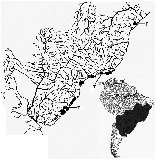
Distribution. Phalloptychus januarius is known from coastal drainages in Rio de Janeiro, São Paulo and Paraná States of Brazil ( Fig. 1 View Fig ).
Remarks. Besides the types examined, the type series includes the lots ZMB 7423 (4) and CAS-SU 1132 (1). It is not absolutely sure, whether the lot ZMB 31497 belongs to the type series, since this number is based secondarily on an old label from the anatomical collection: “No. 25204, R. Janeir [sic]” - the remainder of the label is illegible. This is the collection of the former “Institute of Anatomy of the Humboldt- University” in the 19th century. And this belonged to the Medical Faculty. This is due to the fact that human-anatomists were traditionally doing much research work on comparative anatomy of vertebrates. Thus, collected material was often divided between Zoological (Philosophical) Faculty with the Zoological Collection and the Medical Faculty. Only from approximately 1890 onwards the collections were reunited. But, apparently also R. Virchow, the famous pathologist working at the Charity Hospital at Humboldt-University took over some of the original anatomical collection for study (Paepke & Seegers 1986; P. Bartsch in litt., 2000).As there is no apparent evidence that Hensel has not examined specimens from lot ZMB 31497, it is advisable to label the lot paralectotypes. If future evidence demonstrates that these specimens are not syntypes, they will lose their paralectotype status.
Examined material. Brazil. Lectotype [present designation], ZMB 7422 View Materials , Rio de Janeiro, approximately 22 o 53’S 42 o 21’W, Hensel GoogleMaps . Paralectotypes, ZMB 33219 (1) , same data as lectotype. ZMB 7424 View Materials (1) GoogleMaps , Rio de Janeiro, R . Hensel. ZMB 31497 (20) , Rio de Janeir ... [Sic] . Non-type specimens. Rio de Janeiro: MCP 8493 View Materials (65/4*) , Rio de Janeiro, lagoa de Jacarepaguá, Dec 1979. MCP 9544 View Materials (10) , Rio de Janeiro, lagoa de Jacarepaguá, Oct 1979. MZUSP 38370 View Materials (2) , Araruama, Iguaba Grande, 1984. UFPB 2160 View Materials (9 of 37) , Maricá, lagoa da Barra, 13 Jan 1988. UMMZ 231550 View Materials (10) , Farol de São Tomé, 3 Nov 1989. USNM 331914 View Materials (4) , rio Jundiá , tributary to lago de Saquarema, along road Amaral Peixoto, between Sampaio Correia and Bacaxa, 11 Nov 1982. São Paulo: MZUSP 50018 View Materials (1) , Guarujá, Baía Branca, lagoon near sea. USNM 132422 View Materials (1) , Santos, 13 Sep 1925. Paraná: MNHCI 6174 View Materials (15/4*) , Guaraqueçaba, Ilha de Superagui , 28 Aug 1991. MNHCI 6183 View Materials (27/4*) , Paranaguá, Ilha do Mel, Praia de Brasília , 16 Jul 1991 .
Key to the species of Phalloptychus View in CoL .
1. Females with six pelvic-fin rays; 13 predorsal scales; predorsal distance 64.0-68.6 % SL (rio Catu, Bahia) ............... ................................................. Phalloptychus eigenmanni View in CoL
1’. Females with five pelvic-fin rays; 10-12 predorsal scales; predorsal distance 55.4-62.2 % SL .................................... 2
2. Five to 12 (median = 7) vertical bars along body side in females; 12 or 13 epipleural ribs in adult specimens; 9 gonopodial rays; ninth minute, incompletely ossified gonopodial ray present (Coastal drainages from Rio de Janeiro to Paraná) ..................... Phalloptychus januarius View in CoL
2’. Eight to 18 (median = 12) vertical bars along body sides in females; 9 to 11 epipleural ribs on adult specimens; 8 gonopodial rays (Coastal drainages of Santa Catarina and Rio Grande do Sul) ...................... Phalloptychus iheringii View in CoL
| ZMB |
Museum für Naturkunde Berlin (Zoological Collections) |
| R |
Departamento de Geologia, Universidad de Chile |
No known copyright restrictions apply. See Agosti, D., Egloff, W., 2009. Taxonomic information exchange and copyright: the Plazi approach. BMC Research Notes 2009, 2:53 for further explanation.
|
Kingdom |
|
|
Phylum |
|
|
Class |
|
|
Order |
|
|
Family |
|
|
Genus |
Phalloptychus januarius (Hensel)
| Lucinda, Paulo H. F. 2005 |
Girardinus januarius
| Hensel 1868: 360 |