
Pherania giupponii Kury
|
publication ID |
https://doi.org/ 10.5281/zenodo.156324 |
|
DOI |
https://doi.org/10.5281/zenodo.6276603 |
|
persistent identifier |
https://treatment.plazi.org/id/039B072F-A222-B521-FE81-E81EFD72F8CC |
|
treatment provided by |
Plazi |
|
scientific name |
Pherania giupponii Kury |
| status |
sp. nov. |
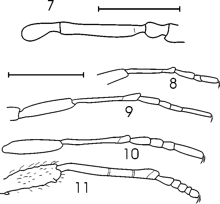
Pherania giupponii Kury View in CoL sp. nov. ( Figs. 1–14 View FIGURES 1 – 3 View FIGURES 4 5 View FIGURE 6 View FIGURES 7 – 11 View FIGURES 12 – 13 View FIGURE 14 )
Type material: Male holotype, 2 male 3 female paratypes ( MNRJ 4494) Brazil. Santa Catarina. Florianópolis, Ilha de Santa Catarina, forest on hill behind buildings of APAE, ÚNICA e SESI (27.61°S, 48.49°W), 15–17 December 1999, leg. A. P. L. Giupponi.
Etymology. Specific name honors Alessandro P. L. Giupponi, member of our team in the Arachnology Laboratory, who discovered the only known representatives of this species.
Diagnosis. Can be distinguished from Pherania pygmaea by: 1) ventroretrolateral apophysis of tibia IV of male anvilshaped (as two tubercles in Ph. pygmaea ); 2) two stout spiniform ventroprolateral apophyses of tibia IV of male (only one in Ph. pygmaea ); 3) tubercle of eye mound rounded (sharp in Ph. pygmaea ); 4) tarsus of leg III tetramerous (pentamerous in Ph. pygmaea ).
Description of male holotype (MNRJ 4494)
Measurements. Carapace 0.8 long, 0.9 wide. Eye mound 0.3 wide. Abdominal scutum 1.1 long, 1.4 wide. Posterior margin of scutum 1.0 wide. Stigmatic area 1.1 wide, 0.8 long, distance between stigmata 0.8. Legs: I Fe 0.7, Ti 0.4, Mt 0.6. II Fe 0.9, Ti 0.8, Mt 1.0. III Fe 0.9, Ti 0.7, Mt 0.9. IV Fe 1.1, Ti 0.8, Mt 1.5.
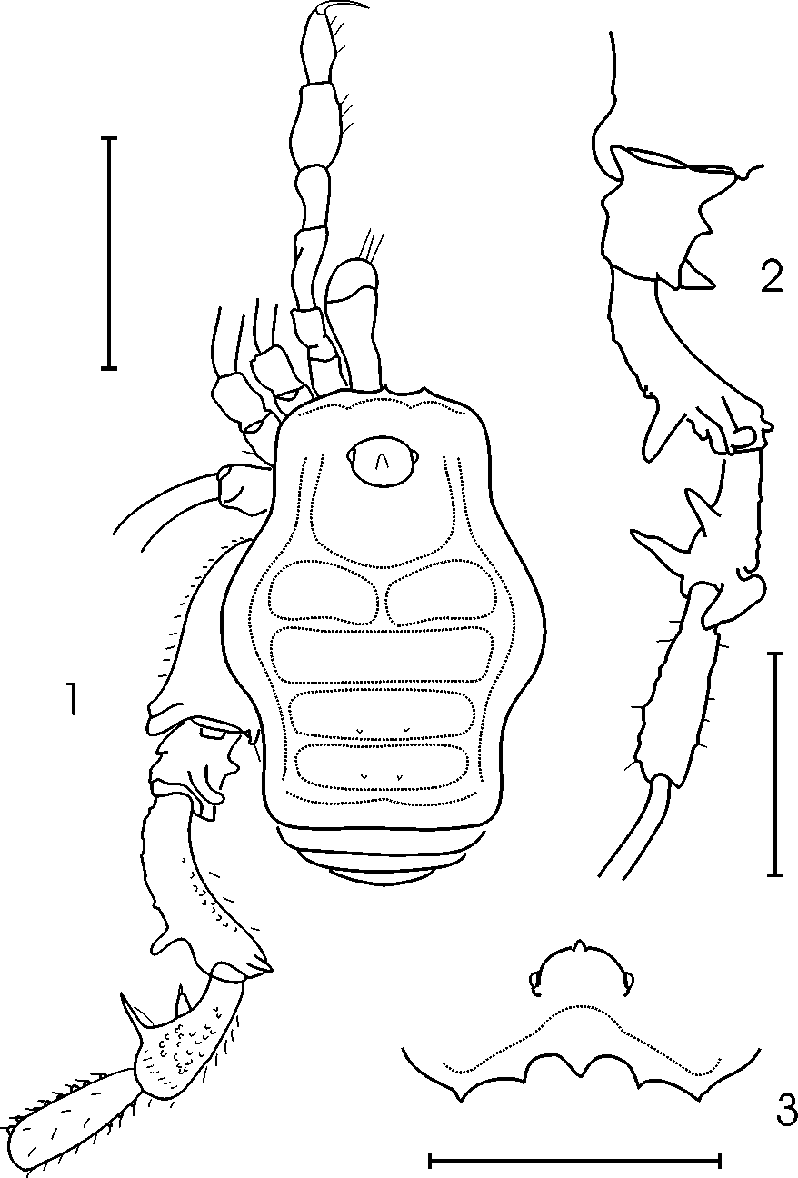
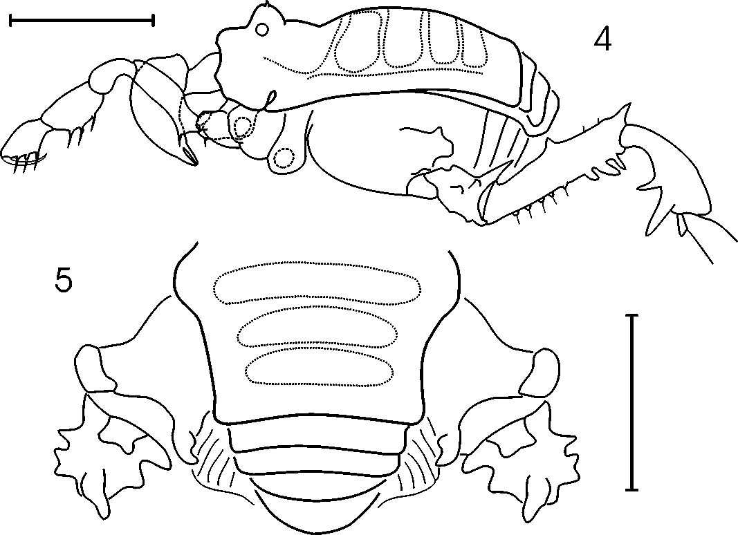
Dorsum ( Figs. 1, 3–5 View FIGURES 1 – 3 View FIGURES 4 5 ): Outline of dorsal scutum sinuous, widest at groove II. Posterior border of scutum straight. Eye mound elliptical, moderately high, well separated from anterior border of carapace, armed with small unpaired median blunt tubercle. Mesotergum divided into 4 welldefined areas by transverse grooves, area I divided into left and right halves by median groove. All areas and tergites smooth and unarmed, except from a pair of very small paramedian granules on each area III and IV.
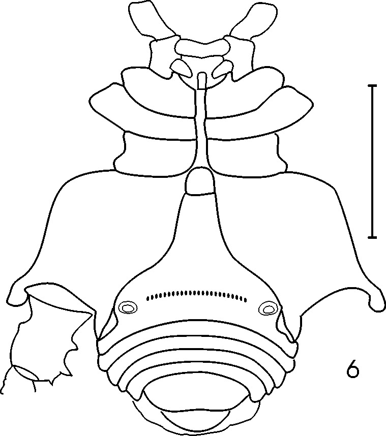
Venter ( Fig 6 View FIGURE 6 ): Coxae I–IV, stigmatic area, sternites and anal opercle finely granular, without remarkable processes. Stigmatic area well separated from coxa IV, Tshaped. Stigmata clearly visible. Coxa IV with ventroapical apophysis applied against free sternite I.
Mouth parts. Chelicerae not swollen. Basichelicerite without welldefined bulla. Pedipalpal trochanter with one basal setiferous tubercle; femur with ventrobasal setiferous tubercle; patella unarmed. Tibia and tarsus armed with weak spines ( Fig 4 View FIGURES 4 5 ). Tibia with 3 (IIi) ectal and mesal spines. Tarsus with 3 mesal (IIi) and 3 ectal (IIi) spines.
Legs. Femur I with a ventral row of delicate setiferous granules. Coxa IV ( Figs. 1 View FIGURES 1 – 3 , 5 View FIGURES 4 5 ) with bifurcate dorsoapical apophysis with two subequal branches and ventroapical apophysis with subdistal secondary branch. Trochanter IV ( Figs. 1–2 View FIGURES 1 – 3 , 4–6 View FIGURES 4 5 View FIGURE 6 ) with welldeveloped square sclerite, a strong subdistal dorsal recurved apophysis, one ventroretrolateral spiniform apophysis and two prolateral spiniform apophyses. Femur IV ( Figs. 1–2 View FIGURES 1 – 3 , 4 View FIGURES 4 5 ) subsigmoid, with strong subdistal prolateral spiniform apophysis and subdistal ventral bifid apophysis. Patella IV ( Figs. 1–2 View FIGURES 1 – 3 ) with stout median ventroprolateral spiniform apophysis and still larger distal prolateral spiniform apophysis, and one ventroapical anvilshaped apophysis. Tibia IV with two rows of small blunt granules. Tarsal joints: 3(2) / 4(3) / 4 / 5. Tarsal claws unpectinate, tarsal process absent.
Color. Body and appendages background dark yellow. Scutal areas IIV are dark brown with slightly lighter areas in the middle. A sharp dark brown band follows outline of scutum on lateral areas and posterior margin. Sparse dark brown mottling on legs IIV and even sparser very faint on pedipalps, chelicerae and venter (coxa + stigmatic area + sternites).
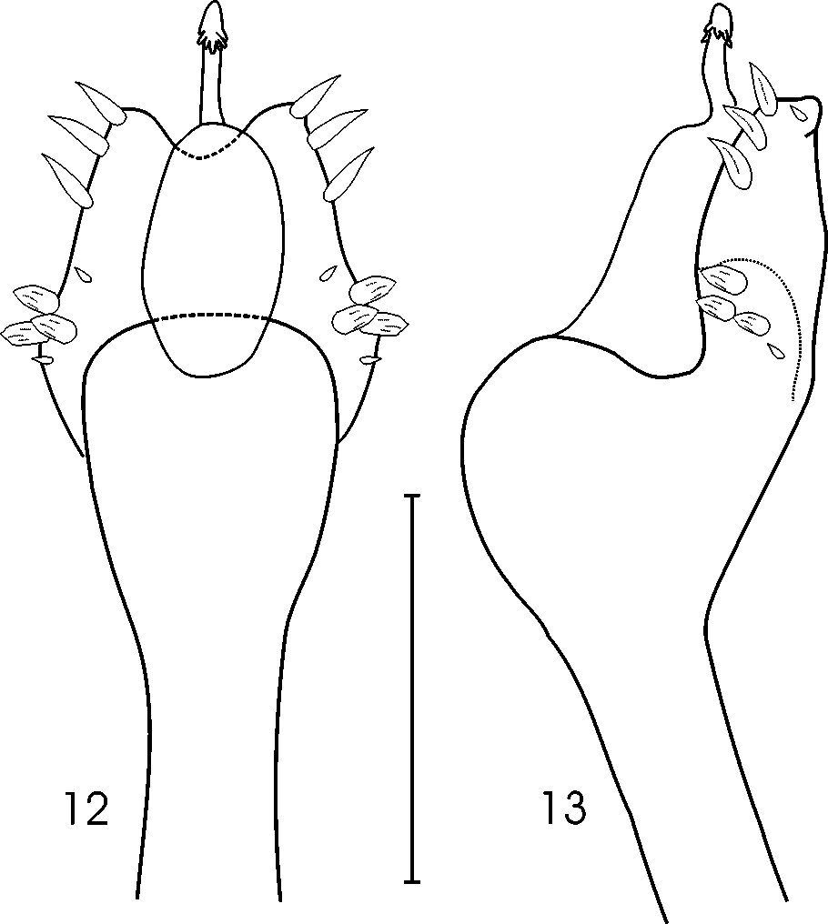
Genitalia. Ventral plate trapezoid narrowing slightly and gradually distally. Distal bor der of penis ventral plate with soft notch. Two groups of setae on ventral plate. Basal group of three short lanceolate setae + two accessory smaller ones. Distal group of three short straight setae + one accessory smaller. Glans without ventral or dorsal processes, stylus with subapical small rounded tubercles.

| MNRJ |
Museu Nacional/Universidade Federal de Rio de Janeiro |
No known copyright restrictions apply. See Agosti, D., Egloff, W., 2009. Taxonomic information exchange and copyright: the Plazi approach. BMC Research Notes 2009, 2:53 for further explanation.