
Phyllium (Phyllium) mabantai Bresseel, Hennemann, Conle & Gottardo, 2009
|
publication ID |
https://doi.org/ 10.11646/zootaxa.2322.1.1 |
|
persistent identifier |
https://treatment.plazi.org/id/4C724261-6C5C-3A63-FF39-FF73370EC168 |
|
treatment provided by |
Felipe |
|
scientific name |
Phyllium (Phyllium) mabantai Bresseel, Hennemann, Conle & Gottardo |
| status |
sp. nov. |
Phyllium (Phyllium) mabantai Bresseel, Hennemann, Conle & Gottardo View in CoL n. sp.
( Figs. 39–41 View FIGURES 39–41 , 58–59 View FIGURES 52–67 , 73, 75 View FIGURES 68–76 , 82 View FIGURES 77–84 , 91 View FIGURES 85–94 , 109–110, 122)
Phyllium siccifolium, Rehn & Rehn, 1933: 414 View in CoL , pl. 17: 1 (in part – only the illustrated ♀ from Butuan, Mindanao ).
[Misidentification] Phyllium woodi, Rehn & Rehn, 1933: 423 View in CoL , pl. 16: 3 (in part – only the ♂ AT from Dapitan, Mindanao).
[Misidentification] HT, ♀: Philippines, Mindanao , Mount Apo, Lake Agko, 16.III.2008, ex coll. JB ( ISNB) PT , ♀ nymph: Philippines, Mindanao , Mount Apo, Lake Agko, 16.III.2008 (coll. JB) PT , 4 ♂♂, 1 ♀, 10 eggs: Philippines, Mindanao , Mount Apo, Lake Agko, captive reared F1 (coll. JB). PT , ♂: Philippines, Mindanao , Mount Apo, Lake Agko, captive reared F1, ex coll. JB ( ISNB) PT , ♂, 2 eggs: Philippines, Mindanao , Mount Apo, Lake Agko, captive reared F1, ex coll. JB (coll. FH, No. 0676-1 &
E). PT, ♀: Butuan, Mindanao, Baker; Phyllium siccifolium L. Rehn & Rehn det. ( ANSP). PT, ♂: Dapitan , Mindanao , Baker ; Phyllium woodi Rehn & Rehn , PARATYPE, Hebard Cln., allotypic ♂ ( ANSP)
Comparison: This new species is closely related to the other Philippine species Ph. bilobatum Gray, 1843 , Ph. woodi Rehn & Rehn, 1933 , Ph. mindorense n. sp. and P. philippinicum n. sp.. From all these species Ph. mabantai n. sp. is well distinguished by the spination of the interior lobe of the profemora, which has a decidedly greater, semi circularly excavated space between the 3 rd and 4 th teeth (roughly equal spaces between all teeth in the other species, Figs. 58–59 View FIGURES 52–67 ).
From Ph. bilobatum ♀ at once differ by: the larger size; lack of lobes of abdominal segments VII and VIII; shorter tegmina which roughly reach the posterior margin of abdominal segment VII (VIII in bilobatum ) and much less prominent teeth of the interior lobe of the profemora ( Fig. 58 View FIGURES 52–67 ).
From Ph. woodi ♀ differ by: the longer tegmina which reach as far as to the posterior margin of abdominal segment VII (VI in woodi ); more elongate and more acutely triangular anal segment; very few and more minute teeth on the outer margin of the exterior lobe of the profemora ( Fig. 58 View FIGURES 52–67 ); more decidedly rounded interior lobe of the protibiae; relatively longer and more slender mesonotum and smaller lateral spines of the mesopleurae ( Fig. 73 View FIGURES 68–76 ).
From Ph. mindorense n. sp. it is distinguished by: the smooth vertex; less distinctly trapezoidal pronotum; more decidedly rounded interior lobe of the protibiae and shape of the profemora of both sexes ( Figs. 58–59 View FIGURES 52–67 ), as well as the rounded cheeks of ♀ (gradually widened towards the posterior in mindorense ) and less rounded or angulate abdominal segment IV of ♂♂. The eggs differ at once by being smaller with the capsule less laterally compressed and much wider than in Ph. mindorense and having the hairy or feather-like appendages very differently structured (Figs. 109–110).
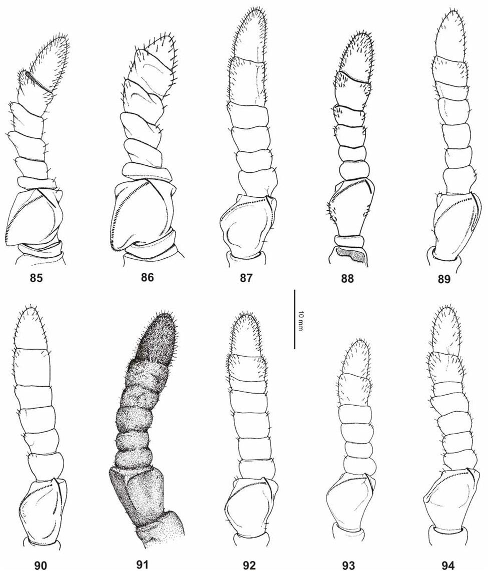
Finally, from Ph. philippinicum n. sp. it can be distinguished by: the slightly larger body dimensions; more decidedly rounded interior lobe of the protibiae; more numerous lateral spines of the mesopleurae ( Fig. 73 View FIGURES 68–76 ) and bright orange marking on the interior surface of the meso- and metacoxae ( Fig. 75 View FIGURES 68–76 , dull orange to brown in philippinicum ) of both sexes. ♀ furthermore differ by: the absence of rounded lobes on abdominal segments VII and VIII; relatively shorter protarsi and structures of the antennae which are relatively shorter, consist of only nine segments (ten in philippinicum ) and have 23–27 teeth on the pars stridens of segment III ( Fig. 91 View FIGURES 85–94 ). The eggs at once differ by having the micropylar plate slightly larger and positioned in the anterior 2/3 of the dorsal egg surface (posterior 2/ 3 in philippinicum ) and much less numerous hairy structures on the lateral surfaces of the capsule (Fig. 109).
Etymology: This new species is dedicated to Benjamin (Benjie) Mabanta (Pasig City, Manila, Luzon Id.) for supporting the fourth author in his study of Philippine Phasmatodea . Furthermore, he discovered the first adult ♀ of this new species during a field trip in Tampakan, South Cotabato in 2007, but unfortunately the specimen could not be preserved.
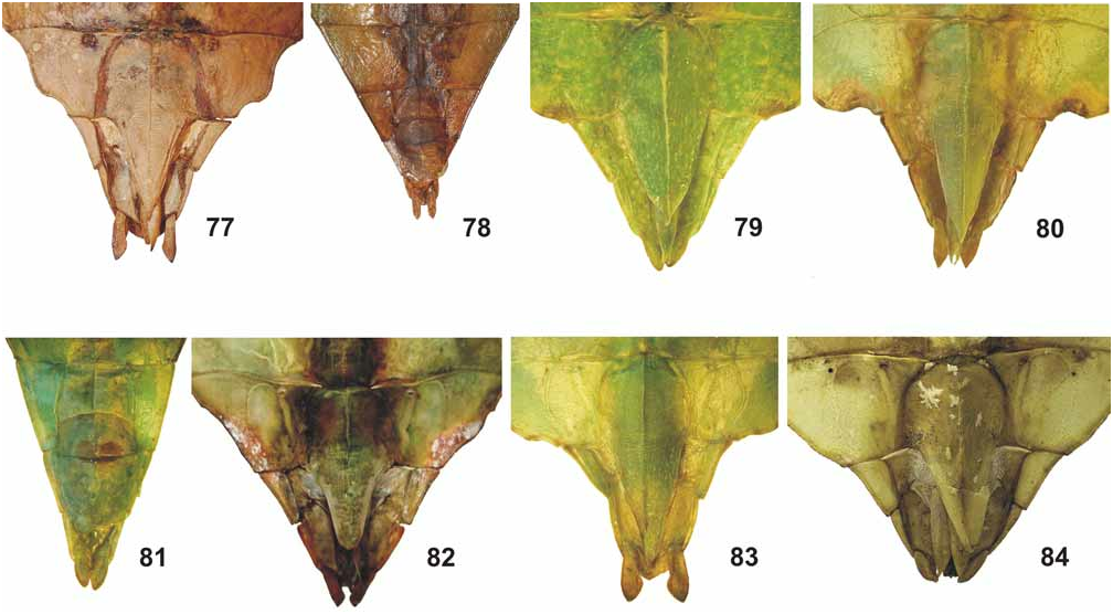
Diagnosis: ♀ ( Figs. 39–40 View FIGURES 39–41 ). Large for the subgenus (body length 91.2–99.4 mm) with a moderately broad abdomen (maximum width 33.4 mm), moderately broad and widely rounded exterior lobe and a roundly triangular interior lobe of the profemora; the interior lobe showing characteristic dentation mentioned above in the differentiation. Colouration quite variable, the HT ( Fig. 39 View FIGURES 39–41 ) being uniform green with a few yellowish speckles on the tegmina and the veins of these organs brown. The F1 captive reared PT ( Fig. 40 View FIGURES 39–41 ) has the legs with several brown speckles or patches and the tegmina plain green. Metasternum often with a conspicuous bold brown marking. Interior surface of meso- and metacoxae coloured bright orange ( Fig. 75 View FIGURES 68–76 ). Head almost oval in dorsal aspect with cheeks gently convex, posteromedially with a small tubercle; vertex either very sparsely and indistinctly granulose (HT) or smooth (PT). Frons with two small rounded impressions just behind the bases of the antennae. Antennae rather slender and consisting of nine segments, apical antennomere (IX) strongly setose, conical and about 1.7x longer than VIII. Pars stridens on antennomere III with 23–27 teeth ( Fig. 91 View FIGURES 85–94 ). Pronotum angulate and distinctly trapezoidal, gradually narrowed towards posterior with anterior margin about 1.8x broader than posterior margin. Meso-praescutum slightly narrowed towards the middle and becoming wider towards the posterior, almost 1.2x longer than wide. Lateral margins with a variable number of distinct but blunt spiniform tubercles and some smaller intercalated nodes; disc with 3–5 blunt spiniform tubercles medially. Anterior margin with a curved and raised transverse ridge which bears a moderately distinct spiniform tubercle medially. Mesopleurae distinctly and gradually diverging; their lateral margins armed with 5–6 prominent spiniform teeth and a few small intercalated nodes ( Fig. 73 View FIGURES 68–76 ). Mesosternum strongly longitudinally convex in median portion and sparsely granulose anteriorly. Tegmina ovate and reaching just over abdominal segment VII. Alae rudimentary. Abdominal segments II–III gradually widening, IV roundly angulate and V–VI sub-parallel. Outline of abdominal segments VII and VIII variable and discussed below. Anal segment (X) slightly wider than long and triangular, apex narrow. Subgenital plate elongate triangular and with a narrow, acutely pointed apex which almost reaches the tip of the anal segment ( Fig. 82 View FIGURES 77–84 ). Profemora with a moderately large, widely rounded exterior lobe; outer margin minutely denticulate. Interior lobe roughly equal in width to exterior lobe, the outer margin in anterior portion armed with five broad teeth and a conspicuous ± semicircularly excavated gap between the 3 rd and 4 th tooth ( Fig. 58 View FIGURES 52–67 ). Interior lobe of protibiae variable, either narrow andgently rounded (HT) or roundly triangular (PT). Dorsal lobe of mesofemora roundly angulate medially. Protarsi long and slender, about 2/3 the length of corresponding tibia. Probasitarsus longer than following three tarsomeres combined.
♁♁ ( Fig. 41 View FIGURES 39–41 ). Moderately sized for the subgenus (body length 55.5–62.5 mm) with a slender and ovate abdomen (maximum width 14.2–16.0 mm). Colour green with a variable degree of brown markings on the front and mid legs. Vertex smooth except for a very small blunt tubercle posteromedially. Antennae consisting of 25 segments, strongly setose and reaching half way segment V. Eyes dark brown. Pronotum and mesothorax structured like in ♀. Meso-praescutum almost 1.4x longer than wide and very slightly narrowing towards the posterior; near anterior margin with a distinct, curved transverse ridge which is protruded into a blunt tubercle medially. Mesopleurae narrow in anterior portion but moderately diverging in posterior, lateral margins armed with rather distinct spiniform tubercles. Between these tubercles always a much smaller intercalated tubercle. Mesosternum roundly tectiform and set with minute granules in anterior portion. Tegmina ovate and slightly constricted apically, ± reaching posterior margin of abdominal segment III. Alae ± reaching posterior margin of abdominal segment IX. Abdominal segment II roughly parallel-sided, III very slightly diverging in the posterior portion. IV distinctly diverging and rounded to ± angular. V–IX oblong-ovate, V slightly diverging with margins gently rounded, VI widest with lateral margins gently rounded. VIII–X faintly more attenuate than previous. Anal segment (X) about 1.5x longer than wide, roundly triangular with the apex rather blunt and rounded. Vomer broadly triangular with a single, straight terminal hook. Poculum moderately convex with a fine longitudinal median carina and slightly projecting over posterior margin of tergite IX. Exterior lobe of profemora low and narrow with the broadest point hardly wider than the shaft of the femur itself; outer margin smooth or with a few very small, hardly visible teeth and very gently rounded. Interior lobe as in ♀ with five rather small but acute teeth and a greater space or gap between the 3 rd and 4 th tooth ( Fig. 59 View FIGURES 52–67 ). Dorsal lobe of mesofemora very narrow in basal 1/3 then gradually expanding and angular 1/3 of the apex. Protibiae with a narrow and gently rounded interior lobe. Protarsus very elongate and roughly equal in length to corresponding tibia.
50: ♀ HT: “Indiis” [ UUZM] © UUZM
51: ♂: Amboina [ BMNH] © Edward Baker, BMNH
Nymphs: Newly hatched nymphs resemble nymphs of Phyllium philippinicum n. sp. and are of moderate size for the subgenus (body length 13.0– 13.5 mm) with a moderately broad abdomen (width 2.9–3.0 mm) and strongly broadened meso- and metafemora. General colour dark brown or almost black with the outer margins of thorax and abdomen, as well as the exterior outer margin of the meso- and metafemora and basitarsi white. The white outer margin of the meso- and metafemora is continued as a transverse band sub-basally. Profemora with two distinct white spots basally, the tibiae all white basally and with two small white spots interiorly. Abdominal segments IV–VII each with a transverse white marking.
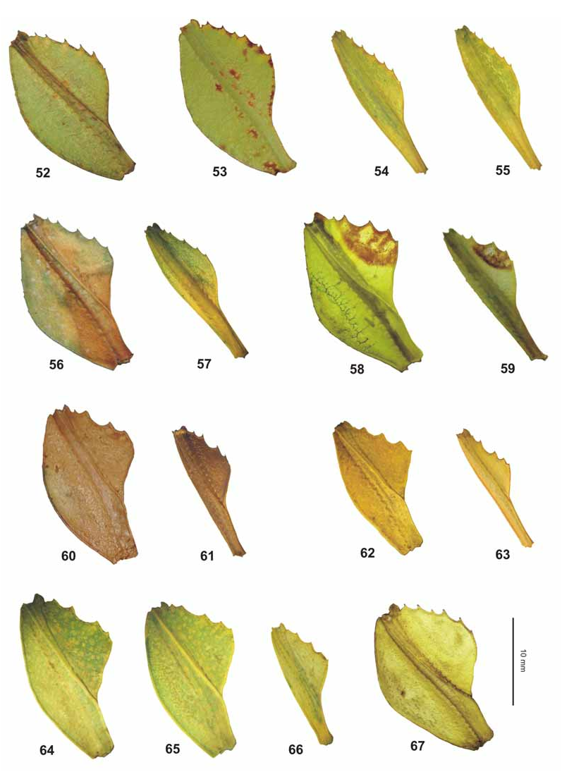
52–53. Ph. (Ph.) philippinicum n. sp., ♀ PT [coll. FH]
54–55. Ph. (Ph.) philippinicum n. sp., ♂♂ PT [coll. FH]
56. Ph. (Ph.) mindorense n. sp., ♀ PT [coll. FH]
57. Ph. (Ph.) mindorense n. sp., ♂ HT [ ZSMC]
58. Ph. (Ph.) mabantai n. sp., ♀ PT [coll. JB]
59. Ph. (Ph.) mabantai n. sp., ♂ PT [coll. JB]
60. Ph. (Ph.) gantungense n. sp., ♀ HT
61. Ph. (Ph.) gantungense n. sp., ♂ PT
62. Ph. (Ph.) jacobsoni Rehn & Rehn, 1933 , ♀ (Java) [coll. FH]
63. Ph. (Ph.) jacobsoni Rehn & Rehn, 1933 , ♂ (Java) [coll. FH]
64–65. Ph. (Ph.) hausleithneri Brock, 1999 , ♀ ( Peninsular Malaysia) [coll. FH]
66. Ph. (Ph.) hausleithneri Brock, 1999 , ♂ ( Peninsular Malaysia) [coll. FH]
67. Ph. (Ph.) siccifolium (Linné, 1758) , ♀ HT (“Indiis”) [ UUZM] UUZM
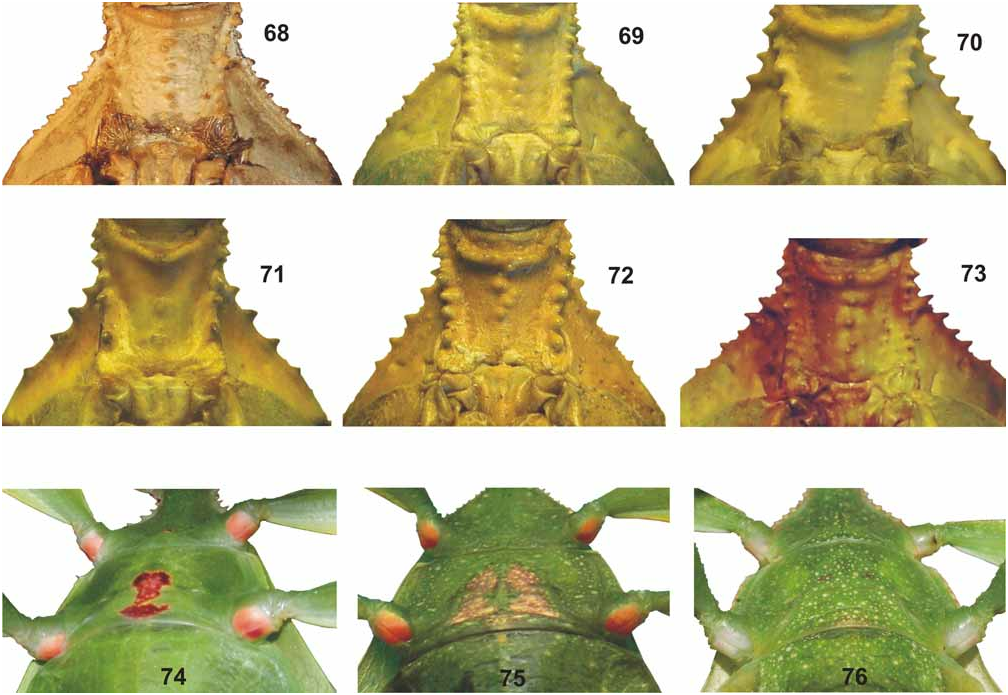
68. Ph. (Ph-.) gantungense n. sp., mesothorax of ♀ HT [ MSNG]
69. Ph. (Ph.) hausleithneri Brock, 1999 , mesothorax of ♀ [coll. FH]
70. Ph. (Ph.) siccifolium (Linné, 1758) , mesothorax of ♀ [coll. FH, No. 0657-2]
71. Ph. (Ph.) philippinicum n. sp., mesothorax of ♀ PT [coll. FH]
72. Ph. (Ph.) mindorense n. sp., mesothorax of ♀ PT [coll. FH]
73. Ph. (Ph.) mabantai n. sp., mesothorax of ♀ PT [coll. JB]
74. Ph. (Ph.) philippinicum n. sp.
75. Ph. (Ph.) mabantai n. sp.
76. Ph. (Ph.) jacobsoni Rehn & Rehn, 1933
Eggs (Figs. 109–110). Of moderate size for the genus with prominent hair or feather-like appendages which do not develop before the egg gets in contact with water. Freshly laid eggs are dark fuchsia and become pale to mid brown after contact with water. General shape of capsule trapezoid with polar-area strongly rounded and laterally surfaces flattened and almost parallel. Lateral longitudinal carinae of capsule each set with a row of moderately long feather-like appendages which reach up to the operculum on the dorsal side and just not reach half way along the capsule on ventral side. In lateral aspect the anterior portion of capsule is narrower than the posterior portion, being only about ¾ the width. Lateral surfaces with two rows of large rounded impressions which roughly form an ellipse, a further short row of rounded impressions between these. Spaces in between appearing like a network of narrow hairy ridges. The outside of the elliptic structure strongly marked with hairy appendages. Micropylar plate positioned in anterior 2/3 of the dorsal egg surface, shape elongate-oval with anterior end gradually narrowed and rounded, and the posterior end pointed. Surface unarmed and outer margin set with moderately long hairy structures. On each side of the micropylar plate a row of rounded impressions, the spaces in between covered with hairy structures. Micropylar cup small and slightly displaced towards the posterior. Operculum almost circular, flat and with the outer margin set with a row of the same long feather-like appendages seen along the longitudinal outer carinae of the egg-capsule.
Measurements including the feather-like structures [mm]: length (including operculum) 5.0– 5.2 mm, length 4.2–4.4, width 2.0– 2.1 mm, height 3.1–3.4 mm, length of micropylar plate 2.4–2.5 mm.
Variation: ♂♂ are quite constant showing some variation only concerning to the size, degree of brown mottling on the legs and absence or presence of transparent eye-like spots on abdominal segment V. These may sometimes only be present on one side of the abdomen and lacking on the other. ♀ however show considerable variation in the colouration, size of the teeth of the pro- and mesofemora, size and number of spines on the mesonotum, shape of the interior lobe on the protibia and shape of abdominal segments VII and VIII. Segments V and VI range from slightly narrowing to almost parallel-sided. VII is either gradually tapered, may have a concave medial excavation (HT, Fig. 39 View FIGURES 39–41 ) or forms a 90° angle (e.g. PT in Fig. 40 View FIGURES 39–41 ). The outer margins of VIII range from very slightly concave to gently convex.
Comments: Both sexes of this new species were misinterpreted by Rehn & Rehn, 1933 as two different species. The ♀ was erroneously referred to as “ Ph. siccifolium ” and the corresponding ♂ was described as Phyllium woodi Rehn & Rehn, 1933 . However, Ph. siccifolium Linné, 1758 does not occur in the Philippines and is distinguished from this species by a number of reliable features (see below). Ph. woodi Rehn & Rehn, 1933 is a rather distinctive species presumed endemic on Sibuyan Island with the ♂ from Mindanao wrongly assigned and here shown to be Ph. mabantai n. sp..
Material of this new species was collected in Tampakan (Mindanao) in 2007 and on Mount Apo in March 2008. Live eggs laid by the HT were imported to Europe from the latter locality and the F2-generation is already being successfully reared by the fourth author. It has proven quite easy to rear in captivity using bramble ( Rubus spp. , Rosaceae ), raspberry ( Rubus idaeus , Rosaceae ), rose ( Rosa spp. , Rosaceae ) and oak ( Quercus robur & Q. ilex , Fagaceae ) as alternative food-plants.
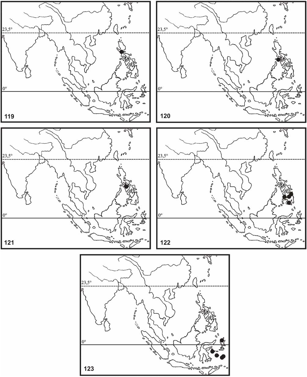
Distribution ( Fig. 122 View FIGURES 119–123 ): Philippines, Mindanao Island (Mount Apo, Butuan, Tampakan & Dapitan).

No known copyright restrictions apply. See Agosti, D., Egloff, W., 2009. Taxonomic information exchange and copyright: the Plazi approach. BMC Research Notes 2009, 2:53 for further explanation.
|
Kingdom |
|
|
Phylum |
|
|
Class |
|
|
Order |
|
|
Family |
|
|
Genus |
Phyllium (Phyllium) mabantai Bresseel, Hennemann, Conle & Gottardo
| Hennemann, Frank H., Conle, Oskar V., Gottardo, Marco & Bresseel, Joachim 2009 |
Phyllium siccifolium
| Rehn, J. A. G. & Rehn, J. W. H. 1933: 414 |