
Pilisaprinus verschureni ( Thérond, 1959 ) Lackner, 2013
|
publication ID |
https://doi.org/ 10.5281/zenodo.5740224 |
|
publication LSID |
lsid:zoobank.org:pub:84292869-A733-4F7F-B2B8-B65A0356127B |
|
persistent identifier |
https://treatment.plazi.org/id/03C4C81D-FFBA-FFE3-244C-E331A0B6FE66 |
|
treatment provided by |
Marcus |
|
scientific name |
Pilisaprinus verschureni ( Thérond, 1959 ) |
| status |
comb. nov. |
Pilisaprinus verschureni ( Thérond, 1959) View in CoL comb. nov.
( Figs 1–19 View Figs 1–9 View Figs 10–12 View Figs 13–19 )
Saprinus verschureni Thérond, 1959: 17 View in CoL ; MAZUR (1984: 62); MAZUR (1997: 231); KANAAR (1996: 130). Styphrus termitophilus Kanaar, 1990: 145 View in CoL . Synonymized by KANAAR (1996: 130). Saprinus (Pilisaprinus) verschureni: MAZUR (2011: 179) View in CoL .
Type locality. Democratic Republic of Congo (former Zairë).
Type specimen examined. Saprinus verschureni . HOLOTYPE: “ Congo Belge, P.N.G. / Miss. H. De Saeger / II/gd/4, 29-v-1952 / J. Verschuren. 3545” (printed); “ HOLOTYPUS ” (orange, red-margined label, printed); “ Saprinus / verschureni nov. sp. / J. Thérond det., 1958” (printed-written); “TYPE” (purple label, printed) ( RMCA). The specimen is of unidentified sex: genitalia lost, sixth abdominal tergite broken in two, left antenna missing, both protibiae missing, right mesotibia broken off glued to the same triangular point with specimen, both mesotarsi missing sidemounted on a triangular point.
Additional material examined. BENIN: 1 J, “BENIN – Niaouli II / February 2002 / in Macrotermes hill / leg. G. Goergen ” ( TLAN) ; 1 J 1 ♀, same label data ( RMNH) ; 1 ♀, “ Benin / Toffo / January 2002 / in Macrotermes hill / leg. G. Goergen ” ( RMNH) . IVORY COAST: 1 J, “ Lamto / 15.iv.1994 / Termitière morte en [= dead termitarium in] / forêt dégradée [= degraded forest] 2 km de / Taabo leg. Cl. Girard” ( RMNH) .
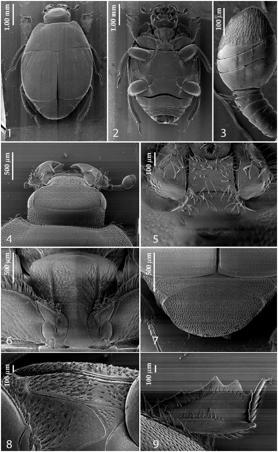
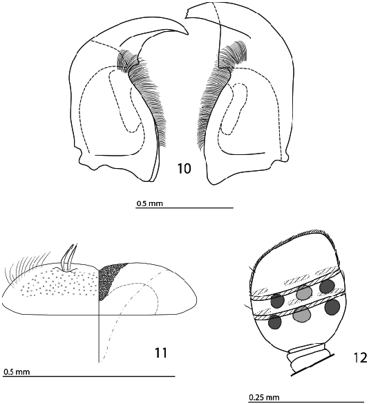
Redescription. Body moderately convex, PEL: 4.50–4.75 mm; APW: 1.85–2.00 mm; PPW: 3.75–4.00 mm; EL: 2.75–3.00 mm; EW: 4.00– 4.25 mm, dorsally with various kinds of punctuation, cuticle black, not metallic; legs, mouthparts and antennae rufopiceous. Antennal scape ( Fig. 4 View Figs 1–9 ) slightly dilated and thickened, densely punctate, with numerous long amber setae; antennal club ( Figs 3 View Figs 1–9 , 12 View Figs 10–12 ) ventrally with two slit-like pits almost encircling it entirely, dorsally with two complementary slit-like pits; lower half (approximately) glabrous or fringed only with few microscopic setae, upper half (approximately) with dense short setae; sensory structures of antennal club ( Fig. 12 View Figs 10–12 ) in form of six vesicles, four of them situated in pairs under ventral slit-like pits and another two of them situated under each-other under dorsal surface medially. Mandibles ( Figs 4 View Figs 1–9 , 10 View Figs 10–12 ) stout, moderately punctate, with evenly rounded outer margin strongly curved inwardly; acutely pointed; sub-apical tooth on inner margin of left mandible moderately large, almost perpendicular; labrum ( Fig. 11 View Figs 10–12 ) slightly convex, slightly depressed medially, finely punctate; labral pits rather deep and large with two short amber setae arising from each; terminal labial palpomere elongate, its width about one-fourth its length; mentum ( Fig. 5 View Figs 1–9 ) square-shaped, anterior angles slightly produced, anterior margin with a shallow notch; disc of mentum covered with numerous short setae that are almost absent along posterior margin; cardo of maxilla laterally with numerous short setae; stipes triangular, with four short setae; terminal maxillary palpomere elongate, its width about one-fifth its length, about three times as long as penultimate; labial palpomeres with dense brush of long setae. Clypeus and frons ( Fig. 4 View Figs 1–9 ) rectangular, broad, with moderate punctures and fine transverse wrinkles; frontal and supraorbital striae absent; eyes slightly flattened, well visible from above. Pronotal sides ( Fig. 1 View Figs 1–9 ) strongly narrowing anteriorly; anterior emargination for head deep; apical angles acute and conspicuous, marginal pronotal stria slightly carinate, complete, slightly weakened behind head; disc entirely covered with coarse round punctuation, punctures separated by about their diameter, laterally coarser than medially; pronotal depressions rather deep and large; scutellum very small; pronotal hypomeron with dense long amber setae ( Fig. 2 View Figs 1–9 ). Elytral epipleuron ( Fig. 8 View Figs 1–9 ) with long amber setae; marginal epipleural stria thin, complete, continued as complete apical stria; marginal elytral stria well impressed, complete, slightly carinate, apically attaining marginal epipleural stria. Humeral elytral stria deeply impressed on basal third, joined with inner subhumeral stria forming a supplementary dorsal elytral stria parallel to first dorsal elytral, reaching about half of elytral length apically; elytral disc with four deeply impressed dorsal elytral striae 1–4, in shallow punctures, all about the same length, reaching about half of elytral length apically, fourth dorsal elytral stria basally connected with sutural elytral stria; sutural elytral stria well impressed, reaching about two-thirds of elytral length apically, otherwise obliterated. Elytral disc entirely with fine small dense punctuation (with the exception of fourth elytral interval and elytral flanks) intermingled with dense and coarse punctures separated by about half their diameters.
Propygidium ( Fig. 7 View Figs 1–9 ) partly covered by elytra; with dense and coarse punctures becoming sparser and finer medially; pygidium ( Fig. 7 View Figs 1–9 ) long, convex, with very coarse and dense punctuation, punctures separated by less than half of their diameter. Anterior margin of median portion of prosternum ( Fig. 6 View Figs 1–9 ) rounded; marginal prosternal stria very thin; prosternal foveae absent; prosternal process compressed, dorso-laterally with irregular coarse to fine punctures with long amber setae; carinal prosternal striae present as tiny fragments on prosternal apophysis, otherwise absent (in some specimens reaching approximately half the length of the prosternum apically, weakened and very approximate); lateral prosternal striae ( Fig. 6 View Figs 1–9 ) well impressed, carinate, somewhat divergent anteriorly, united in front by anterior stria.
Anterior margin of mesoventrite medially emarginate; discal marginal mesoventral stria well impressed, carinate; disc of mesoventrite with dense shallow punctures separated by about their own to twice their diameters; meso-metaventral suture weakly impressed, curved outwardly.
Intercoxal disc of metaventrite with long median excavation (male), with punctuation similar to that of mesoventrite, punctures becoming sparser and finer medially; lateral metaventral stria well impressed, carinate, straight, curved outwardly, connected to metaventral-metepisternal suture; lateral disc of metaventrite ( Fig. 8 View Figs 1–9 ) excavated, with large deep setiferous punctures; metepisternum on basal two-thirds with even coarser and denser punctuation with long amber setae, punctuation on apical third becoming much sparser and finer, metepisternum ( Fig. 8 View Figs 1–9 ) laterally with an additional straight, well-impressed metepisternal stria.
Intercoxal disc of first abdominal sternite almost completely striate laterally, with punctuation similar to that of metaventrite; lateral disc of all visible abdominal sternites setose laterally.
Protibia ( Fig. 9 View Figs 1–9 ) dilated, outer margin with two low triangular distal teeth topped by tiny denticle, followed by two tiny denticles rather distant from the two distal teeth and another two microscopic denticles visible only from ventral view; setae of outer row regular, diminishing in size apically; protarsal groove deep; anterior protibial stria present on basal two-thirds, otherwise obliterated; setae of median row similar in length to those of outer row, present only along the shortened anterior protibial stria; tarsal denticles absent; protibial spur well developed, bent, inserted near protarsal insertion; apical margin of protibia posteriorly with about nine minuscule denticles abutting each other; outer part of posterior surface obscurely variolate-punctate, delimitation of outer and median parts of posterior surface non-existent; posterior protibial stria absent; inner row of setae double, setae dense, increasing in length apically.
Mesotibia slender, outer margin with several closely-set thin denticles near mesotarsal insertion; setae of outer row shifted to approximately middle of posterior mesotibial surface, very regular and dense; setae of median row situated right below the setae of outer row, much shorter; posterior mesotibial stria present only on basal third, otherwise (or apically) obliterated; anterior surface of mesotibia with dense row of thin closely-set denticles near outer margin, rest of surface of anterior mesotibial stria imbricate-punctate; anterior mesotibial stria almost complete; inner anterior denticles absent; mesotibial spur short, thick; apical margin of mesotibia anteriorly with two widely-spaced short denticles; inner margin of mesotibia with sparse row of long thin setae; claws of apical tarsomere bent, shorter than half its length; each mesotarsomere with a single long seta ventrally; dorsally without setae; metatibia more slender and longer than mesotibia, in all aspects similar to it.
Male genitalia. Eighth sternite ( Figs 13–14 View Figs 13–19 ) separated longitudinally; vela absent; apex of eighth sternite laterally with dense brush of short setae ( Figs 13–15 View Figs 13–19 ); eighth tergite and eighth sternite fused laterally ( Fig. 15 View Figs 13–19 ). Ninth tergite ( Figs 16–17 View Figs 13–19 ) longitudinally fused medially; spiculum gastrale ( Fig. 16 View Figs 13–19 ) gradually dilated from middle in most of apical half, basal end only slightly triangularly dilated. Aedeagus ( Figs 18–19 View Figs 13–19 ) slender, almost parallel-sided; basal piece of aedeagus short, ratio of its length: length of parameres 1: 5; parameres of aedeagus fused almost along their basal three-fourths; aedeagus only slightly curved ventrad, apex of aedeagus flattened dorso-ventrally ( Fig. 19 View Figs 13–19 ).
Sexual dimorphism. Females are devoid of longitudinal metaventral depression.

No known copyright restrictions apply. See Agosti, D., Egloff, W., 2009. Taxonomic information exchange and copyright: the Plazi approach. BMC Research Notes 2009, 2:53 for further explanation.
|
Kingdom |
|
|
Phylum |
|
|
Class |
|
|
Order |
|
|
Family |
|
|
Genus |
Pilisaprinus verschureni ( Thérond, 1959 )
| Lackner, Tomáš 2013 |
Saprinus verschureni Thérond, 1959: 17
| MAZUR S. 2011: ) |
| MAZUR S. 1997: 231 |
| KANAAR P. 1996: 130 |
| KANAAR P. 1996: 130 |
| KANAAR P. 1990: 145 |
| THEROND J. 1959: 17 |