
Pinodytes tibialis ( Hatch, 1957 ) Peck & Cook, 2011
|
publication ID |
https://doi.org/ 10.11646/zootaxa.3077.1.1 |
|
DOI |
https://doi.org/10.5281/zenodo.5243853 |
|
persistent identifier |
https://treatment.plazi.org/id/03B37553-3D5E-A158-9DC1-FF10FC760B92 |
|
treatment provided by |
Felipe |
|
scientific name |
Pinodytes tibialis ( Hatch, 1957 ) |
| status |
comb. nov. |
Pinodytes tibialis ( Hatch, 1957) View in CoL , new combination
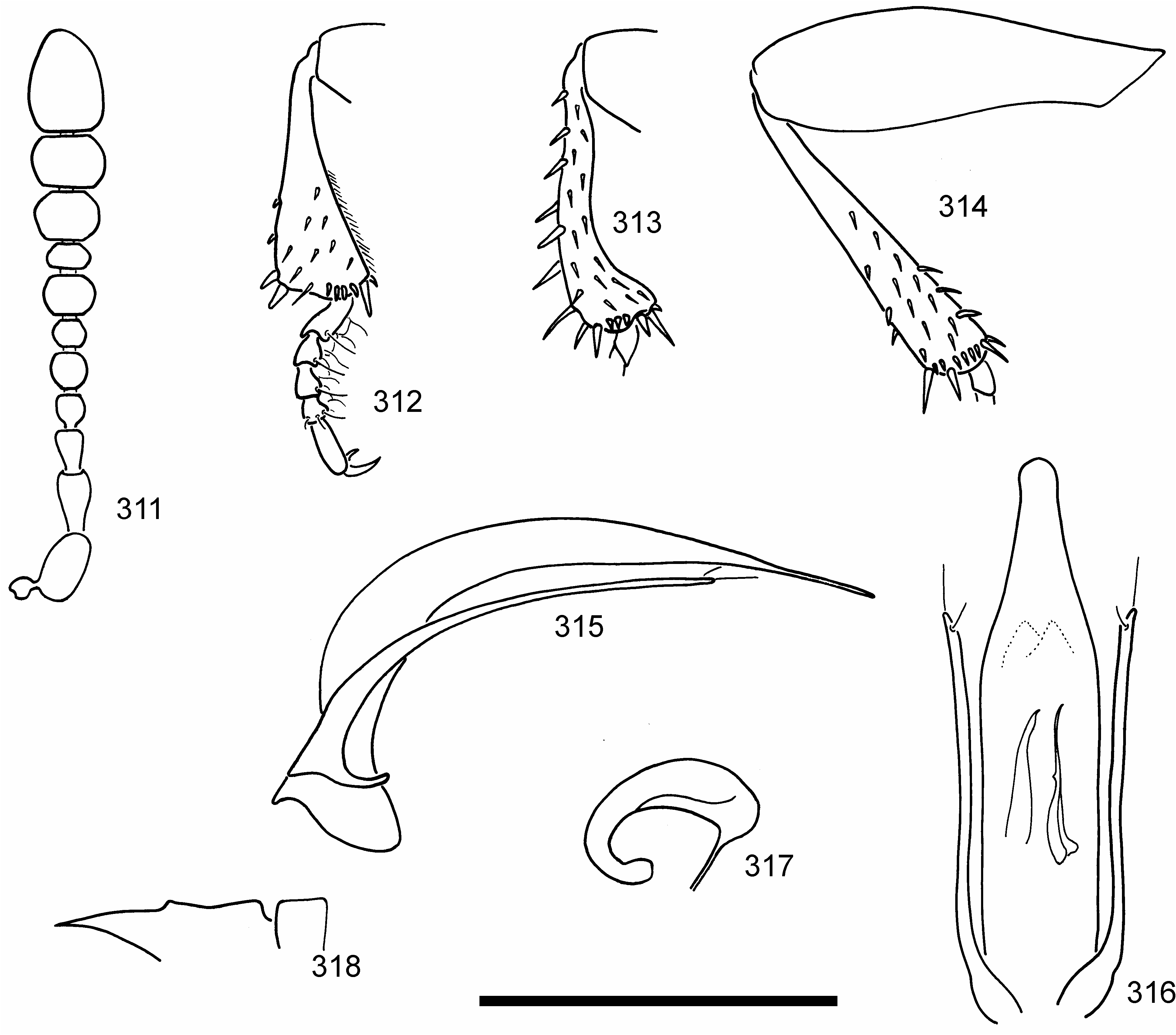
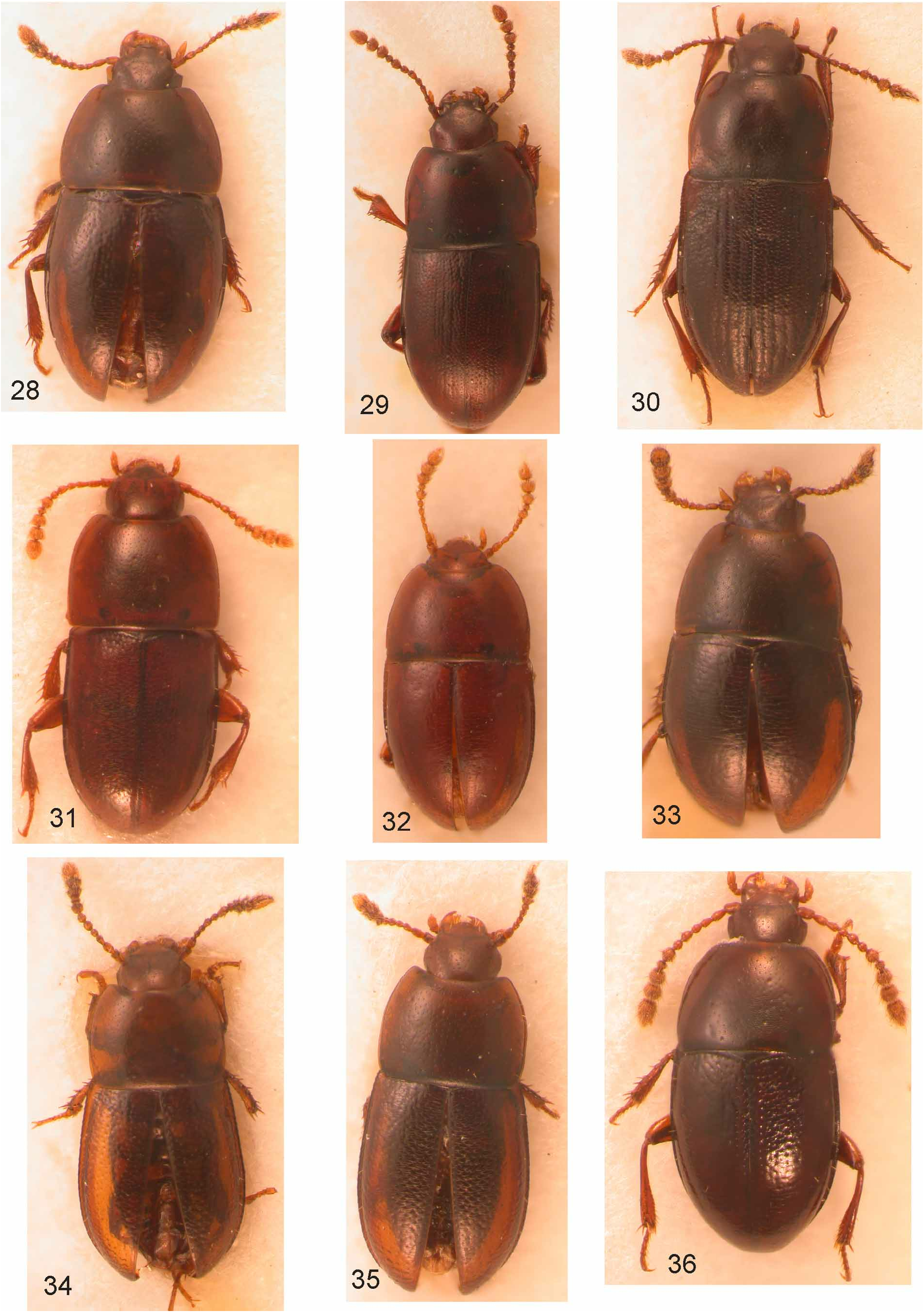
( Figs. 34 View FIGURES 28–36 , 310–318 View FIGURE 310 View FIGURES 311–318 )
Catopocerus tibialis Hatch 1957: 20 View in CoL .
Type material. Type male in USNM, seen. Type label data: Gold Beach , Curry County, Oregon ; 21.IV.1955, J. Capizzi.
Additional material examined. We examined 90 additional specimens (see Appendix) for a total of 91 specimens.
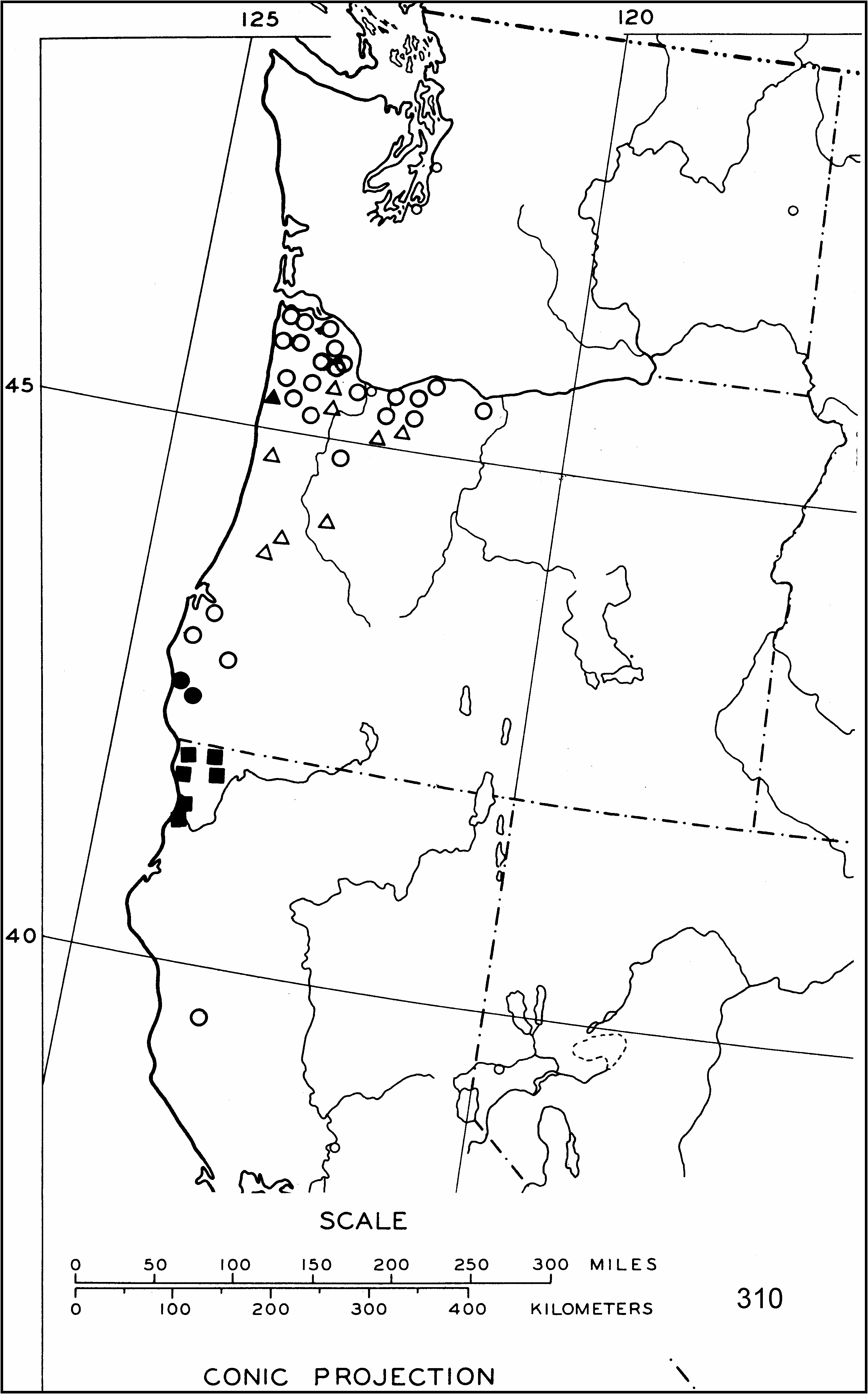
Distribution. Specimens ( Fig. 310 View FIGURE 310 ) are known only from Curry County in southwestern Oregon.
Diagnosis. Total length 1.40–1.60 mm; greatest width 0.65–0.80 mm. Reddish brown; elongate-oval in shape ( Fig. 34 View FIGURES 28–36 ). Head. Moderately finely, irregularly punctate; with reticulate microsculpture. Eyes absent. Antenna ( Fig. 311 View FIGURES 311–318 ) with antennomere 2 longer than 3; antennomere 5 slightly larger than 4 and 6; antennomere 7 clearly larger than 8; antennomeres 9 and 10 lack visible sensory vesicles. Pronotum. Finely punctate, punctures separated by 3– 4 diameters; with reticulate microsculpture. Disc with three pairs of larger punctures, positioned subapically, posteromedially and subbasally. Slightly wider at middle than at base, narrowing to apex; apical margin weakly emarginate, basal margin straight; apical angles rounded, basal angles weakly obtuse. Elytra. Moderately coarsely punctate; a few striae weakly indicated adjacent to suture; punctures joined by fine strioles that create a weakly imbricate pattern basally. Joined elytra slightly wider than pronotum; sides subparallel in basal one-half, narrowing to apex. Legs. Protibia ( Fig. 312 View FIGURES 311–318 ) narrow at base, apical one-half strongly widened in male, less so in female; two curved spines at apex of outer margin; apical one-half of inner margin with fine, dense spines. Male mesotibia ( Fig. 313 View FIGURES 311–318 ) with basal two-thirds sinuate, angulate at apical one-third; unmodified in female; strong spines on outer margin in both sexes. Metatibia ( Fig. 314 View FIGURES 311–318 ) elongate, narrow, straight in both sexes. Metafemur ( Fig. 314 View FIGURES 311–318 ) slender in both sexes. Male protarsomeres ( Fig. 312 View FIGURES 311–318 ) bearing elongate setae laterally and thin, colorless, broad phanerae ventrally. Mesotarsomeres without phanerae. Venter. Mesoventrite ( Fig. 318 View FIGURES 311–318 ) carinate, with a small median tooth; excavation behind transverse carina. Male genitalia. Median lobe of aedeagus ( Figs. 315, 316 View FIGURES 311–318 ) cylindrical, curved, with flattened apex narrowing to weak rounded lobe. Inverted internal sac ( Fig. 316 View FIGURES 311–318 ) with elongate sclerotized structures. Parameres ( Figs. 315, 316 View FIGURES 311–318 ) narrow, about three-fourths length of median lobe; each paramere with one apical and one subapical seta. Spermatheca. Tubular ( Fig. 317 View FIGURES 311–318 ), curved.
No known copyright restrictions apply. See Agosti, D., Egloff, W., 2009. Taxonomic information exchange and copyright: the Plazi approach. BMC Research Notes 2009, 2:53 for further explanation.
|
Kingdom |
|
|
Phylum |
|
|
Class |
|
|
Order |
|
|
Family |
|
|
Genus |
Pinodytes tibialis ( Hatch, 1957 )
| Peck, Stewart B. & Cook, Joyce 2011 |
Catopocerus tibialis
| Hatch, M. H. 1957: 20 |