
Polycentropus vernus Hamilton, Harris, and Lago, 1990
|
publication ID |
https://doi.org/ 10.1080/00222933.2023.2271609 |
|
DOI |
https://doi.org/10.5281/zenodo.10498698 |
|
persistent identifier |
https://treatment.plazi.org/id/03BF3616-8B1C-2654-FE89-2721FB27FB3A |
|
treatment provided by |
Plazi |
|
scientific name |
Polycentropus vernus Hamilton, Harris, and Lago |
| status |
|
Polycentropus vernus Hamilton, Harris, and Lago View in CoL
( Figures 35 View Figure 35 , 54 View Figure 54 )
Polycentropus vernus Hamilton, Harris, and Lago, 1990: 365–367 View in CoL , fig. 2, J; type locality ′ Alabama: Fayette County: small intermittent stream entering Wallace Branch at headwaters, 5.5 mi SE Berry ̍ ( USNM, lost) .
Polycentropus View in CoL n. sp. (nr. chelatus View in CoL ) Lago and Harris, 1987: 258.
Diagnosis. The diminutive size of the male of Polycentropus vernus (2.8–3.6 mm forewing length) distinguishes this species from other members of the P. confusus species group. The next smallest measured male of the group was a specimen of P. chelatus , possessing a forewing length of 4.0 mm. This species is most similar to the P. confusus species group members with the more elongate head or enlargement on the basodorsal arm of the inferior appendage. These species are P. chelatus , P. confusus , P. floridensis , P. neiswanderi , P. pentus , P. stephani and P. thaxtoni . Polycentropus vernus can be distinguished from the above-mentioned species through a combination of several characters including the shape of the body of the pre-anal appendage, the size and shape of the dorsal arm of the inferior appendage, the length and shape of the ventral arm of the inferior appendage, and the shape of the phallus. On P. vernus the emargination of the body of the pre-anal appendage is very shallow compared to P. floridensis , P. stephani and P. thaxtoni , and it is also different from that of other members of the confusus group. The phallus of P. vernus with its basoventral swelling is similar only to that of P. dinkinsorum , P. floridensis and P. pentus . The shape of the inferior appendage can be used to distinguish these three species from P. vernus . In P. pentus the basodorsal arm of the inferior appendage is strongly curved, almost sickle-shaped, while in P. vernus it is triangular. Also, in P. pentus the mesobasal tooth on the basal arm of the inferior appendage is little developed, vs well developed in both P. vernus and P. floridensis . While the basodorsal arm of P. floridensis is similar to that of P. vernus , the lateral margin of the basal arm is more excavated, exposing the relatively prominent mesobasal tooth. In P. floridensis and P. dinkinsorum , which have a ventral swelling of the phallus, the basodorsal process of the inferior appendage of these two species form a round head vs the elongate head of the basodorsal process of the inferior appendage of P. vernus .
Adult description
General. Forewing length of male 2.8–3.6 mm.
Male genitalia ( Figure 35A–E View Figure 35 ). Abdominal segment VIII annular. Terga IX and X fused, membranous, extended caudad over bases of intermediate appendages. Sternum IX semicircular in lateral view, posterior margin slightly sinuous. Intermediate appendages originating beneath terga IX+X and extending beyond them, curved ventrad apically, their apices each bearing 3 small setae; in dorsal view apices proximate, subtly divergent. Bodies of pre-anal appendages in lateral view each with round caudal margin, dorsomesal emargination at about mid-height, dorsal process long, curved ventrad, extending below apex of head of basodorsal process of corresponding inferior appendage, acute apically; in dorsal view slender, elongate, subparallel, apices incurvate. Inferior appendages in lateral view each with prominent basodorsal process erect, neck short, head large, shaped like oblong in-turned blade, apex curved slightly posterad, mesoventral protuberance small, main body of appendage with surface dorsomesal excavation to about mid-length, dorsal margin curved ventrad from base to 1/4 length, subparallel with ventral margin beyond 1/4 length, ventral margin slightly convex along length, tapering evenly to round apex extended posterad, terminating slightly anterad to intermediate appendages; in ventral view subparallel along length, each wide basally, tapering evenly to round apex, basodorsal process mostly hidden by main body of inferior appendage, anterior apex projecting beyond medial margins, in cleared specimen broad, round, anterior apex subtriangular, in caudal view stout, subtriangular, with medial projection subtriangular. Phallus tubular, in lateral view moderately decurved, with basal ventral swelling, apex slightly pointed, internal spinules absent; internal phallic sclerite distal in lateral view, moderately elongate, oblong and uniform in depth along length.
Female genitalia. Unknown.
Larva. Unknown.
Pupa. Unknown.
Notes. The type series represented all known material of this species. Unfortunately, the type series is lost and numerous collecting efforts over multiple years and using several methods at the type locations and nearby streams by the author yielded no new material. During those collection efforts, abundant coal mining and clearcut logging activity was observed at and around the stream sites. Due to the lack of available material, the description and illustrations presented here are based on the original description by Hamilton et al. (1990).
Biology. Polycentropus vernus has been collected most often in small intermittent and headwater streams of the Cumberland Plateau physiographic region from March to May.
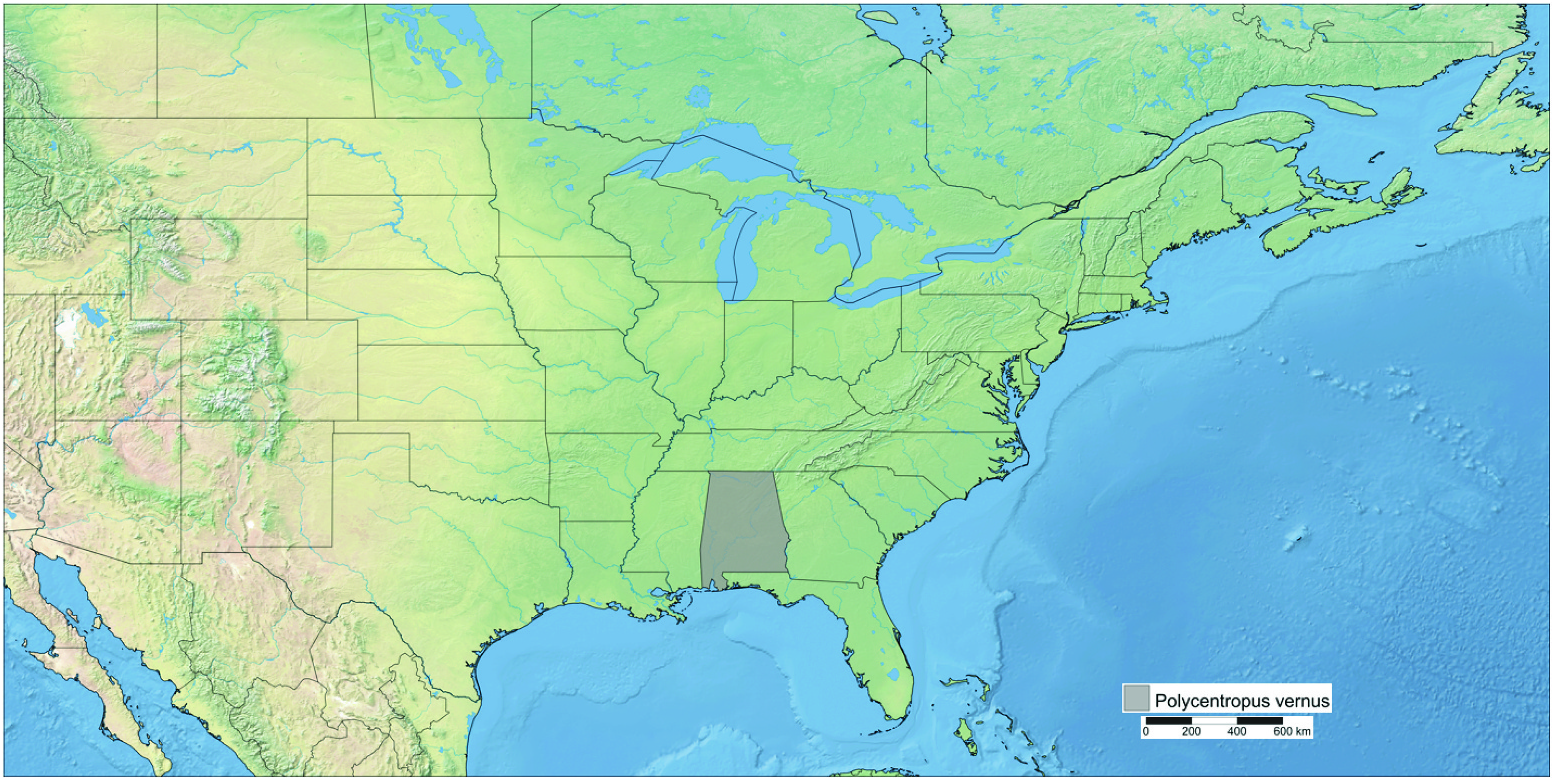
Distribution ( Figure 54 View Figure 54 ). USA: Alabama.
Material examined. None (see Notes).
Material listed from the original publication is as follows:
Holotype. United States: Alabama: Fayette County: small intermittent stream entering Wallace Branch at headwaters, 5.5 mi SE Berry, T16 S-R10- Sec. 36, sweep net, 16.v.1982, S.C. Harris . Paratypes. Fayette Co., same data as holotype, 10 males; same data except 26 .iv .1983, 1 male; same data except 26 .iv .1983, 1 male metamorphotype; same data except 15 .iii .1984, 1 male; same data except 11 .iv .1984. 9 males; same data except Wallace Branch at headwaters, 5 mi SE Berry, 16 .v .1984, 2 males; Tuscaloosa Co ., Wallace Branch , 5 mi S Berry, T17 S-R10-Sec . 10, 28 .iv .1982, 1 male, S.C. Harris; same data except 25.iv .1982, 1 male; Little Tyro Creek , 6 mi SE Berry, T17 S-R10W-Sec . 11, 16 .v .1984, 2 males, P.E. O̍Neil and R.L. Smith; same data except 19 .iii .1982, 1 male; small intermittent stream entering Little Tyro Creek , 6 mi SE Berry, T17 S-R10- Sec . 11, 26 .iv .1983, 1 male, S.C. Harris; Tyro Creek on unmarked Co . Rd., 3.5 mi E New Lexington , T17 S-R10-Sec . 15, 28 .iv .1982 , 1 male; Walker County: Wolf Creek off Hwy 102, 7 mi S Carbon Hill, 26 .iv .1983, 1 male, S.C. Harris and P. E. O̍Neil.
Dichotomous keys
Key to males of the Polycentropus confusus species group
Although not required, it is recommended to clear male genitalia for ease of use and best results.
1 Apices of ventral portions of inferior appendages curved strongly inward in ventral view (e.g. figs. 5C; 9C) .................................................................................................. 2
1̍ Apices of ventral portions of inferior appendages not curved strongly inward in ventral view (e.g. figs. 3C; 7C) .................................................................................................. 5
2(1) Body of pre-anal appendage with acute caudoventral projection in lateral view (e. g. figs. 5A; 20A) .............................................................................................................................. 3
2̍ Body of pre-anal appendage broad, blunt in lateral view (e.g. figs. 9A; 11A) ..... 4
3(2) Phallus with ventral swelling (fig. 20D); head of dorsobasal process of inferior appendage blunt in lateral view (figs. 20A) P ....... dinkinsorum Orfinger and Etnier
3̍ Phallus lacking ventral swelling (fig. 5D); head of dorsobasal process of inferior appendage pointed in lateral view (fig. 5A)... P ................................................................... .................................................................................. alabamensis Hamilton, Harris, and Lago View in CoL
4(2̍) Dorsobasal process of inferior appendage short, deflexed, with broad head in lateral view (fig. 9A) ........................................... ........................................... P. carlsoni Morse View in CoL
4̍ Dorsobasal process of inferior appendage longer, thumb-like, with smaller, rounded head (fig. 11A) ........................................................................ P. carolinensis Banks View in CoL
5(1̍) Ventral portion of inferior appendage abruptly narrowing distally in ventral view (figs. 28C; 32C) ................................................................................................................................ 6
5̍ Ventral portion of inferior appendage gradually narrowing distally or not narrowing distally in ventral view (e.g. figs. 17C; 26C) .............................................................. 12
6(5) Phallus with ventral swelling or spur (e.g. figs. 23D; 32D) ........................................... 7
6̍ Phallus lacking ventral swelling or spur (e.g. figs. 3D; 7D) .......................................... 9
7(6) Phallus with rounded ventral swelling (e.g. fig. 23D) .................................................... 8
7̍ Phallus with basoventral spur (fig. 32D) P. stephani Bowles, Mathis, and Hamilton View in CoL
8(7) Dorsobasal process of inferior appendage narrow with sharp mesal point in lateral view (fig. 28A); body of pre-anal appendage bifid in lateral view (fig. 28A) ............... ...................................................................................................................................... P. pentus Ross View in CoL
8̍ Dorsobasal process of inferior appendage broad with rounded mesal lobe in lateral view (fig. 23A); body of pre-anal appendage broad, rounded in lateral view (fig. 23A).. ................................... .................................. P. floridensis Lago and Harris View in CoL
9(6̍) Phallus curved strongly ventrad beyond middle in lateral view (e.g. figs. 18D; 24D) .................................................................................................................................................. 10
9̍ Phallus curved only slightly ventrad beyond middle in lateral view (e.g. figs. 3D; 21D) .................................................................................................................................................. 11
10(9) Posterior apex of phallus broad, adorned with microspicules (fig. 18D); dorsobasal process of inferior appendage extending far beyond outside margins of ventral portion of inferior appendage in ventral view (fig. 18C) .............. P. confusus Hagen View in CoL
10̍ Posterior of apex of phallus not broad, lacking microspicules (fig. 24D); dorsobasal process of inferior appendage not extending beyond margins of ventral portion of inferior appendage in ventral view (fig. 24C) ...................................... P. maculatus Banks View in CoL
11(9̍) Dorsal processes of pre-anal appendages strongly incurved in dorsal view (fig. 3B; body of pre-anal appendage shallow, rounded in lateral view (fig. 3A); phallic sclerite restricted to anterior half of phallus (fig. 3D) .............................................................. ..................................................................................................... P. aileenae Orfinger and Moulton
11̍ Dorsal processes of the pre-anal appendages subparallel, apices only slightly incurved in dorsal view (fig. 21B); body of pre-anal appendage more produced, acute in lateral view (fig. 21A); phallic sclerite along entire length of phallus (fig. 21D) .................................................................................................................................................... P. elarus Ross View in CoL
12(5̍) Head of dorsobasal process of inferior appendage acute in lateral view (e.g. figs. 7A; 17A) .................................................................................................................................................. 13
12̍ Head of dorsobasal process of inferior appendage blunt in lateral view (e.g. figs. 30a; 34A) ................................................................................................................................................ 15
13(12) Dorsobasal process of inferior appendage erect (e.g. figs. 7A; 13A); intermediate appendages parallel to subparallel in dorsal view, curved ventrad in lateral view (e. g. figs. 7A, B; 13A, B) ........................................................................................................................ 14
13̍ Dorsobasal process of inferior appendage strongly deflexed (fig. 17A); intermediate appendages curved dorsad, inflected inward, crossing (fig. 17A, B) ....................... ..................................................................................................... P. chenoides Ross and Yamamoto
14(13) Phallus nearly straight in lateral view; phallic sclerite large, occupying posterior 2/3 of phallus length in lateral view (fig. 13E); dorsal processes of pre-anal appendages extending posteriorly as far as termination of segment IX+X in dorsal view (fig. 13C) .................................................................................................................................... P. centralis Banks View in CoL
14̍ Phallus curved slightly ventral beyond middle in lateral view (fig. 7D); phallic sclerite small, restricted to ventral margin of posterior ¼ of phallus in lateral view (fig. 7D); dorsal processes of pre-anal appendage extending posteriorly well past segment IX+X in dorsal view (fig. 7B) .......... P. blicklei Ross and Yamamoto View in CoL
15(12̍) Dorsobasal process of the inferior appendage not digitiform, with ventral emargination in lateral view (e.g. figs. 30A; 34A) ............................................................................ 16
15̍ Dorsobasal process of the inferior appendage digitiform, roughly uniform depth along length in lateral view (e.g. figs. 15A; 26A) ............................................................... 18
16(15) Body of pre-anal appendage with shallow or no emargination in lateral view (figs. 30A; 35A); dorsobasal process of inferior appendage not extending beyond outside margins of ventral portion of inferior appendage in ventral view (e.g. figs. 30C; 35C) .......................................................................................................................................................... 17
16̍ Body of pre-anal appendage with deep emargination in lateral view (fig. 34A); dorsobasal process of inferior appendage extending far beyond outside margins of ventral portion of inferior appendage in ventral view (fig. 34C) ................................. ............................................................................................... P. thaxtoni Hamilton and Holzenthal View in CoL
17(16) Phallus with basoventral swelling (fig. 35D); whole animal small in size, forewing length not exceeding 3.6 mm .............................. P. vernus Hamilton, Harris, and Lago View in CoL
17̍ Phallus lacking basoventral swelling (fig. 30D); whole animal larger in size, forewing length at least 5.5 mm .......................................................................................... P. pixi Ross View in CoL
18(15̍) Body of pre-anal appendage with prominent dorsal point, deep emargination in lateral view (fig. 26A); phallus strongly curved ventrad apically in lateral view fig. 26D); dorsobasal process of inferior appendage approximately straight along length (fig. 26A) ............................................... ............................................... P. neiswanderi Ross View in CoL
18̍ Body of pre-anal appendage lacking dorsal point, not emarginate in lateral view (fig. 15A); phallus only moderately curved ventrad apically (fig. 15D); dorsobasal process of inferior appendage curved caudad beyond middle (fig. 15A) ..................... ........................................................................................................ P. chelatus Ross and Yamamoto View in CoL
Key to the known females of Eastern Nearctic Polycentropus Curtis, 1835 View in CoL
Accurate identifications using this key are dependent upon viewing cleared genitalia. While some species might be identifiable without clearing genitalia, it is strongly recommended that users first clear the genitalia to best visualise internal morphology.
The females of the following eastern Nearctic Polycentropus species remain unknown: P. chenoides Ross and Yamamoto , P. dinkinsorum Orfinger and Etnier , P. floridensis Lago and Harris , P. thaxtoni Hamilton and Holzenthal , and P. vernus Hamilton, Harris, and Lago of the Polycentropus confusus species group, and P. barri Ross and Yamamoto and P. colei Ross of the Polycentropus colei species group.
1 Posterior apex of the external parts of gonopods VIII broad and rounded, slightly triangular, or bifid, but not narrow and acute (e.g. figs. 4B; 6B; 8B; 10B) ...................... ...................................................................................... 2 ( Polycentropus confusus View in CoL species group)
1̍ Posterior apex of the external parts of gonopods VIII terminating in narrow, acute, thorn-like process in ventral view (fig. 1E by Yamamoto (1966))...................................... ................................................................................................................................. P. rickeri Yamamoto View in CoL
2(1) Internal parts of gonopods VIII appearing smooth (e.g. figs. 16B; 19B) .................. 3
2̍ Internal parts of gonopods VIII appearing wrinkled (e.g. figs. 4B, 6B) ..................... 8
3(1) Anterior part of genital chamber well formed, sclerotised (e.g. figs. 14B; 22B) ... 4
3̍ Anterior part of genital chamber poorly defined or absent (e.g. figs. 19B; 33B) .... 6
4(3) Anterior part of genital chamber smooth, not appearing cushioned (e.g. figs. 22B; 31B) ............................................................................................................................................................ 5
4̍ Anterior part of genital chamber appearing cushioned (fig. 14B) P. centralis Banks View in CoL
5(4) External parts of gonopods VIII terminating posteriorly in tapered, acute, slightly recurved apex in lateral view (fig. 22A) ........................................................ P. elarus Ross View in CoL
5̍ External parts of gonopods VIII terminating posteriorly in blunt, rounded apex in lateral view (fig. 31A) ................................................................................................ P. pixi Ross View in CoL
6(3̍) External parts of gonopods VIII digitiform in lateral view with rounded posterior apex (e.g. figs. 16A; 33A; ventral plates elongate, posterior apex rounded (e.g. figs. 16A, B; 33A, B) ................................................................................................................................ 7
6̍ External parts of gonopods VIII not digitiform, terminating posteriorly in tapered, pointed apex in lateral view (fig. 19A); ventral plates truncate, posterior apex sinuous in lateral view (fig. 19A) ........................................................................ P. confusus Hagen View in CoL
7(6) Processus spermathecae subovoid, lacking accompanying process in ventral view (fig. 33B); ventral plates partially overlaying internal parts of gonopods VIII in ventral view (fig. 33B) .............................................. P. stephani Bowles, Mathis, and Hamilton View in CoL
7̍ Processus spermathecae subtriangular, possessing ensiform process projected posteriorly in ventral view (fig. 16B); ventral plates not overlaying internal parts of gonopods VIII in ventral view (fig. 16B) .............. P. chelatus Ross and Yamamoto View in CoL
8(2̍) Ventral plates convergent in ventral view (e.g. figs. 10B; 25B) .................................. 9
8̍ Ventral plates parallel to subparallel in ventral view (e.g. figs. 6B; 27B) .............. 13 9(8) Anterior parts of genital chamber semicircular (e.g. figs. 4B; 10B) ......................... 10
9̍ Anterior parts of genital chamber sinuous or apparently absent (e.g. 25B; 29B) ................................................................................................................................................... 12
10(9) Internal parts of gonopods VIII semielliptical or blade-like in ventral view (e.g. figs. 4B; 8B) .............................................................................................................................................. 11
10̍ Internal parts of gonopods VIII subrectangular in ventral view (fig. 10B) .................... ................................................................................................................................. P. carlsoni Morse View in CoL
11(10) Internal parts of gonopods VIII semielliptical in ventral view (fig. 4B) .......................... ............................................................................................... P. aileenae Orfinger and Moulton
11̍ Internal parts of gonopods VIII blade-like in ventral view (fig. 8B) ................................. ..................................................................................................... P. blicklei Ross and Yamamoto View in CoL
12(9̍) Anterior parts of genital chamber sinuous, elaborate (fig. 25B); apex of external parts of gonopods VIII ventrally recurved in lateral view (fig. 25A, B) ........................... ............................................................................................................................ P. maculatus Banks View in CoL
12̍ Anterior parts of genital chamber apparently absent (fig. 29B); apex of external parts of gonopods VIII straight, oriented caudally (fig. 29A, B) ........ P. pentus Ross View in CoL
13(8̍) Posterior apex of external parts of gonopods VIII rounded (e.g. figs. 12B; 27B) ................................................................................................................................................... 14
13̍ Posterior apex of external parts of gonopods VIII bifid (fig. 6) ......................................... ............................................................................. P. alabamensis Hamilton, Harris, and Lago View in CoL
14(13) Apex of external parts of gonopods VIII blunt, ventrally recurved in lateral view (fig. 27A); anterior parts of genital chamber semicircular in ventral view (fig. 27B) ............................................................................................................................ P. neiswanderi Ross View in CoL
14̍ Apex of external parts of gonopods VIII acute, curved slightly dorsally in lateral view (fig. 12A; anterior parts of genital chamber semitrapezoidal in ventral view (fig. 12B) ...................................................................................................... P. carolinensis Banks View in CoL

| USNM |
Smithsonian Institution, National Museum of Natural History |
| R |
Departamento de Geologia, Universidad de Chile |
No known copyright restrictions apply. See Agosti, D., Egloff, W., 2009. Taxonomic information exchange and copyright: the Plazi approach. BMC Research Notes 2009, 2:53 for further explanation.
|
Kingdom |
|
|
Phylum |
|
|
Class |
|
|
Order |
|
|
Family |
|
|
Genus |
Polycentropus vernus Hamilton, Harris, and Lago
| Orfinger, Alexander Benjamin 2023 |
Polycentropus vernus
| Hamilton SW & Harris SC & Lago PK 1990: 367 |
Polycentropus
| Lago PK & Harris SC 1987: 258 |