
Prionospio cf. tridentata Blake & Kudenov, 1978
|
publication ID |
https://doi.org/ 10.11646/zootaxa.4019.1.22 |
|
publication LSID |
lsid:zoobank.org:pub:88F2DB05-58C4-4726-89D5-99302FABB908 |
|
DOI |
https://doi.org/10.5281/zenodo.4658192 |
|
persistent identifier |
https://treatment.plazi.org/id/5E51D737-FFF7-FF8F-FF4A-A0AA1D3AFDDA |
|
treatment provided by |
Plazi |
|
scientific name |
Prionospio cf. tridentata Blake & Kudenov, 1978 |
| status |
|
Prionospio cf. tridentata Blake & Kudenov, 1978 View in CoL
( Figs 27–29 View FIGURE 27 View FIGURE 28 View FIGURE 29 )
Prionospio (Prionospio) tridentata Blake & Kudenov, 1978: 219 View in CoL , fig. 23. Prionospio tridentata View in CoL .— Hutchings & Murray 1984: 61; Wilson 1990: 246.
Material examined. Queensland: AM W.45259, MI QLD 2330b (1); AM W.45379, MI QLD 2440 (2); AM W.47869, MI QLD 2431 (2). Northern Territory: AM W.47868 (5), NTM W025903 (4), MIMB 28130 (3), Dudley Point, Fanny Bay, Darwin, 12.435600°S, 130.832300°E, muddy sand intertidal, 5 Sep 2013.
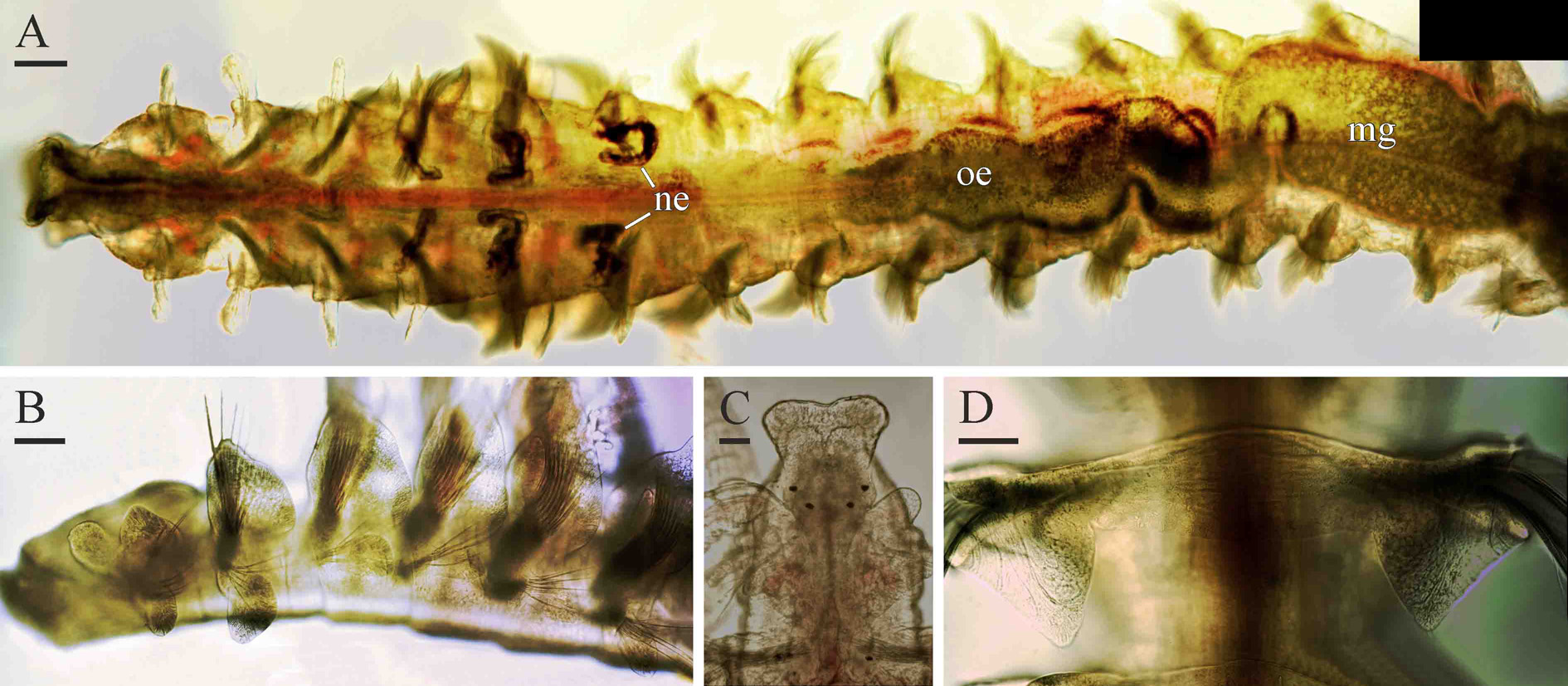
Adult morphology. Up to 14 mm long, 0.55 mm wide for 62 chaetigers. Pigmentation in life absent. Prostomium bell-shaped, anteriorly wide, blunt to concave, extending posteriorly to end of chaetiger 1 as a prominent caruncle ( Fig. 27 View FIGURE 27 C). Five small knobs with short non-motile sensory cilia present on frontal and frontolateral edges of prostomium. Occipital antenna absent. Two pairs of small red eyes arranged trapezoidally. Nuchal organs U-shaped ciliary bands on lateral sides of caruncle. Posterior dorsal parts of peristomium fused to notopodial lamellae of chaetiger 1 forming prominent ear-shaped structures. Palps as long as 10–15 chaetigers, with frontal longitudinal groove lined with fine cilia, short transverse bands of cilia regularly arranged on inner surface, and short compound motile cilia on fronto-lateral surfaces along frontal groove; cilia of inner transverse bands beating towards distal end of palp, while compound fronto-lateral cilia beating perpendicular palp axis towards frontal groove. Longitudinal band of cilia absent on outer fronto-lateral side along frontal groove.
Chaetiger 1 with capillaries and postchaetal lamellae in both rami; notopodial lamellae fused to posterior dorsal parts of peristomium forming prominent ear-shaped structures. Notopodial lamellae of chaetigers 2–6 largest, triangular; lamellae becoming gradually smaller and rounded on following chaetigers. Lower part of neuropodial postchaetal lamellae of chaetiger 2 rounded to pointed, elongated ventrally ( Fig. 27 View FIGURE 27 A).
Prominent dorsal crest with straight upper edge joining notopodial postchaetal lamellae of chaetiger 7 ( Fig. 27 View FIGURE 27 D); no crests or folds on other chaetigers. Lateral pouches and ventral flaps absent.
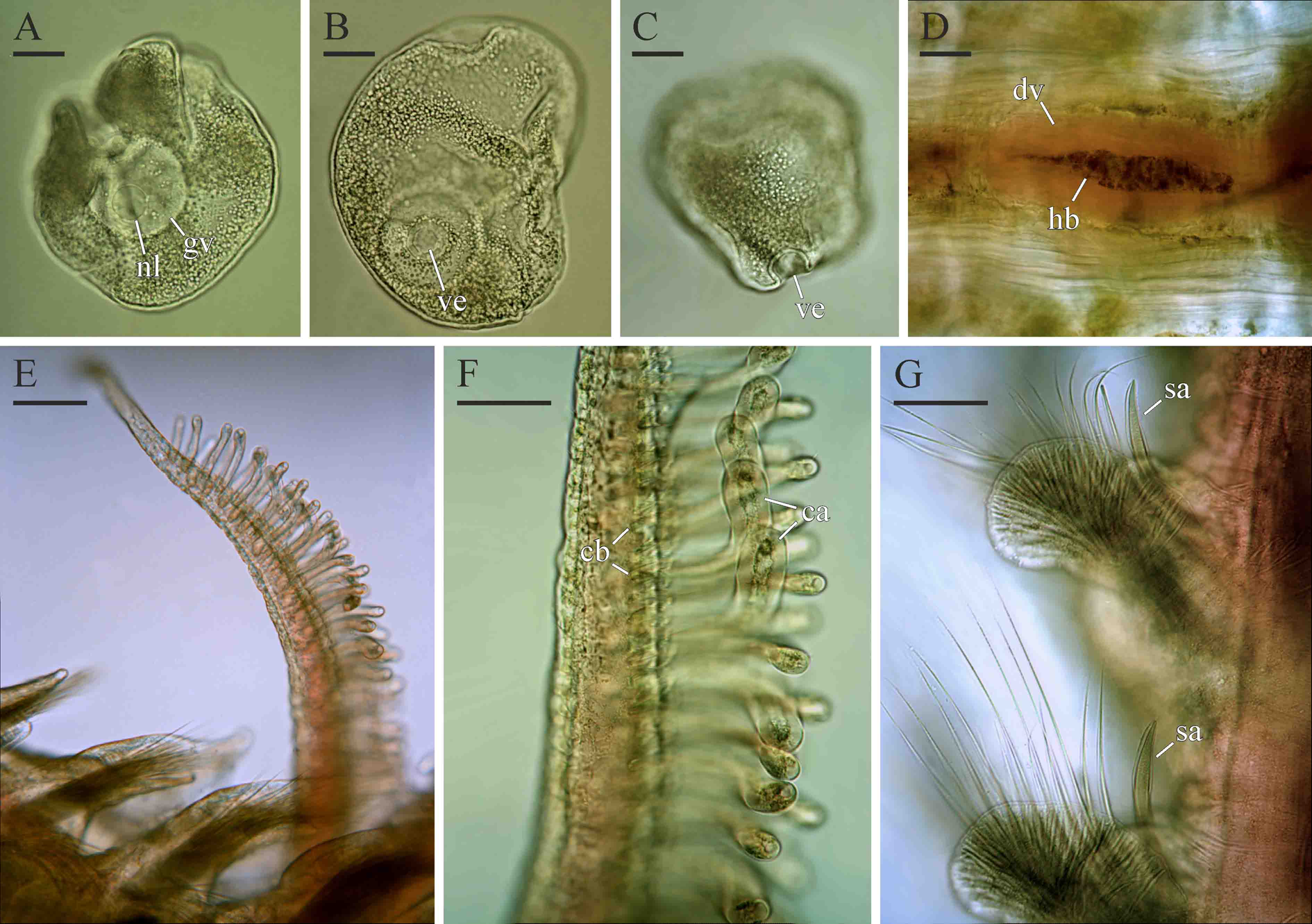
Sabre chaetae in neuropodia from chaetiger 11, thick, robust, with narrow limbation and fine granulation on distal end of shaft ( Fig. 28 View FIGURE 28 G); chaetae largest in chaetiger 11 ( Fig. 29 View FIGURE 29 C) and gradually diminishing in size on succeeding chaetigers ( Fig. 29 View FIGURE 29 D) towards midbody, then gradually increasing again.
Hooks in notopodia from chaetigers 33–41, up to six in a series among capillaries. Hooks in neuropodia from chaetigers 19–26, up to ten in a series, accompanied by inferior sabre chaetae and alternating capillaries throughout. Alternating capillaries thin, alimbate, about twice as long as hooks. Hooks bidentate, with small median upper tooth above main fang, with outer and inner hoods; shaft slightly curved ( Fig. 29 View FIGURE 29 A, B).
Four pairs of branchiae on chaetigers 2–5. Branchiae on chaetiger 2 shortest, as long as notopodial lamellae; branchiae on chaetigers 3 and 4 slightly longer than notopodial lamellae; all three pairs apinnate, robust, flattened, with surfaces oriented perpendicular to body axis. Branchiae on chaetiger 5 longest, extending posteriorly to end of chaetiger 9 (shorter in small individuals), cylindrical, with digitiform pinnae regularly arranged along lateral and posterior sides ( Fig. 28 View FIGURE 28 E). All branchiae with longitudinal bands of cilia running along inner and outer sides; afferent and efferent blood vessels interconnected by numerous circular blood capillaries giving branchiae annulate appearance; circular capillaries forming loops inside pinnae on chaetiger 5 branchiae ( Fig. 28 View FIGURE 28 F).
Short nototrochs present between branchial bases on chaetigers 2–4, each composed of one row of short cilia. Dorso-lateral longitudinal ciliation present on chaetigers 3–6 as short bands of cilia extending between successive notopodia. Intersegmental transverse ciliation absent.
Pygidium with one long middorsal cirrus and a pair of shorter and thicker ventral cirri; middorsal cirrus 3–4 times as long as ventral cirri ( Fig. 29 View FIGURE 29 E).
Oesophagus extending through 7–13 anterior chaetigers ( Fig. 27 View FIGURE 27 A). Ventral buccal bulb below oesophagus extending to end of chaetiger 1. Gizzard-like structure in digestive tract absent.
Main dorsal blood vessel transformed into gut sinus in anterior part of midgut. Soft heart body up to 35 µm in diameter extending inside main dorsal vessel from level of chaetigers 3–4 through chaetigers 11–12 ( Fig. 28 View FIGURE 28 D). Nephridia in chaetigers 4–6, greenish in life ( Fig. 27 View FIGURE 27 A).
Reproduction. Prionospio cf. tridentata is gonochoristic. Both in female and males gametes develop from chaetiger 17 to chaetigers 50–55. Oogenesis is intraovarian. Vitellogenic oocytes develop in ovaries attached to segmental blood vessels. Intraovarian oocytes were up to 90 µm in diameter, with germinal vesicle about 50 µm and single nucleolus 18 µm in diameter. Oocyte envelope is 2–3 µm thick, with smooth external surface. Developed coelomic oocytes were about 140 µm in diameter, with germinal vesicle about 60 µm and single nucleolus 20 µm in diameter ( Fig. 28 View FIGURE 28 A–C). The oocyte envelope has single semi-spherical depression about 20 µm in diameter and 15 µm deep ( Fig. 28 View FIGURE 28 B, C). Spermatogonia proliferate in testes; spermatogenesis occurs in the coelomic cavity. Spermatids are joined in tetrads. Spermatozoa are ect-aquasperm with small acrosome, spherical nucleus 2–3 µm in diameter, spherical mitochondria probably four in number, and a long flagellum.
Remarks. Prionospio tridentata was originally described based on two individuals collected from Burwood Beach, Newcastle, New South Wales, Australia, by Blake & Kudenov (1978). Two individuals from Botany Bay, New South Wales were also mentioned in the original description but not designated as types of the species. The species was characterized by the bell-shaped prostomium, two pairs of small red eyes, short and smooth branchiae on chaetiger 2, thick and smooth branchiae on chaetigers 3 and 4, and long pinnate branchiae on chaetiger 5, prominent dorsal crest on chaetiger 7, sabre chaetae in neuropodia from chaetiger 11, and tridentate hooks in notopodia from chaetiger 28 and in neuropodia from chaetiger 19.
Imajima (1990b) reported P. tridentata from the Ryukyu Islands, Japan. This record may, however, be misidentification of Prionospio nova Annenkova, 1938 originally described from the Russian part of the Sea of Japan.
Prionospio from Lizard Island and Darwin fit major diagnostic characteristics of P. tridentata but differ from the description of the species in having bidentate instead of tridentate hooks. Blake & Kudenov (1978: 219, fig. 23d) described and illustrated two upper teeth situated in line above main fang. Tridentate hooks with teeth arranged in a vertical line are unusual for Prionospio where adults usually have multidentate hooks with paired upper teeth arranged in two vertical rows above main fang. Bidentate hooks are present in Prionospio caspersi Laubier, 1962 originally described from Venice, Mediterranean, Italy, and Prionospio saldanha Day, 1961 originally described from Saldanha Bay, South Africa 1. Adult Prionospio cf. tridentata from Lizard Island and Darwin differ from those of P. saldanha by having sabre chaetae in neuropodia from chaetiger 11 instead of chaetiger 12. They also differ from P. caspersi by more posterior start of hooks in neuropodia, from chaetigers 19– 26 instead of chaetigers 18–19. If the tridentate dentition of hooks will be confirmed in P. tridentata from New South Wales, Prionospio from Queensland and Northern Territory should be recognized as a new species.
Habitat. In this study, adults of Prionospio cf. tridentata were found in fine coral sand or muddy sand from intertidal to 16 m depth.
Distribution. Australia: New South Wales,? Queensland, Northern Territory.? Ryukyu Islands, Japan.

No known copyright restrictions apply. See Agosti, D., Egloff, W., 2009. Taxonomic information exchange and copyright: the Plazi approach. BMC Research Notes 2009, 2:53 for further explanation.
|
Kingdom |
|
|
Phylum |
|
|
Class |
|
|
Order |
|
|
Family |
|
|
Genus |
Prionospio cf. tridentata Blake & Kudenov, 1978
| Radashevsky, Vasily I. 2015 |
Prionospio (Prionospio) tridentata
| Wilson 1990: 246 |
| Hutchings 1984: 61 |
| Blake 1978: 219 |