
Proteinus sp.
|
publication ID |
https://doi.org/ 10.1080/00222933.2023.2231569 |
|
DOI |
https://doi.org/10.5281/zenodo.8274018 |
|
persistent identifier |
https://treatment.plazi.org/id/03C80B33-1820-FF9C-FDB1-FC8DF64FFC67 |
|
treatment provided by |
Plazi |
|
scientific name |
Proteinus sp. |
| status |
|
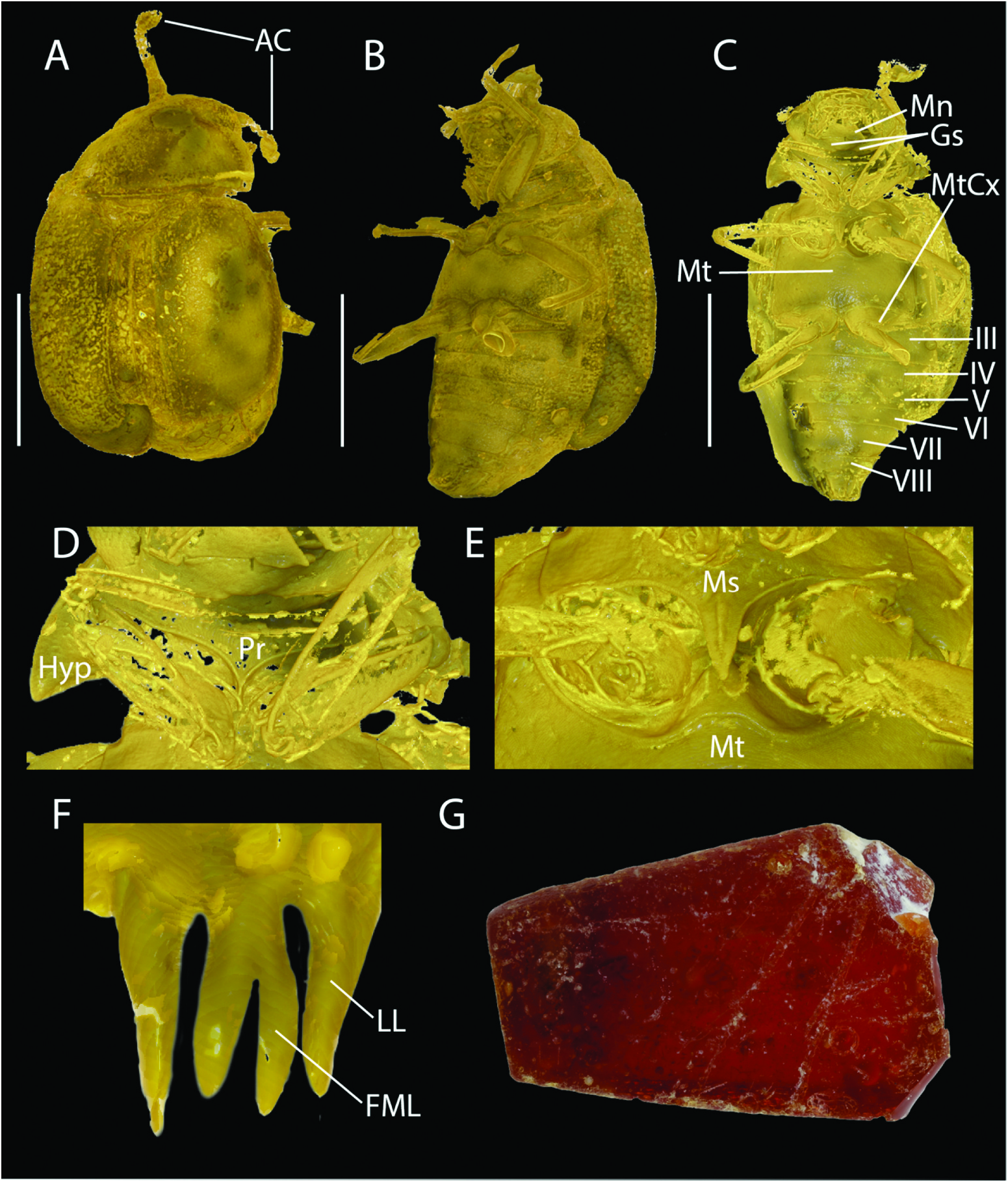
( Figure 1 View Figure 1 (A–F))
Material. 1 female, NHMD-36909, Baltic amber ( Figure 1 View Figure 1 (G)). Apparently previously glued to a piece of card with the number 83 written below. With the following labels ′Dr. J. Ipsen, Esbjerg /: STAPHYLINIDAE PROTEININAE det. R.A. Crowson: Proteininae ? A. Solodovnikov det. 2007: Proteinini : Proteinus sp. ♀ det. A.Newton 2007̍.
Origin and horizon. The piece of Baltic amber was collected from Esbjerg, Denmark. Baltic amber is dated as 45.0–38.0 Ma (Bartonian to Priabonian, mid-late Eocene).
Description. Measurements (all in millimetres): TL: 1.4; HW: 0.39; PW: 0.5; PL: 0.27; EL: 0.64. Head. Antennae with first two antennomeres distinctly larger than following antennomeres before distinct club( Figure 1 View Figure 1 (A,AC)).Eyes occupying almost entire side of head ( Figure 1 View Figure 1 (B, C)); mentum trapezoidal ( Figure 1 View Figure 1 (C, Mn)); gular sutures apparently separated ( Figure 1 View Figure 1 (C, Gs)). Prothorax: widest posteriorly; posterior portion of right hypomeron visible in ventral view (Figure (1D, Hyp)); prosternal process acutely pointed (Figure (1D, Pr)). Procoxae and profemora approximately equal in length; protibiae weakly expanded towards middle, parallelsided from middle to apex. Mesothorax: mesosternal process acutely pointed with distinct ridge along its length (Figure (1E, Ms)). Mesocoxae rather circular;mesofemora and mesotibae with weakly curved outer edge. Metasternum with weak and sparse punctation ( Figure 1 View Figure 1 (C, Mt)). Elytra rather long but not covering entire abdomen; apical three abdominal segments exposed ( Figure 1 View Figure 1 (B, C)). Metacoxae wide, anterior portion occupying almost entire posterior edge of metathorax ( Figure 1 View Figure 1 (C)), slightly expanded into lateral lobe (Figure (1C, MtCx)). Abdomen consisting of clearly visible sternites III to VIII ( Figure 1 View Figure 1 (C)). Apical segment of abdomen with prong-shaped sternite X and long styli on either side ( Figure 1 View Figure 1 (F)). Gonocoxal plate as in Figure 1 View Figure 1 (F), consisting of forked median lobe ( Figure 1 View Figure 1 (F, FML)) and the lateral lobes ( Figure 1 View Figure 1 (F, LL)). The styli are not visible.
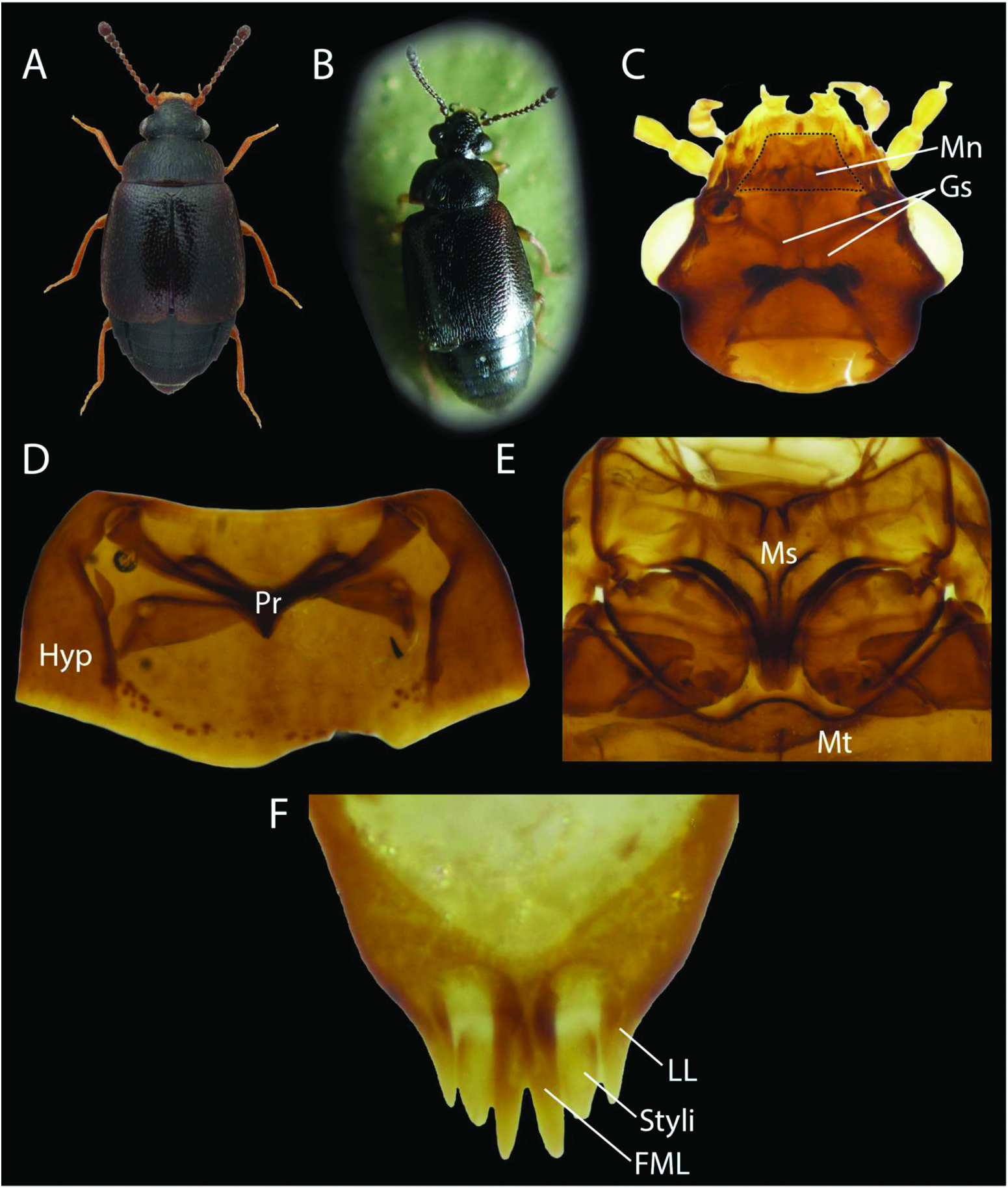
Remarks. Placement of the fossil in the subfamily Proteininae is supported by the clubbed antennae; rather widely separated gular sutures; broadly transverse pronotum; and moderately long elytra leaving abdominal segments VII, VIII and genital segments exposed. Other characters that support placement in Proteininae are unfortunately not visible – for example, absence of ocelli on the dorsal side of the head. The specimen can be assigned to the tribe Proteinini based on the simple pro-tibiae (excised internally before apex in Anepiini) and distinctive morphology of the apical abdominal segment in females (see fig. 40 in Steel 1966). It is placed in the genus Proteinus based on the clubbed antennae; trapezoidal mentum; smooth sides of pronotum (often crenulate in Megarthrus and Metopsia ); short prosternum with acutely pointed prosternal process; mesosternal process with ridge; wide metacoxae occupying almost entire posterior edge of metathorax; and shortened elytra exposing abdominal segments VI to X. The specimen can be identified as a female based on the lack of any emargination on sternite VIII, which occurs in males of Proteinus , and the structure of the gonocoxal plate ( Figure 1 View Figure 1 (F)), which is very similar to females of extant Proteinus ( Figure 2 View Figure 2 (F, FML, Styli, LL)) and is characteristic of the tribe Proteinini ( Proteinus , Metopsia , Megarthrus ) ( Steel 1966). The gonocoxal plate consists of a distinctly forked median lobe ( Figure 1 View Figure 1 (F, FML)) and lateral lobes ( Figure 1 View Figure 1 (F, LL)). In the fossil, the styli are either not visible ( Figure 1 View Figure 1 (F)) despite µ-CT scanning or absent, most likely the former. In extant species the styli can be observed between the forked median lobe and lateral lobes ( Figure 2 View Figure 2 (F)).
Since the taxonomy of the genus Proteinus is poorly known and many species are very similar to each other ( Figure 2 View Figure 2 (A, B)), a sound species identification of this fossil would require a through dissection and comparison of all extant congeners. Since species delimitation within Proteinus is largely aided by examination of the male genitalia, attempting such work based on females only was not feasible. It is likely that this fossil is an extinct species to be described as new to science if more material and thus characters become available. From the common and widespread West Palaearctic species Proteinus laevigatus ( Figure 2 View Figure 2 (C–F)), the amber fossil differs in the shorter mesothorax, much more pointed apex of the weakly notched mesosternal process, and longer styli. Such characters may be informative at the species level and worth further consideration in future works.
| R |
Departamento de Geologia, Universidad de Chile |
No known copyright restrictions apply. See Agosti, D., Egloff, W., 2009. Taxonomic information exchange and copyright: the Plazi approach. BMC Research Notes 2009, 2:53 for further explanation.