
Pseudoglyptodon chilensis, McKENNA & WYSS & FLYNN, 2006
|
publication ID |
https://doi.org/ 10.1206/0003-0082(2006)3536[1:PPXFCC]2.0.CO;2 |
|
persistent identifier |
https://treatment.plazi.org/id/ED2D8785-3114-8B31-9CE2-FD08FC2AFA93 |
|
treatment provided by |
Carolina |
|
scientific name |
Pseudoglyptodon chilensis |
| status |
sp. nov. |
Pseudoglyptodon chilensis , new species
Pseudoglyptodon sp. : Wyss et al. (1990: fig. 4 View Fig ).
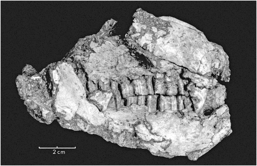
TYPE SPECIMEN: SGO PV 2995, damaged skull and mandibles with seemingly complete dentition .
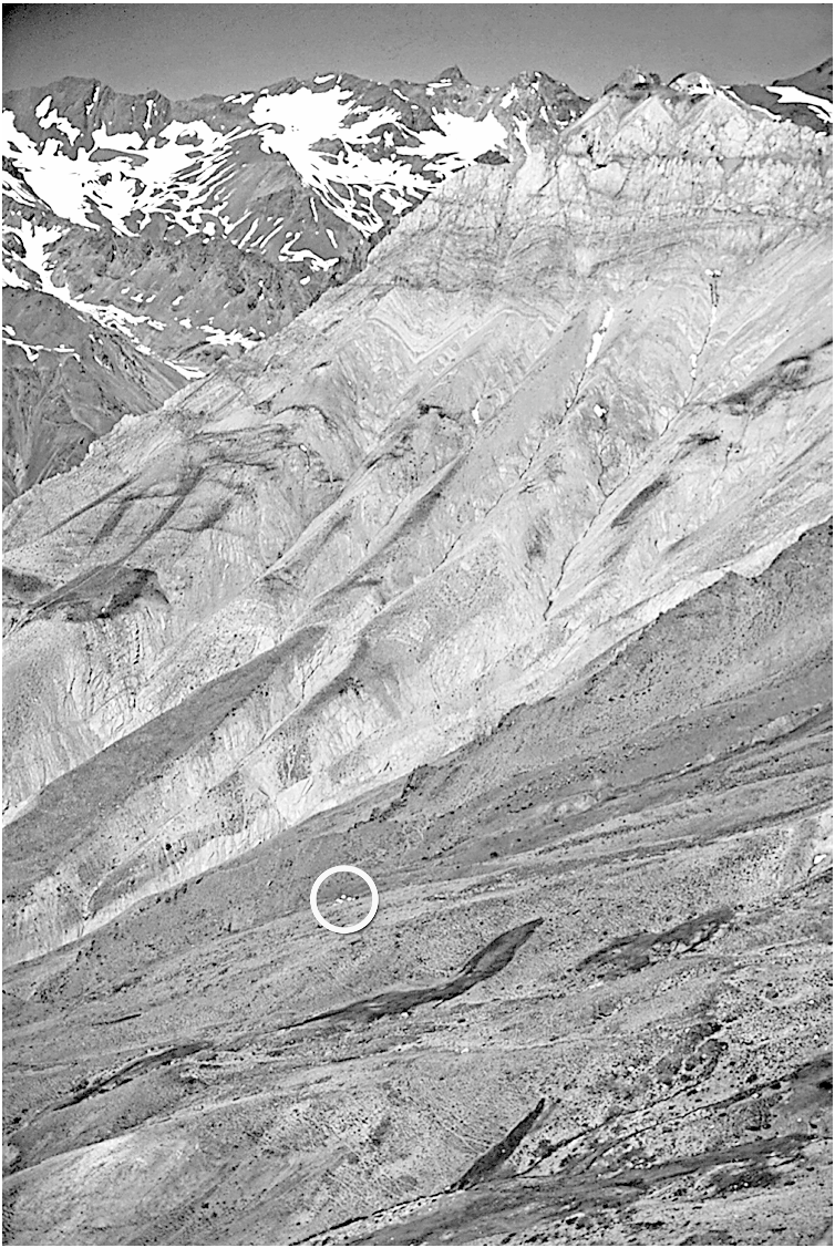
TYPE LOCALITY: The type and referred Chilean specimens are from the Tinguiririca River valley (, 35 ° S) in the Cordillera Principal of the Central Andes, approximately 7 km west of the Argentine border. They are derived from a steep set of exposures north of an unnamed pass (the latter of which is identified by its 2738 m elevation on the topographic sheet [ Anonymous, 1985]), approximately 3 km south of the summer resort town of Termas del Flaco. Pseudoglyptodon chilensis and its associated fauna occur in 35–50 ° westward-dipping volcaniclastic deposits of various colors, dominantly brownish red, interbedded with volcanic flows and tuffs ( fig. 2 View Fig ). Prior to discovery of fossil mammals in the region (Novacek et al., 1989) these deposits were mapped as pertaining to the Colimapu Formation of poorly constrained Aptian–Albian age (e.g., Klohn, 1960). More recent detailed mapping and associated geochronologic studies (Wyss et al., 1993; Charrier et al., 1996) indicate that the mammal-producing unit pertains to the Abanico Formation (5 Coya-Machalí Formation), a widespread and stratigraphically important unit in this region of the Andes. Fossils occur most abundantly in a massive, purplish brown, volcano-sedimentary horizon, near the apparent local base of the formation. Owing to structural complexity, it has not been possible to establish the relative stratigraphic position of the fossiliferous horizon within the approximately 2-kmthick Abanico Formation. A second, substantially older fauna has been recovered from volcaniclastic sediments of the Tinguiririca Valley some 15 km west of those hosting P. chilensis ( Flynn et al., 1991; Wyss et al., 1996), indicating that the Pseudoglytodon - producing beds do not correspond to anything approaching the lowest stratigraphic levels in the formation. Still further to the west (, 20 km), but still at the same latitude (35 ° S), thick exposures of the Abanico Formation have yielded three stratigraphically superposed fossil mammal faunas, the lowest of which also clearly predates the Tinguirirican SALMA (Wyss et al., 2004).
AGE: Tinguirirican SALMA. The diverse fauna co-occurring with Pseudoglyptodon at Termas del Flaco allows unambiguous correlation with the SALMA sequence. The absence of such diagnostic taxa as Pyrotherium , primates, mesotheres (which is problematic, because this group occurs in the Divisaderan), Archaeohyrax , Plagiarthrus , hegetotheres, and Morphippus (Marshall et al., 1983) indicates a pre-Deseadan age for this Chilean fauna. Co-occurrence of taxa known elsewhere only from Mustersan and older deposits (notostylopids, notopithecines, and polydolopids) with taxa previously known only from younger beds (a clade of notohippids diagnosed by hypsodont incisors, interatheriine interatheres, and rodents) identifies the fauna as representing a biochronologic interval interposed between the Deseadan and Mustersan SALMAs, the Tinguirirican ( Flynn et al., 2003). In this connection, the Pseudoglyptodon -containing fauna from Chile bears little resemblance to the problematic Divisaderan assemblage (known from a single locality some 250 km to the northeast, in western Argentina). Whatever the still uncertain relative temporal relationship of the faunas from Termas del Flaco and Divisadero Largo may be, the two are undoubtedly distinct.
That the Tinguririca Fauna derives from a thick volcanic and volcaniclastic sequence is fortuitous from the standpoint of radioisotopic dating. Multiple single-crystal laser fusion 40 Ar/ 39 Ar dates (Wyss et al., 1993) and fewer conventional 40 K/ 40 Ar analyses (Wyss et al., 1990) constrain the absolute age of Pseudoglyptodon chilensis . Dates from immediately above the fossiliferous horizon indicate P. chilensis to be minimally,31.5 Ma (early Oligocene) in age ( Flynn et al., 2003). Levels immediately below the fossiliferous horizon (but within the same stratigraphic unit) have been dated ( Flynn et al., 2003) at a locality producing a fauna indistinguishable from the one associated with P. chilensis , but this second locality has not yet produced P. chilensis itself. Present evidence suggests, albeit indirectly, that the Tinguirirican SALMA likely extends no further back in time than an additional 1–2 Ma ( Flynn et al., 2003), i.e., very near the Eo-Oligocene transition.
REFERRED SPECIMENS: A second specimen from Termas del Flaco, SGO PV 2999, consists of a badly damaged anterior end of a right mandibular ramus and part of the fused symphysis. Only alveoli and fragmentary tooth bases remain. One alveolus suggests a trilobed cheek tooth like that of the type specimen of P. chilensis . Unfortunately, the referred specimen from Termas del Flaco provides little useful information. A Deseadan cheek tooth from Patagonia referred by Florentino Ameghino (1897: 507) to Glyptatelus tatusinus , not demonstrably associated with the (lecto) type specimen and not a glyptodont in any case, may belong here as well. It provides limited information but is less certainly conspecific with P. chilensis than is SGO PV 2999.
DIAGNOSIS: Pseudoglyptodon chilensis differs from P. sallaensis and the unnamed species discussed above in the former’s much larger size, thinner cheek-tooth wall of hard dentine, and more sharply angular cheektooth lobes.
DESCRIPTION: Skull: The skull of Pseudoglyptodon chilensis ( fig. 3 View Fig ) is evidently short, as suggested by the small number of teeth, the position of the jaw articulation, and the position in the matrix of the two petrosal bones. The distance between the front of the skull and the anterior edge of the orbit (as judged by the position of the lacrimal foramen) is truncated, a condition generally seen in sloths but even more marked in glyptodonts. The little of the orbit that may be discerned occurs not far above the roots of the upper cheek teeth, which, although almost certainly hypselodont (no evidence of closed roots is seen on the CT scans—which are frontal sections), are not highly elongate prisms requiring a deep maxilla. Because all upper teeth appear to originate in the maxilla and no suture is evident at the anterior end of the maxillary wall of the rostrum, the premaxilla was either lost postmortem or is completely fused to the maxilla in the specimen at hand. The two nasal bones are fused to each other but not to the maxilla. They extend posteriorly to at least a position over the posterior end of the first molariform upper cheek tooth, but damage obscures their full posterior extent and whether they widened in the rear. The nasals are thus quite long and thin, contrasting with the short wide nasal judged to be typical—and ambiguously synapomorphic—of tardigrades ( Gaudin, 2004 — his character 100); foreshortened nasals are also typical of glyptodonts. In SGO PV 2995 the right nasal is, 5 mm wide at the midpoint of its preserved portion, while the element was at least 30 mm long and quite likely reached twice that length originally. Striking features of the otherwise already bizarre dentition of P. chilensis are the massive upper and lower ‘‘canines.’’ The oval, upper caniniform tooth base is housed in a prominent bulge in the maxillary bone on the side of the snout. The snout is too damaged to provide information about the anterior end of the palate, housing
+
west-dipping strata. Fossil mammal localities occur in the dark band of volcaniclastic sediments of the Abanico (5 Coya Machalı´) Formation in the mid-foreground (straddled by the top half of the circle). A cluster of tents in the circle gives a sense of scale. The light-colored strata in the middle distance are of the Baños del Flaco Formation (Neocomian), with the snow-covered rocks in the distance belonging to the Río Damas Formation (Kimmeridgian). The horizon approximates the border with Argentina.
for the organ of Jacobson, septomaxillary bone if any, or other anterior structures. The narial opening was large, but apparently little flared. Details of the orbit are lacking due to damage, but the orbit was probably not large. An apparent lacrimal bone occurs on the right side, where it appears to be fused to the maxilla. Its large lacrimal foramen lies anteri- or to the orbital rim. The anterior end of a possibly deep, posteroventrally descending wing of the anterior part of the zygomatic arch arises between the lacrimal foramen and the anterior end of the second of the three molariform upper cheek teeth. Although the maxillary part of the arch does not appear to have been especially strong (judged from its broken cross section), there is circumstantial evidence of a strong descending process of the jugal. A thin, triangular fragment of bone is appressed against the dorsoexterior border of the right mandibular ramus near the base of the coronoid process, and outboard of the last upper and lower cheek teeth. This element (obviously not part of the mandible) sits at a considerable distance from broken base of the anterior root of the zyomatic arch. Nevertheless, if this element is in anything close to its life position, it can only represent a distal portion of an elongated ventral process of the jugal. The leading edge of this element is seemingly smooth and unbroken, its orientation consistent with that expected for a descending process of the jugal, as seen in many sloths. The possibility that this element represents a displaced element from the skull roof or orbit cannot be completely excluded, however. If this element is indeed a portion of the zygomatic arch, it resembles much more the condition seen in tardigrades than in glyptodontids (wherein the descending process is much more anteriorly situated).
The zygomatic arch was probably not continuous with the squamosal, but evidence is weak. We have not seen the infraorbital foramen, but it may be obscured by breakage and unremoved matrix. No traces of the frontals remain, unless one of several fragments of bone above the right lacrimal foramen represents the anterolateral corner of the right frontal. The parietals, squamosals, occipitals, alisphenoids, basisphenoid, basioccipital, vomer, pterygoids, and certainly any possible mesethmoid are all now absent, but the rear of the palate, presumably involving the palatine bones, extends to the rear past and around the posterior lobe of the last upper molariform cheek tooth, forming an indented torus of sorts that may incorporate a part of the palatine as well as the maxilla. The palate is unusually narrow between the cheek-tooth rows (best seen on CT scan images). At the gumline the two upper tooth rows are nearly parallel centrally but diverge slightly anteriorly (particularly from the first molariform tooth forward) as well as posteriorly (particularly m3).
The lower jaw is massive, especially in the symphyseal area, which is fused but shows traces of the suture on SGO PV 2995 but not on SGO PV 2999. The horizontal rami bulge laterally beginning below the second molariform tooth, extending and becoming more pronounced posteriorly. This results in a,5- mm-wide shelflike area lateral to the third molariform tooth. The anterior end of the jaw supports a short upturned ‘‘spout,’’ below which lie one large and several smaller mental foramina on each ramus. Immediately behind the ‘‘spout’’ is the massive base of the lower caniniform tooth, which is followed by the three molariform lower cheek teeth (which are set off from each other by short diastemata). The lower cheek-tooth rows diverge posteriorly, especially deep within the alveoli. However, their occlusal surfaces meet those of the upper cheek-tooth row with less posterior divergence.
The ascending process of the mandible arises from the side of the horizontal ramus lateral to the last lower molariform cheek tooth, slanting up at an angle of about 135 ° to the plane of occlusion. The junction of the ascending and horizontal rami of the mandible occurs near the midpoint of the third lower molariform tooth; this, coupled with the slow rate of climb of the ascending process, results in the cheek teeth being exposed in lateral view (i.e., not covered by the ascending process), save for the posterior third of the last lower teeth and the posteroventral corner of last upper teeth. Importantly, there is no evidence of an external opening of the posterior mandibular canal near the horizontal–ascending ramus junction. Although the inferior portion of the horizontal ramus is broken on the right side of SGO PV 2995, enough is preserved to demonstrate that no such foramen was present. The occurrence of a foramen in this region uniquely characterizes tardigrades among xenarthrans ( Gaudin, 2004).
Owing to the shelf of bone lateral to the third lower molariform mentioned previously, the ascending process occupies a plane substantially lateral to the cheek-tooth row. The ascending process appears to be small, unexcavated either laterally or medially, evidently not projecting upward or rearward very far. Breakage of the dorsal, posterior, and ventral borders of the process, however, obscures its original size and shape. A small, detached knob of bone floating in the matrix near the posteroventral corner of the preserved part of the ascending ramus may be a remnant of the right mandibular condyle. If so, the condyle is positioned low, near the plane of occlusion, just in front of and lateral to the right petrosal. This contrasts with the primitive condition seen in most sloths (except mylodontids and Choloepus ) and dasypodids (glyptodontids included), wherein the condyle is positioned well dorsal to the tooth row ( Gaudin, 2004). Nothing can be said of the posteroventral parts of the mandible. A trace of a robust hyoid bone may possibly be represented by a bone fragment in the matrix at the appropriate position anterior to the right petrosal and medial to the presumptive mandibular condyle.
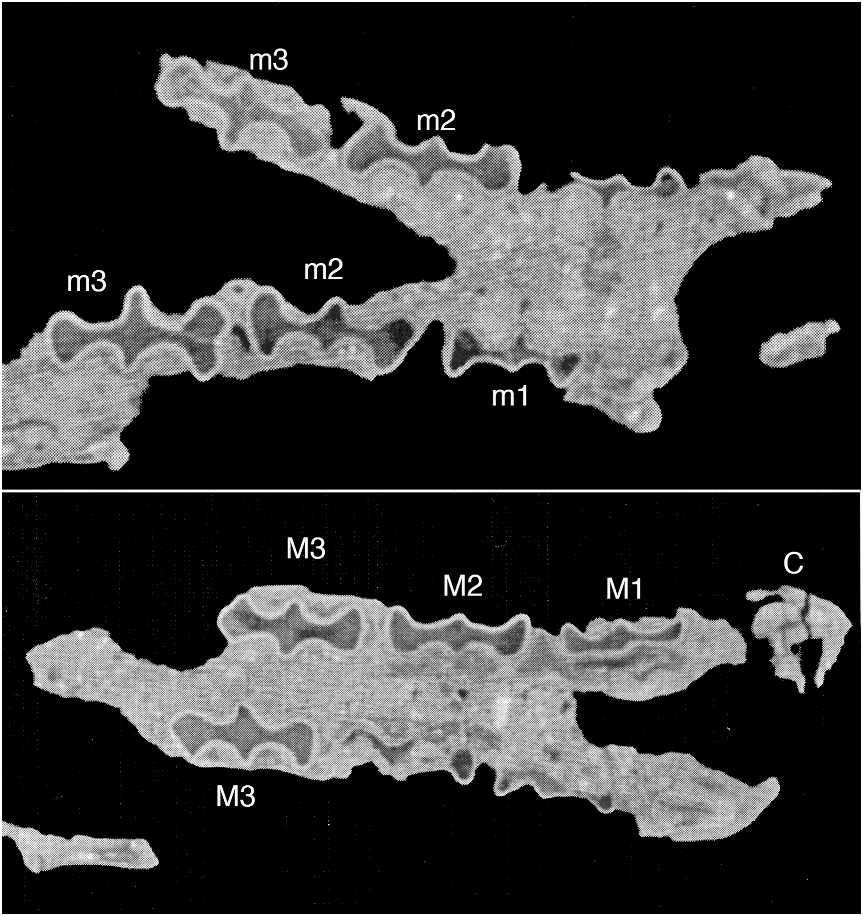
Dentition: The significance of SGO PV 2995 was revealed on the outcrop when its melonsized encasing nodule was delicately cleaved with a sledge hammer; just the surface of the anterior end of the left mandible was visible initially. Mechanical preparation revealed the labial faces of the teeth. Because the mandibles are tightly clenched, however, it has not been possible to disengage the upper and lower dentitions. Computerized tomography was used to more fully elucidate the dental morphology of SGO PV 2995. Twenty CT cross sections, taken as parallel to the occlusal plane as possible (CT scan nos. 563-11 through 563-30) were generated by Scientific Measurement Systems, Inc., (Austin, Texas) using a 420-kV 3-mA X-ray source. This stack of slices ranges from the bases of the lower cheek teeth to above the roots of the upper molariform cheek teeth. Distance in the x direction is 128.8 mm (i.e., preserved skull length), and distance in the y direction is 61.8 mm (i.e., preserved skull width). Each slice is 0.25 mm thick. Separation of the slices is 2.5 mm. The following description is based largely on this CT imagery. It must be cautioned that although the CT scans roughly parallel the occlusal plane, because the vertical axes of the high-crowned teeth are not consistently normal to this plane, the tooth outlines seen on the scans are distorted by the progressively more oblique angle at which they were sectioned ( fig. 4 View Fig ). This is particularly true for sections taken the greatest distance from the occlusal plane, especially for the posterior postcanines, whose apparent buccolingual dimensions are exaggerated near the tooth bases owing to canted and slightly bowed vertical axes of these teeth.
The number of teeth in Pseudoglyptodon is unusual, probably just four above and four below on each side, all fairly closely spaced with only short gaps between them. A more substantial gap behind the upper caniniform tooth on the specimen’s left side is likely artifactual, as a large crack disrupts the specimen in this region. Additionally, the degree to which the upper and lower left caniniforms are compressed into each other anteroposteriorly suggests a small degree of postmortem distortion in this region of the specimen. It is uncertain whether teeth occurred anterior of the upper caniniforms, because that region of the rostrum is missing.
The caniniform teeth of Pseudoglyptodon may or may not be true canines. Grassé (1955) regarded the anterior teeth in sloths to represent the true canine of the upper tooth row and the first premolar of the lower. Nevertheless, as with other xenarthrans, until detailed embryological work is carried out, the homology of these teeth remains uncertain. It seems plausible, however, that either the upper or the lower caniniform tooth in Pseudoglyptodon is not a true canine, because the occlusion of these teeth, as in sloths, differs from that seen in other mammals. This conclusion assumes that an anterior premolar can be more readily transformed into a canine imposter, than the position of true canines can be shifted anteriorly or posteriorly relative to the opposing tooth. Regardless of whether phyllophagan ‘‘canines’’ are C1/p1, C1/c1, or some other permutation, tardigrades are unique among xenarthrans in having upper tooth rows extend anterior to the lower tooth rows. On the damaged left side of SGO PV 2995 the lower caniniform appears to occlude behind the upper, but on the better preserved right side the upper and lower caniniforms sit side by side. Thus, the upper and lower tooth rows of Pseudoglyptodon terminate at nearly the same level anteriorly. Pseudoglyptodon is therefore alone among xenarthrans in this regard, bearing an apomorphic resemblance to the condition seen in tardigrades (where the upper tooth row extends anterior to the lower). Some artiodactyls convert an anterior lower premolar into a caniniform tooth that occludes behind the upper canine, but the resemblance to sloths is not as close as that seen in Pseudoglyptodon .
The lower caniniform tooth of Pseudoglyptodon occludes with the sloping posteromedial wear facet of the upper caniniform tooth, much as in Choloepus except that, in Pseudoglyptodon , the lower tooth is more medially placed relative to the upper. The occlusal relationship of these teeth is best exhibited on the right side of SGO PV 2995 ( fig. 3 View Fig ), because postmortem deformation has damaged the left pair of caniniforms. On the right side, the upper and lower caniniforms sit in a more normal orientation, directly side by side, the medial surface of the upper tooth occluding against the lateral side of the lower.
Judged from the CT scans, the tip of the lower caniniform tooth was not accommodat- ed by an excavation in the palate. The three molariform cheek teeth following the caniniform tooth on both the upper and lower tooth rows are anteroposteriorly elongate and trilobed, being about as high as they are long. It cannot be established whether any of the teeth is deciduous, or had replaced a precursor.
The upper caniniform tooth is enormous and is supported by a large oval base (not really a root in the usual sense) that extends high into the maxilla. At its dorsal extremity the base of the upper caniniform tooth opens widely rather than closing to a blunt tip. CT sections of the unworn parts of the tooth are narrower at the front than the rear, and the labial wall of the tooth is relatively convex, whereas the lingual wall is flatter. A rather flat transverse wear facet has been created by action with the lower caniniform tooth, from the recurved anterior tip of the upper caniniform tooth diagonally upward until the facet reaches the broad rear of the tooth’s base at the gum line.
The lower caniniform tooth differs in shape from its upper counterpart. CT scans show that its massive open base is wide in front and nearly flat on the anterior face within the alveolus. It then narrows, followed by a narrower rearprojecting lobe. The cross sections within the alveolus thus have a ‘‘pinched,’’ triangular shape. Near the gum line, the broad anterior face of the lower caniniform tooth becomes more rounded, and the posterior lobe becomes even narrower. Above the gum line, the anterior face is transversely worn by the action of the upper caniniform tooth. The diagonal (anteroventral–posterodorsal) slope of the transverse wear facet is guaranteed by the initial wear that would have occurred when these curiously shaped teeth first made contact.
As with the caniniforms, the homologies of the molariform teeth in Pseudoglyptodon are uncertain. All three molariform cheek teeth in both the upper and lower dentition have essentially the same trilobed external shape of glyptodont teeth, in outline reminiscent of a bat in flight seen from directly below. The long axes of these teeth parallel the long axis of the tooth row. As in glyptodontids, the crowns of these teeth are worn nearly flat except for the anterolabial lobe of the right third lower molariform cheek tooth, which projects somewhat between the second and third upper molariform cheek teeth labially in a manner reminiscent of the anterior ends of the crowns of rear lower molariform cheek teeth of Orophodon and Octodontotherium (Hoffstetter, 1958: fig. 42). The various molariform cheek teeth change slightly in shape with wear, as seen in their various cross sections, but they do not change significantly in dimensions throughout the various levels of each tooth. The base of each molariform cheek tooth is open, as in most tooth-bearing xenarthrans. Unlike glyptodont molariform cheek teeth, there is no central figure in the dentine of teeth of Pseudoglyptodon .
The first of the three upper molariform cheek teeth is the smallest of the upper postcaniniform series and is the narrowest transversely. Its anterior lobe is flattened and oriented normal to the tooth’s anteroposterior axis deep within the alveolus. Near the occlusal surface the flat anterior surface faces more linguad. The isthmus between the anterior and medial lobes is narrower than in the two more posterior upper cheek teeth. The medial lobe is blunter and projects less than those of the succeeding teeth, and the posterior wall of the posterior lobe is more flattened. The indentations demarcating the lobes of the molariform teeth are less pronounced on the lingual walls of the teeth than they are labially. Both the anterior and posterior labial lobes diverge from one another strongly, in contrast to those of the succeeding teeth.
The second of the molariform upper cheek teeth is more symmetric about its medial lobes than the first, although the labial reentrant between the anterior and medial lobes seems to have a small secondary fold high above the present occlusal surface (at least on the specimen’s right side). The anterior wall of the anterior lobe and the posterior wall of the posterior lobe are gently convex. The medial lobe is smaller and less acute than that of the third molariform tooth. As on the first molariform tooth, the anterior and posterior lobes of the second diverge labially more than lingually, contrasting with the orientation of the lobes of the posterior tooth.
The third (and last) upper molariform cheek tooth is the largest of the upper series. The anterior wall of its anterior lobe is gently convex and is not subdivided by an anterior indentation, as is its lower counterpart. The posterior lobe is broad, with a flattened, posterolabially facing wall that is indented slightly on the animal’s left tooth but not on the right one. Both the anterior and posterior lobes are more acute labially than lingually, but the prominent medial lobe is acute both lingually and labially, forming the widest part of the tooth. Breakage of the maxilla posterior to the last left molariform reveals that this tooth is implanted such that its vertical axis slopes labially from top to bottom.
The symphysis of SGO PV 2995 is well enough preserved that the presence of any lower teeth anterior of the caniniforms seems unlikely. Confoundingly, SGO PV 2999 exhibits the broken stubs of two small teeth floating in matrix above the symphyseal region. Both consist of little more than broken, ovoid cross sections 2–3 mm in diameter. Nevertheless, these tooth remnants are positioned symmetrically (one on either side of the symphysis, and about 1 cm apart from one another), so it must be assumed that they are preserved in life position. What are these teeth? Two explanations seem credible. The bone–matrix interface on SGO PV 2999 is indistinct anteriorly, making it difficult to determine what these teeth were originally attached to. Nevertheless, there is a distinct rim of bone immediately lateral of the right tooth—this rim is clearly the medial margin of an alveolous for an enlarged anterior lower tooth, probably the caniniform. The medial position of this tooth relative to the mandibular alveolus suggests that both tooth remnants are likely the tips of the upper caniniforms (which were clenched), the remainder of the upper dentition having been broken away. Alternatively (but less likely), these tooth remnants could represent small anterior teeth of the lower dentition, elements which simply are not preserved in SGO PV 2995.
The first lower molariform is the smallest cheek tooth, and has the narrowest isthmuses between lobes. The anterior lobe lies mainly anterolabial to its isthmus, with the result that there is little or no anteroposterior curvature of the lingual wall of the tooth anterior to the medial lobe. The medial lobe projects slightly labially, but forms a larger and more acute projection on the lingual side, limiting the anterior end of a deep reentrant behind it. The posterior lobe is more symmetrical than the offset anterior one, and it is slightly indented at the rear. It is the widest and most massive lobe.
The second lower molariform tooth is larger than the first and bears a large posterolabially–anterolingually oriented anterior lobe, the anterolabial wall of which is nearly flat. The lingual part of the lobe is larger and less acute than the labial part. The posterior lobe is even larger than the anterior one but is oriented somewhat posterolingually–anterolabially. It too is larger and less acute lingually than labially. The medial lobe is smaller and, like the other lobes, more pronounced lingually than labially.
The third molariform, the largest lower cheek tooth, is distinguished by a very large and transverse anterior lobe that is markedly indented anteriorly, resulting in a large, rounded lingual sublobe and a smaller, somewhat more acute labial one. The medial lobe is also very large, transverse, and acute on both sides of the tooth, but it is especially prominent lingually. The posterior lobe is somewhat asymmetrically placed, lying mainly posterolabially, somewhat the mirror image of the anterior lobe of the first molariform tooth. As in the other lower molariform cheek teeth, it is more acute labially than lingually. Breakage of the dentary posterior to molariform 3 on the left side of SGO PV 2995 shows this tooth to be inclined lingually. It also shows clearly that the root of this tooth is quite short compared with the second molariform of P. sallensis . In the latter, the second molariform reaches the base of the mandible, and the tooth is nearly 3 cm high (i.e., the height is triple the length). In the Chilean form the molariforms are subequal in height and width.
Petrosals: Although both petrosals are preserved (indeed, they constitute nearly the entirety of the preserved portion of the skull posterior to the upper dentition), neither reveals much anatomical or phylogenetically informative data. The left petrosal consists of a badly damaged, featureless lump. The right petrosal preserves perfectly ordinary-looking oval and round windows, and an unremarkable promontorium.
No known copyright restrictions apply. See Agosti, D., Egloff, W., 2009. Taxonomic information exchange and copyright: the Plazi approach. BMC Research Notes 2009, 2:53 for further explanation.
|
Kingdom |
|
|
Phylum |
|
|
Class |
|
|
Order |
|
|
Family |
|
|
Genus |