
Pseudonereis kihawensis, Hsueh, 2021
|
publication ID |
https://doi.org/ 10.11646/zootaxa.4996.3.4 |
|
publication LSID |
lsid:zoobank.org:pub:50282C10-075C-4B2C-B61C-3A772D5BC2F2 |
|
persistent identifier |
https://treatment.plazi.org/id/A3DBE7D1-9A60-4949-8174-BA12A1BC6174 |
|
taxon LSID |
lsid:zoobank.org:act:A3DBE7D1-9A60-4949-8174-BA12A1BC6174 |
|
treatment provided by |
Plazi |
|
scientific name |
Pseudonereis kihawensis |
| status |
sp. nov. |
Pseudonereis kihawensis View in CoL n. sp.
Figs 5A–C View FIGURE 5 , 6A–F View FIGURE 6 , 7A–F View FIGURE 7
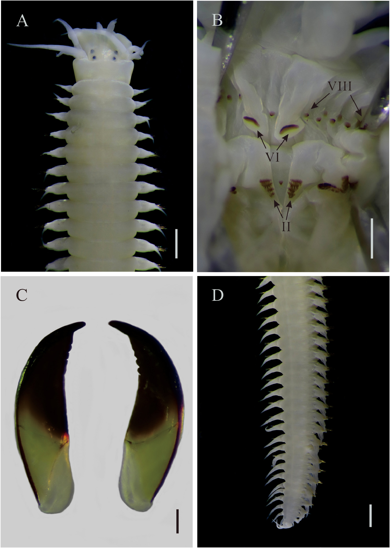
Material examined. Holotype ( NMNS 8383-25 View Materials ), atoke, Jihuei (23°6.97'N 121°24.31'E), Taitung County, Taiwan, rocky intertidal habitats, 27 March 2014. GoogleMaps
Description. Holotype, complete, body length 49.5 mm with 95 chaetigers, chaetiger 10 width 1.5 mm, excluding parapodia; beige in alcohol ( Fig. 5A View FIGURE 5 ). Prostomium wider than long, lateral antennae antero-lateral, longer than palps, palpophores globose, palpostyles conical. Four pairs of tentacular cirri, longest one reaching chaetiger 3. Two pairs of eyes, in trapezoidal arrangement, subequal in size. Apodous anterior segment about 1.3 times longer than chaetiger 1. Pharynx with dark brown jaws, each with 3 lateral teeth; paragnath pattern: I=1; II=35 (left), 33 (right), in 5 comb-like rows; III=58, in 4 transverse comb-like rows; IV=71 (left), 67 (right), in 5 comb-like rows with addi- tional cones and p-bars in sigmoid towards the jaws; V=1; VI=1 (left), 1 (right), crescent-shaped bars; VII–VIII=19, p-bars and cones interspaced, p-bar row slightly behind cone row, one paragnath on each furrow and ridge regions ( Fig. 5B–C View FIGURE 5 ). Ridge pattern of areas VI–V–VI, λ-shaped ( Fig. 5B View FIGURE 5 ).
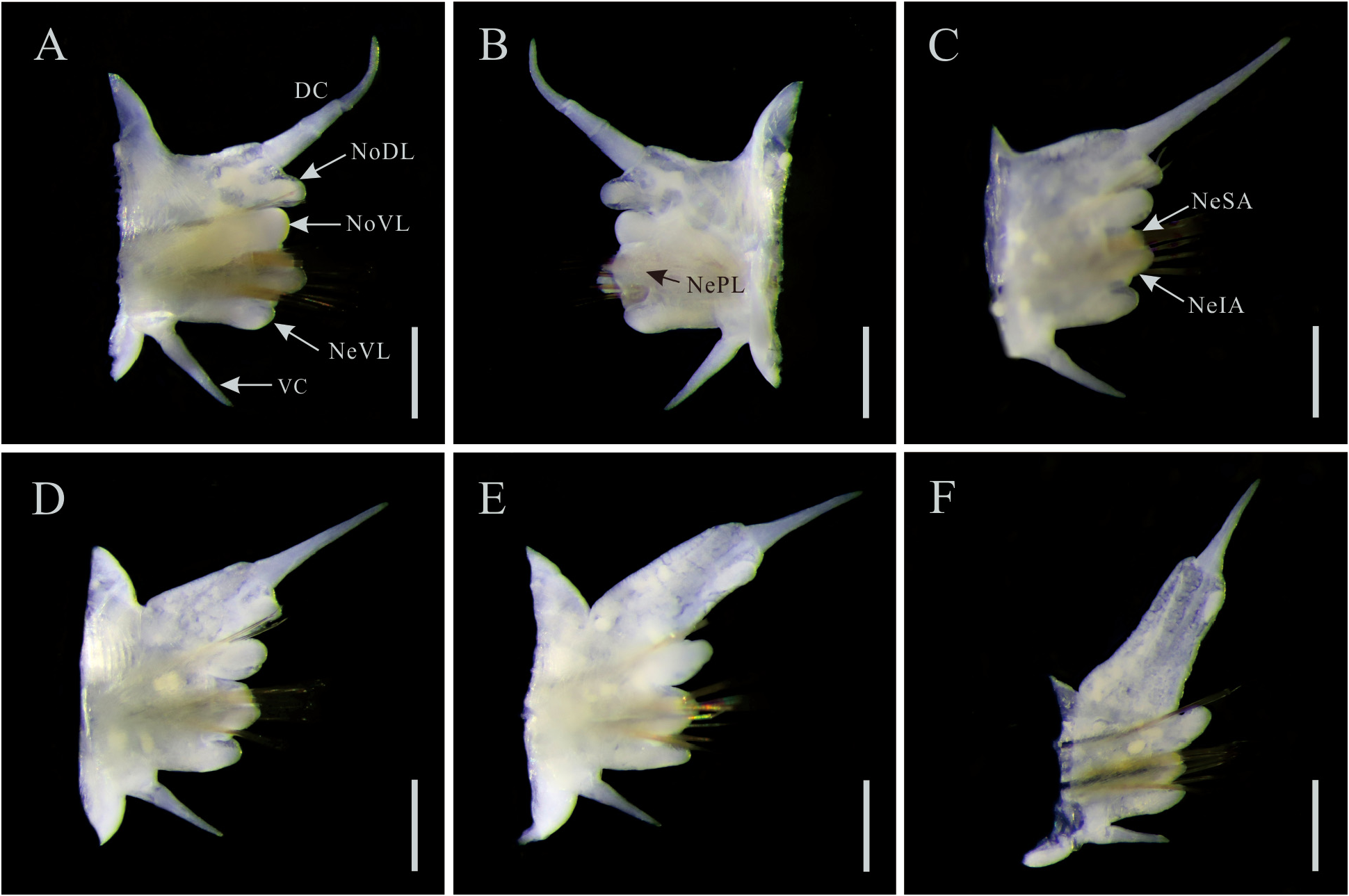
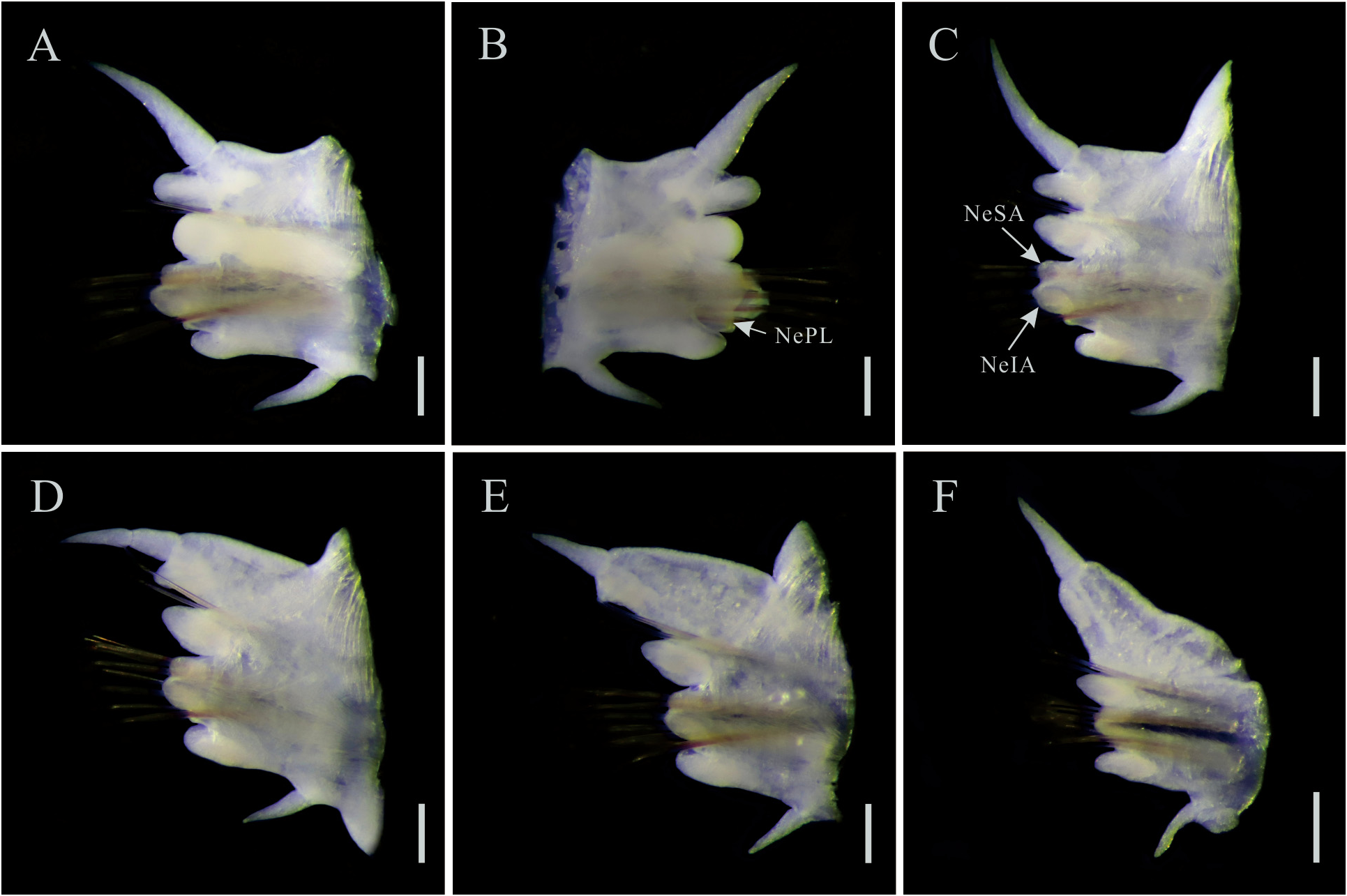
Dorsal cirri digitiform, basally attached to dorsal ligule on anterior chaetigers, about as long as dorsal ligule ( Fig. 6A–B View FIGURE 6 ), subdistally attached to dorsal ligule on mid-body to posterior chaetigers, about 0.8 times as long as dorsal ligule on mid-body chaetigers ( Fig. 6C–D View FIGURE 6 ), about 0.4–0.5 times as long as dorsal ligule on posterior chaetigers ( Fig. 6E View FIGURE 6 ), distally attached to dorsal ligule on last 13 chaetigers, about 0.5 times as long as dorsal ligule ( Fig. 6F View FIGURE 6 ).
Dorsal ligules subconical on anterior chaetigers ( Fig. 6A–B View FIGURE 6 ), becoming small pennant-like lobe on middle of mid-body to anterior half of posterior chaetigers ( Fig. 6C–E View FIGURE 6 ); base of dorsal ligule slightly elongate and broader on mid-body chaetigers ( Fig. 6D View FIGURE 6 ), markedly elongate and broader on posterior chaetigers ( Fig. 6E–F View FIGURE 6 ). Notopodial prechaetal lobe absent.
Median ligule conical on anterior-most chaetigers, shorter than neuroacicular ligule ( Fig. 6A–B View FIGURE 6 ), subconical thereafter and longer than neuroacicular ligule ( Fig. 6C–F View FIGURE 6 ).
Neuroacicular ligule with predominant inferior lobe on anterior to anterior half of posterior chaetigers ( Fig. 6A–E View FIGURE 6 ), inferior and superior lobes subequal in length on last 13 chaetigers ( Fig. 6F View FIGURE 6 ), about 1.3 times longer than ventral ligule on anterior to mid-body chaetigers, about as long as ventral ligule on posterior chaetigers ( Fig. 6A–F View FIGURE 6 ). Neuropodial postchaetal lobe present throughout. Ventral ligule subconical throughout. Ventral cirri mid-ventrally attached to ventral edge of parapodia, about 0.8 times as long as ventral ligule on anterior chaetigers, about 0.7 times as long as ventral ligule on mid-body chaetigers, about 0.5 times as long as ventral ligule on posterior chaetigers ( Fig. 6A–F View FIGURE 6 ).
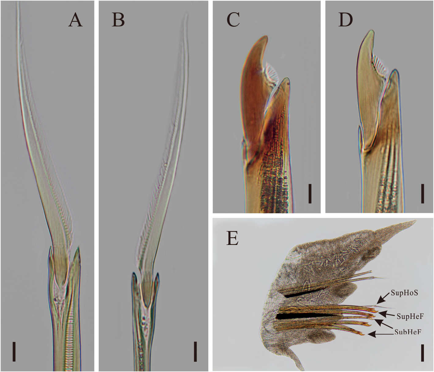
Notochaetae present from chaetiger 3 to posterior chaetigers, homogomph spinigers present throughout ( Fig. 7A View FIGURE 7 ). Supra-acicular fascicle of neuropodia: homogomph spinigers and short-bladed heterogomph falcigers with serrations present throughout ( Fig. 7B–C View FIGURE 7 ). Subacicular fascicle of neuropodia: short-bladed heterogomph falcigers with serrations present throughout ( Fig. 7D View FIGURE 7 ), heterogomph spinigers absent throughout ( Fig. 7E View FIGURE 7 ).
Pygidium crenulated; anal cirri cirriform, as long as last 2 chaetigers.
Etymology. The name is derived from “Kihaw”, the local Amis aboriginal language for the location of the sampling site.
Type locality. Jihuei , Taitung County, Taiwan .
Distribution. Known only from type locality.
Remarks. Of all known Pseudonereis , only P. formosa , P. jihueiensis n. sp., P. kihawensis n. sp., and P. podocirra are noted without heterogomph spinigers in the subacicular fascicle of the neuropodia ( Fig. 7E View FIGURE 7 , Table 1). However, P. kihawensis n. sp. can be readily distinguished from the other three congeners by having crescentshaped bars in Area VI of the pharynx ( Fig. 5B View FIGURE 5 , Table 1). Pseudonereis kihawensis n. sp. has additional features that are not seen in P. formosa , which are having: 1) smaller ratio of dorsal cirrus to dorsal ligule on anterior chaetigers (about 1.0 versus 2.0); 2) tip of dorsal ligule becoming small pennant-like lobe on mid-body to the anterior half of posterior chaetigers (versus rounded); 3) greater length to width ratio of dorsal ligule on chaetigers 30, 50, and 70 (2.1, 1.8, 2.3 versus 1.3, 1.4 and 1.4, respectively); and 4) greater number of chaetigers with dorsal cirri distally attached to notopodial dorsal ligules (last 13 chaetigers versus posterior-most chaetigers only) ( Bakken 2007: 158, fig. 7A–E; Conde-Vela 2018: 270; Fig. 6A–F View FIGURE 6 , Table 1). Also, P. kihawensis n. sp. can be further differentiated from P. jihueiensis n. sp. by having: 1) three lateral teeth on cutting edge of the jaws (versus none); 2) λ-shaped ridge pattern of areas VI–V–VI (versus χ-shaped); 3) smaller ratio of dorsal cirrus to dorsal ligule on anterior and mid-body chaetigers (1.0 and 0.8 versus 1.3 and 1.0, respectively); 4) greater length to width ratio of dorsal ligule on chaetigers 30, 50, and 70 (2.1, 1.8, 2.3 versus 1.3, 1.4 and 1.4, respectively); and 5) shorter anal cirri (as long as last 2 chaetigers versus as long as 4–7 chaetigers) ( Figs 1D–E View FIGURE 1 , 2A–F View FIGURE 2 , 5B–D View FIGURE 5 , 6A–F View FIGURE 6 , Table 1). Lastly, P. kihawensis n. sp. can be further distinguished from P. podocirra by having: 1) fewer number and arrangement of paragnaths in Areas VII–VIII (19 in two rows with one paragnath on each furrow and ridge regions versus 40 in 2–4 rows with 2–3 paragnaths on each furrow and ridge regions in Area VIII); 3) smaller ratio of dorsal cirrus to notopodial dorsal ligule on chaetigers 30, 50 but a larger ratio of dorsal cirrus to notopodial dorsal ligule on chaetiger 70 (1.0, 0.8, 0.4 and 0.5 versus 1.7, 2.5, 0.8 and 0.4, respectively); 4) greater length to width ratio of dorsal ligule on chaetigers 30, 50, and 70 (2.1, 1.8, and 2.3 versus 0.9, 1.3 and 2.2, respectively); and 5) greater number of chaetigers with dorsal cirri distally attached to notopodial dorsal ligules (last 13 chaetigers versus the entire posterior chaetigers) ( Kara et al. 2018: 1287–1288, figs 3C–I, 4A–C; Figs 5B View FIGURE 5 , 6A–F View FIGURE 6 , Table 1).
Of the 20 known species of the genus, only P. ferox (Hansen, 1882) and P. variegata (Grube & Kröyer in Grube, 1858) have crescent-shaped paragnaths in Area VI of the pharynx as in P. kihawensis n. sp. ( Conde-Vela 2018: 268–269, fig. 10D; Fig, 5B, Table 1). However, P. kihawensis n. sp. can be distinguished from P. ferox by having: 1) more rows of comb-like paragnaths in Areas II & IV (5 and 5 versus 3 and 4, respectively); 2) λ-shaped ridge pattern of areas VI–V–VI (versus ɔc-shaped); 3) dorsal ligule longer than median ligule on anterior chaetigers (versus subequal); 4) dorsal ligule becoming small pennant-like lobe on mid-body to the anterior half of posterior chaetigers (versus rounded); 5) dorsal ligule broaden basally on posterior-most chaetigers (versus broaden in the middle); 6) greater number of dorsal cirrus distally attached to dorsal ligule (last 13 chaetigers versus posterior-most chaetigers); and 7) no heterogomph spinigers in the subacicular fascicle of the neuropodia (versus present from about chaetiger 40) ( Bakken 2007: 116–170, figs 14A, 15A–F; Conde-Vela 2018: 269; Figs 5B View FIGURE 5 , 6A–F View FIGURE 6 , Table 1). Pseudonereis kihawensis n. sp. differs from by P. variegata by having: 1) fewer number of paragnaths and a single band arrangement of paragnaths in Area VII–VIII (versus 43 and 2 bands, respectively); 2) λ-shaped ridge pattern of areas VI–V–VI (versus ɔc-shaped); 3) dorsal cirrus as long as dorsal ligule on anterior chaetigers (versus longer than dorsal ligule); 4) dorsal ligule longer than median ligule on anterior chaetigers (versus subequal); 5) dorsal cirrus distally attached to dorsal ligule from last 13 chaetigers (versus posterior-most chaetigers); and 6) no heterogomph spinigers in the subacicular fascicle of the neuropodia (versus present) ( Conde-Vela 2018: 267–269, fig. 10C–I; Figs 5B View FIGURE 5 , 6A–F View FIGURE 6 , Table 1).

No known copyright restrictions apply. See Agosti, D., Egloff, W., 2009. Taxonomic information exchange and copyright: the Plazi approach. BMC Research Notes 2009, 2:53 for further explanation.
|
Kingdom |
|
|
Phylum |
|
|
Class |
|
|
Order |
|
|
Family |
|
|
Genus |