
Psyllaphorura jiangsuensis, Yan & Huang & Chen, 2007
|
publication ID |
https://doi.org/ 10.11646/zootaxa.1510.1.5 |
|
DOI |
https://doi.org/10.5281/zenodo.5087935 |
|
persistent identifier |
https://treatment.plazi.org/id/03AD87F2-FFE5-934A-5CD7-FD1DE85CFDFF |
|
treatment provided by |
Felipe |
|
scientific name |
Psyllaphorura jiangsuensis |
| status |
sp. nov. |
Psyllaphorura jiangsuensis sp. nov.
Figs 1–12 View FIGURES 1–6 View FIGURES 7–12 , Tab. 1
Type material. Holotype: female, China, Jiangsu Province, Nanjing: Qixiashan Park, under leaf litter, stones and bricks in deciduous forest; 14-X-1994; collection number C8420, coll. Jian-xiu Chen; paratypes: 3 females, same data as holotype. 1 female, same locality as holotype, 25-V-2005, C9251, coll. Hai-juan Yan & Jun-li Jia; in the collection of the Laboratory of Zoology , Nanjing University, China.
Diagnosis. Antennal III sense organ with 4 guard papillae. Postantennal organ composed of 20–22 simple or bilobed vesicles. Furca reduced to 2 mamelons, each with 3 distal setae. Pseudocellar formula dorsally as 20/000/00013, pseudocelli absent on ventral side and subcoxae. Cephalic chaetotaxy without seta d 0. Unguiculus basal lamella absent. Ventral tube with 5+5 setae. Anal spines set on very distinct papillae.
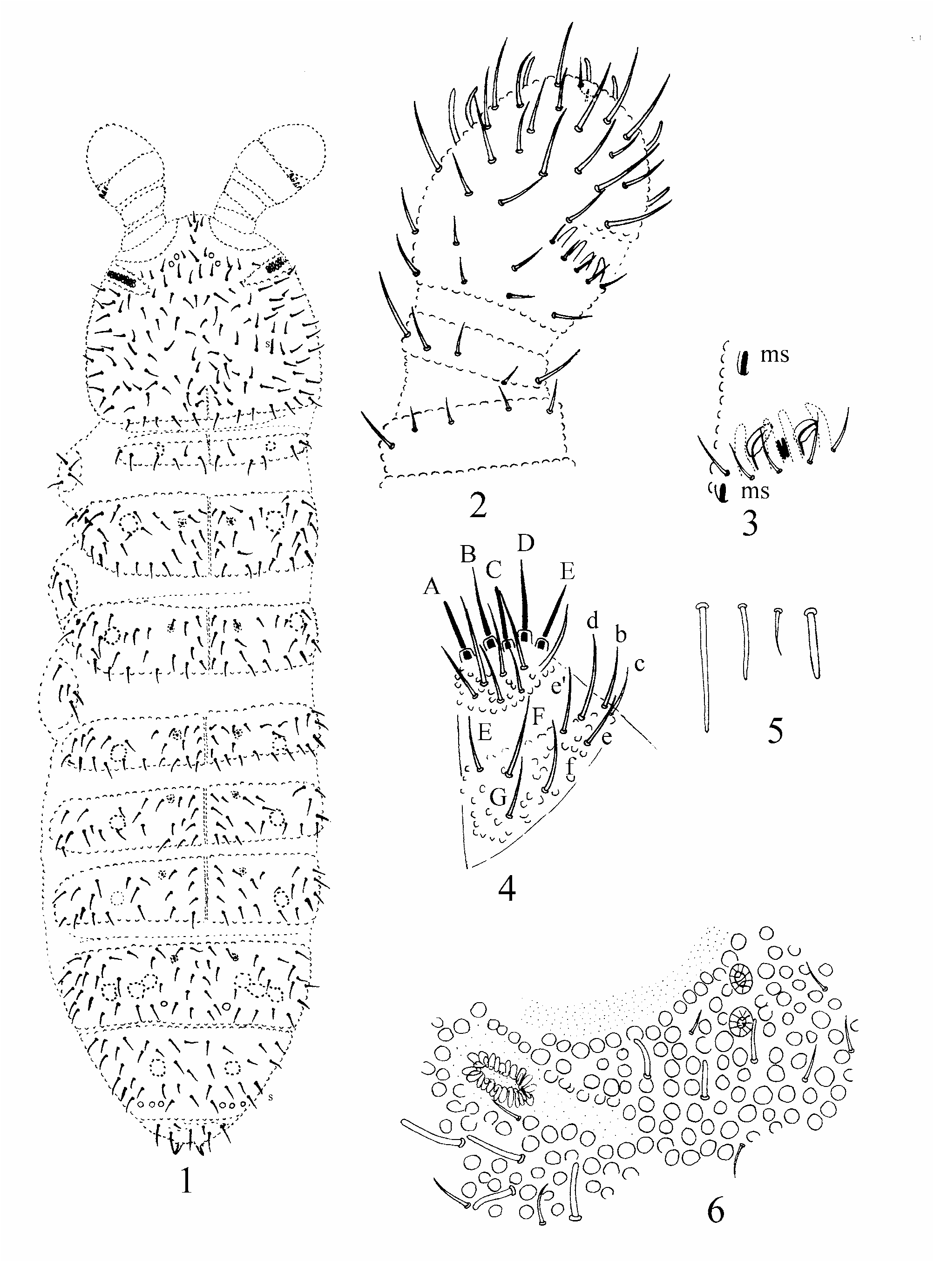
Description. Maximum body length: females 1.52 mm. Habitus typical for the genus: oval, plump, dorsoventrally flattened with very short abdominal segment VI ( Fig. 1 View FIGURES 1–6 ). Body color milky white. Integument coarse. Granules up to 5 µm in diameter dorsally on body, 3 µm on antennae and much smaller ventrally on body and antennae. Antennal base well marked with very fine granules. Body segments distinctly defined. Thoracic terga I–III and abdominal terga I–III with two rows of smaller granules along medial line.
Pseudocelli mostly with margin poorly demarcated and hardly distinguished, dorsally arranged as 20/000/ 00013, absent on ventral side and subcoxae. Parapseudocelli invisible. Thoracic terga II–III and abdominal terga I–IV with1+1 pseudopores ( Fig. 1 View FIGURES 1–6 ).
Body setae: macrosetae and mesosetae thick and apically rounded, microsetae short and apically pointed. Sensory setae (s) thick and short ( Fig. 5 View FIGURES 1–6 ), microsensillum (ms) tiny. S formula dorsally as 1/011/11111 ( Fig. 1 View FIGURES 1–6 ).
Head with seta d 0 absent. 2+2 anterior cephalic pseudocelli, located outside area of fine granulation at antennal base ( Figs. 1 & 6 View FIGURES 1–6 ). Posterior pseudocelli absent. Postantennal organ composed of 20–22 simple or bilobed vesicles perpendicular to the long axis of the organ. Antennae cylindrical, 0.6–0.7 times as long as head. Antennal segments III and IV fused into one club. Length ratio of antennal segments I–IV as 1: 1.0–1.2: 1.4–2.0: 2.1–3.6. Antennal segment IV with 6–7 distinct short and thick sensory setae, 1 small subapical peg in shallow pit, and 1 baso-lateral microsensillum. Ant III organ with 4 papillae, 5 guard setae, 2 small rods and 2 sensory clubs; club smooth, curved, each with one rib. Lateral microsensillum on antennal segment III slightly behind sense organ ( Fig. 3 View FIGURES 1–6 ). Other antennal setae all acuminate. Labium of AC type ( Fjellberg, 1999), labial proximal setae 6. Basomedian field with 4 setae (E, F, G, and f), basolateral–with 5 (b, c, d, e, e’) (D’ Haese, 2003) ( Fig. 4 View FIGURES 1–6 ). Postlabial setae 3+3 present along ventral groove.
Dorsal thoracic chaetotaxy as in Fig. 1 View FIGURES 1–6 . Thoracic terga II and III each with 1+1 lateral microsensillum. Tibiotarsal chaetotaxy symmetrical, with 11 setae in whorl 1 (distal whorl), 7 & 1 respectively in whorl 2 and 3 ( Fig. 8 View FIGURES 7–12 ). Unguis without teeth. Unguiculus narrow and pointed, without basal inner lamella, apical filament 0.8–0.9 as long as inner edge of unguis. All setae on legs acuminate.
Abdominal terga I–IV without axial setae. Abdominal tergum V with m 0 and p 0 setae. Abdominal tergum VI with one unpaired seta m 0. Anal spines short, about 0.3–0.4 times length of inner edge of hind unguis, set on very distinct papillae ( Fig. 1 View FIGURES 1–6 ). Ventral tube with 5+5 distal setae, without setae at base ( Fig. 10 View FIGURES 7–12 ). Furcula as 2 mamelons, each bearing 3 setae ( Fig. 11 View FIGURES 7–12 ). Female genital plate with 26 setae (only seen in one paratype). Anal valves with numerous acuminate setae, each lateral valve with a 0, 2 a 1 setae, upper valve with a 0, 2 a 1, 2 a 2, 2 b 1, 2b 2, c 0, and 2 c 1 setae ( Fig. 12 View FIGURES 7–12 ).
Etymology. The new species is named after the type locality: Jiangsu Province.
Discussion. Habitus, coarse granulation, absence of posterior cephalic pseudocelli and the structure of furcal remnant show that the new species belongs to the genus Psyllaphorura . Within the genus, the new species distinctly resembles the South Siberian P. martynovae in the pseudocellar formula and the number of papillae on antennal III sense organ. However, it differs from the latter by the characters shown in table 1.

No known copyright restrictions apply. See Agosti, D., Egloff, W., 2009. Taxonomic information exchange and copyright: the Plazi approach. BMC Research Notes 2009, 2:53 for further explanation.
|
Kingdom |
|
|
Phylum |
|
|
Class |
|
|
Order |
|
|
Family |
|
|
Genus |