
Rhyacophila zwickorum Malicky 1972
|
publication ID |
https://doi.org/ 10.11646/zootaxa.3669.1.5 |
|
publication LSID |
lsid:zoobank.org:pub:11FE7DFD-788C-44B7-9392-4D3ED1BE4ED7 |
|
DOI |
https://doi.org/10.5281/zenodo.6151349 |
|
persistent identifier |
https://treatment.plazi.org/id/C75A8795-FF91-FFEB-FF74-FC5AD1AB36F6 |
|
treatment provided by |
Plazi |
|
scientific name |
Rhyacophila zwickorum Malicky 1972 |
| status |
|
Rhyacophila zwickorum Malicky 1972 View in CoL
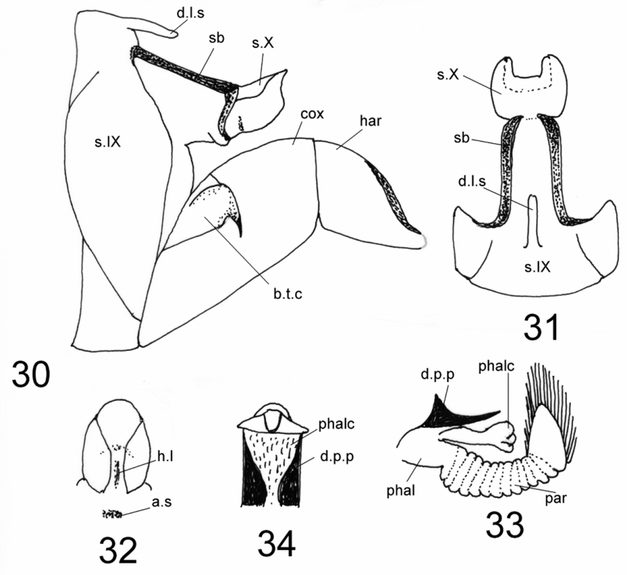
( Figures 30–39 View FIGURES 30 – 34 View FIGURES 35 – 39 )
Material examined. Turkey, Rize, Çamlıhemşin, Holovit, 22.viii.1983, 4 males, 1 female (CD: A-41); Rize, Ayder, 1100 m, 5.ix.1988, 2 males (CD: A-86); same place, 1.viii.1989, 1400-1600 m, 2 males (CD: A-108); same place except Aşaġı Kavron, 2000 m, 8.viii.1989, 3 males (CD: A-110); Rize, Çamlıhemşin, Zilkale, Tozkapan, 850 m, 20.viii.1992, (light), 3 males (CD: A-294); all leg. and coll. Sipahiler.
Rhyacophila zwickorum Malicky 1972 was described based on material collected from Ardeşen (Rize); the redescription given below is also based on material collected from Rize Province.
General description. Antennae, maxillary palps, legs, and wings in alcohol brown. Length of each anterior wing of male 7.5– 8 mm (n=5).
Male genitalia ( Figures 30–34 View FIGURES 30 – 34 ): Segment IX with apicodorsal lobe ( Figs. 30, 31 View FIGURES 30 – 34 ). Sclerotized bands articulating on segment X rather long ( Figs. 30, 31 View FIGURES 30 – 34 ); in lateral view each with dorsal apex pointed and posterior edge rounded ( Fig. 31 View FIGURES 30 – 34 ); in caudal view segment X with posterolateral edges convex laterally and mesally, with narrow, vertical sclerotized tubercle in middle ( Fig. 32 View FIGURES 30 – 34 ). Coxopodite of each inferior appendage having apical edge of sclerotized basal tendon with hook-shaped projection on ventral corner; harpago somewhat dilated on posterodorsal edge, tip blunt ( Fig. 30 View FIGURES 30 – 34 ). In lateral view, dorsolateral processes of phallotheca long, almost reaching apex of phallicata, each with basal 1/3rd three-four times as tall as distal 2/3rds, triangular, acute at dorsal tip, distal 2/3rds tapered to sharp point ( Fig. 33 View FIGURES 30 – 34 ); in dorsal view, these processes very broad at their bases, tapering to acute apices ( Fig. 34 View FIGURES 30 – 34 ); large dorsomesal band of phallicata covered with small tubercles ( Fig. 34 View FIGURES 30 – 34 ); apical portion of phallicata with narrow sclerotized band ( Fig. 34 View FIGURES 30 – 34 ); paramere slender, dilated at oval apex, its crown densely fringed with about 34 long, plumose spines ( Fig. 33 View FIGURES 30 – 34 ).
Female genitalia ( Figs. 35–39 View FIGURES 35 – 39 ): In lateral view, sclerotized part of segment VIII rather short, about 1.1 times as long ventrally as tall anteriorly, and posterolateral edges each irregularly crenulate and slightly concave above mesal posteroventral lobe ( Fig. 35 View FIGURES 35 – 39 ); in dorsal view, fully sclerotized and posterior edge convex ( Fig. 36 View FIGURES 35 – 39 ); in ventral view, median lobe of posterior edge ~4 times as broad as long, evenly rounded ( Fig. 37 View FIGURES 35 – 39 ). Anterior process of bursa copulatrix oval and its posteroventral edge smooth in lateral view ( Fig. 38 View FIGURES 35 – 39 ); posterior process broad subdistally and becoming very narrow towards tip in lateral and ventral views ( Figs. 38, 39 View FIGURES 35 – 39 ).

No known copyright restrictions apply. See Agosti, D., Egloff, W., 2009. Taxonomic information exchange and copyright: the Plazi approach. BMC Research Notes 2009, 2:53 for further explanation.