
Ribautia lewisi, Pereira, Luis Alberto, 2013
|
publication ID |
https://doi.org/ 10.11646/zootaxa.3630.2.2 |
|
publication LSID |
lsid:zoobank.org:pub:5CDCBF9E-9A49-4C92-94FD-DD9584933E48 |
|
DOI |
https://doi.org/10.5281/zenodo.5695493 |
|
persistent identifier |
https://treatment.plazi.org/id/03DE8797-1172-FFED-3EB8-F9E5A447FE47 |
|
treatment provided by |
Plazi |
|
scientific name |
Ribautia lewisi |
| status |
sp. nov. |
Ribautia lewisi sp. nov.
( Figs. 1–61 View FIGURES 1 – 10 View FIGURES 11 – 14 View FIGURES 15 – 19 View FIGURES 20 – 26 View FIGURES 27 – 39 View FIGURES 40 – 47 View FIGURES 48 – 50 View FIGURES 51 – 56 View FIGURES 57 – 59 View FIGURES 60 – 61 )
Diagnosis. A Neotropical species of Ribautia characterized by the presence of one cluster of coxal organs in each coxopleuron of the ultimate leg-bearing segment and a claw-like pretarsus in the ultimate legs. Among the Neotropical taxa currently included in the genus, only the present new species, R. combinata Pereira, Uliana & Minelli, 2006 (from Peru), and R. jakulicai Pereira, 2007 (from Argentina) share these two combined features. R. lewisi sp. nov. differs from the two latter by having the following unique traits: female with 45, 47 leg-bearing segments, male with (43?), 45, 47 leg-bearing segments; lateral margins of cephalic plate with a small concavity located anteriorly ( Figs. 11, 12 View FIGURES 11 – 14 : a); internal limbs of tentorium bearing an unusual conspicuous tooth-shaped sclerotized process directed inwards; first and second article of telopodites of second maxillae with a very small distoectal process ( Figs. 14 View FIGURES 11 – 14 , 15 View FIGURES 15 – 19 : b); pore-fields absent on some sternites of mid-body; sternite of female ultimate leg-bearing segment provided with a narrow band of numerous very small setae near the posterior edge ( Fig. 61 View FIGURES 60 – 61 ).
Other morphological traits included in Table 1 differentiate R. lewisi sp. nov. from R. combinata and R. jakulicai .
Remarks. For characters differentiating R. lewisi sp. nov. from other Neotropical species of Ribautia , see Discussion below.
Type material examined. ARGENTINA: Entre Ríos Province: Concordia Department: Concordia, 9 December 2007, L. A. Pereira legit: holotype 3, 45 l.-b.s., b.l. 20 mm; paratype A (Ƥ), 45 l. -b.s., b.l. 15 mm; paratype B (Ƥ), 45 l. -b.s., b.l. 16 mm; paratype C (Ƥ), 45 l. -b.s., b.l. 16 mm; paratype D (Ƥ), 45 l. -b.s., b.l. 16 mm; paratype E (Ƥ), 45 l. -b.s., b.l. 16 mm; paratype F (Ƥ), 45 l. -b.s., b.l. 17 mm; paratype G (Ƥ), 45 l. -b.s., b.l. 17.5 mm; paratype H (Ƥ), 45 l. -b.s., b.l. 17.5 mm; paratype I (Ƥ), 47 l. -b.s., b.l. 17 mm; paratype J (Ƥ), 47 l. -b.s., b.l. 19 mm; paratype K (Ƥ), 47 l. -b.s., b.l. 20 mm; paratype L (Ƥ), 47 l. -b.s., b.l. 21 mm; paratype M (Ƥ), 47 l. -b.s., b.l. 26 mm; paratype N (3), 45 l. -b.s., b.l. 17 mm; paratype O (3), 47 l. -b.s., b.l. 16 mm; paratype P (3), 47 l. -b.s., b.l. 17 mm.
Depository of types: MLP.
Other material examined. All specimens from the same locality, date and collector as the type series: 2 ƤƤ juv., 45 l. -b.s., b.l. 12 mm (with 9-10 coxal organs in each cluster), and 13 mm (with 11-12 coxal organs in each cluster); 1 3 subadult, 45 l. -b.s., b.l. 14 mm (with 9 coxal organs in each cluster); 2 3 juv., 45 l. -b.s., b.l. 10 mm (in moulting process), and 12 mm (with 9-10 coxal organs in each cluster); 1 3 juv., 47 l. -b.s., b.l. 11 mm (with 6 coxal organs in each cluster); 4 juv. (sex unknown), 43 l. -b.s., b.l. 4.5 mm (Specimen A) (with 1+1 coxal organs only), 4.5 mm (with 1 + 1 coxal organs only), 10 mm (with 5 coxal organs in each cluster), and 10 mm (with 5–6 coxal organs in each cluster); 1 juv. (sex unknown), 47 l. -b.s., b.l. 4.5 mm (with 1 + 1 coxal organs only). (MLP).
Description. Male holotype. Forty-five leg-bearing segments, body length 19 mm, maximum body width 0.65 mm, length of cephalic plate 0.67 mm, width of forcipular coxosternite 0.56 mm. Colour (of preserved specimen in alcohol) pale yellow, forcipular segment a little darker (pale ochreous).
Antennae. Relatively long, ca. 4.0 times as long as the cephalic plate, distally attenuate, ratio of width of a.a. I/ width of a.a. XIV ca. 1.68: 1. A.a. I nearly as long as wide, remaining a.a. longer than wide. Ventral chaetotaxy: setae on a.a. I–VII of various lengths and relatively few in number; those of a.a. VIII–XIV progressively shorter and more numerous towards the tip of the appendage ( Figs. 1, 2 View FIGURES 1 – 10 ). Dorsal chaetotaxy: similar to the ventral side. A.a. XIV with ca. 4–5 claviform sensilla on the external border and ca. 2–3 on the internal border; distal end of this a.a. with ca. 5–6 very small hyaline specialized sensilla apparently not split apically. Ventral and dorsal surface of a.a. II, V, IX and XIII with very small specialized sensilla. On the ventral side these sensilla are restricted to an internal latero-apical area and are represented by two different types: a and b. Type a sensilla are very thin and not split apically ( Fig. 5 View FIGURES 1 – 10 : a); type b sensilla ( Fig. 5 View FIGURES 1 – 10 : b) are very similar to those on the apex of a.a. XIV. Specialized sensilla on dorsal side are restricted to an external latero-apical area and are represented by similar type a and b sensilla of ventral side ( Fig. 9 View FIGURES 1 – 10 : a, b). Number and distribution of specialized sensilla on ventral and dorsal sides of a.a. II, V, IX and XIII, as in Table 2 View TABLE 2 .
Cephalic plate. Distinctly longer than wide (length/width ratio ca. 1.64: 1). Lateral margins convergent towards the posterior region, showing anteriorly a small concavity ( Figs. 11, 12 View FIGURES 11 – 14 : a). Anterior margin slightly concave on the middle; posterior margin straight. Shape and chaetotaxy as in Figure 11 View FIGURES 11 – 14 .
Clypeus. With four setae located on the clypeal area; 1 + 1 anterior-lateral setae, posterior to the latter; and two setae in the middle ( Fig. 12 View FIGURES 11 – 14 ). Clypeal area with surface very densely areolated ( Fig. 13 View FIGURES 11 – 14 ).
Labrum. Mid-piece well developed, with ca. 7 sharp pointed teeth. Side pieces with ca. 9 + 10 filaments of different size. (Compare with Figure 54 View FIGURES 51 – 56 , illustrating the labrum of the female paratype M).
Tentorium. Internal limb bearing a conspicuous tooth-shaped sclerotized process directed inwards. (Compare with Figures 54–56 View FIGURES 51 – 56 , illustrating the tentoria of female paratype M).
Mandible. Pectinate lamella with ca. 22 hyaline teeth. (Compare with Figure 57 View FIGURES 57 – 59 , illustrating a mandible of female paratype M).
First maxillae. Coxosternite without lappets, telopodites with very small lappets. (Compare with Figure 58 View FIGURES 57 – 59 illustrating left side of first maxillae of female paratype M). Coxosternite devoid of setae; coxal projections subtriangular, round tipped and provided with 6 + 5 setae ( Fig. 14 View FIGURES 11 – 14 ). Article II of telopodites with 3 + 4 large setae on ventral side, and 2 + 2 small sensilla on the external edge ( Fig. 14 View FIGURES 11 – 14 ).
Second maxillae. Coxites medially joined through a narrow, hyaline and non-areolate membranous isthmus and provided with 6 + 7 setae near the internal margin and 2 + 2 small sensilla near the external margin ( Fig. 14 View FIGURES 11 – 14 ). Process of antero-internal corners of coxosternite well developed with shape and relative size as in Figures 14 View FIGURES 11 – 14 , 15 View FIGURES 15 – 19 : a. Telopodites with setae of uniform thickness; first and second article with a very small distoectal process ( Figs. 14 View FIGURES 11 – 14 , 15 View FIGURES 15 – 19 : b); apical claw of telopodite well developed, tip curved inward ( Figs. 14 View FIGURES 11 – 14 , 16 View FIGURES 15 – 19 ). Chaetotaxy of coxosternites and telopodites as in Figures 14–16 View FIGURES 11 – 14 View FIGURES 15 – 19 .
Forcipular segment. When closed, the telopodites project slightly beyond the anterior margin of the head. Forcipular tergite trapeziform; chaetotaxy represented by an irregular transverse row of ca. 7 setae of different lengths near the posterior margin and a few smaller setae dispersed on the remaining surface ( Fig. 11 View FIGURES 11 – 14 ). Coxosternite with incomplete chitin-lines ( Fig. 17 View FIGURES 15 – 19 ); middle part of anterior border bearing 1 + 1 small unpigmented denticles, each provided with an apical seta, aspect and relative size as in Figures 17, 18 View FIGURES 15 – 19 . Telopodites: medial edge of trochanteropraefemur apically with a conspicuous unpigmented round-tipped tooth; proximally near the vestigial suture between trochanter and praefemur with a rudimentary unpigmented round-pointed projection ( Figs. 17–19 View FIGURES 15 – 19 ). Femur and tibia without denticles. Tarsungulum basally with a well-developed and pigmented subtriangular denticle ( Figs. 17, 19 View FIGURES 15 – 19 , 20 View FIGURES 20 – 26 ); medial ventral edge of tarsungulum slightly serrate ( Figs. 17, 19 View FIGURES 15 – 19 , 20 View FIGURES 20 – 26 ). Relative size of poison glands as in Figures 17, 19 View FIGURES 15 – 19 , calyx of poison gland subtriangular ( Fig. 21 View FIGURES 20 – 26 ). Chaetotaxy of coxosternite and telopodites as in Figures 11 View FIGURES 11 – 14 , 17 View FIGURES 15 – 19 .
Sternites of leg-bearing segments 1 to penultimate. With a shallow median longitudinal sulcus along all the body length, areolation of its surface as in Figures 25 View FIGURES 20 – 26 , 27–30 View FIGURES 27 – 39 ). Pore-fields present on sternites 2–22, 26, 30, and 36–44 (penultimate); totally absent on sternites 1, 23–25, 27–29 and 31–35. Sternites 2–12 with undivided porefields, subcircular in shape ( Figs. 22–25 View FIGURES 20 – 26 , 33–39 View FIGURES 27 – 39 ); sternites 13 ( Fig. 26 View FIGURES 20 – 26 ), 14–16, 19–21, 37 ( Fig. 27 View FIGURES 27 – 39 ), 38, and 40–44 ( Figs. 28–32 View FIGURES 27 – 39 ) with pore-fields subdivided in two subsymmetrical areas; sternites 17, 18, 22, 26 with pores on the left side only; sternites 30, 36, 39 with pores on the right side only. Shape of pore-fields changes along the trunk as in Figures 22–39 View FIGURES 20 – 26 View FIGURES 27 – 39 . Number of pores as follows: sternite 2 (23); 3 (32); 4 (38); 5 (44); 6 (44); 7 (45); 8 (45); 9 (44); 10 (38); 11 (38); 12 (29); 13 (7 + 8); 14 (3 + 2); 15 (1 + 2); 16 (1 + 1); 17 (0 + 2); 18 (0 + 1); 19 (1 + 3); 20 (2 + 4); 21 (1 + 2); 22 (0 + 1); 26 (0 + 1); 30 (3 + 0); 36 (2 + 0); 37 (1 + 1); 38 (1 + 1); 39 (1 + 0); 40 (3 + 3); 41 (7 + 7); 42 (7 + 8); 43 (6 + 6); 44 (7 + 5).
Legs (pair 1 to penultimate). First pair shorter than the second (ratio ca. 0.85: 1). Legs with setae of uniform thickness, chaetotaxy similar throughout the entire body length. Distribution, number, and relative size of setae as in Figures 40-45 View FIGURES 40 – 47 . Claws with two thin and pale accessory spines ventrobasally, one anterior and one posterior, of similar size ( Fig. 46 View FIGURES 40 – 47 : a, b).
Ultimate leg-bearing segment. Intercalary pleurites absent at both sides of the ultimate pretergite ( Fig. 47 View FIGURES 40 – 47 ); ultimate presternite divided along the sagittal plane ( Fig. 48 View FIGURES 48 – 50 ). Length/width ratio of tergite, ca. 0.80: 1; length/ width ratio of sternite, ca. 0.74: 1. Shape and chaetotaxy of tergite and sternite as in Figures 47 View FIGURES 40 – 47 , 48 View FIGURES 48 – 50 . Coxopleura very slightly protruding at their distal-internal ventral ends, setae small and numerous on the distal-internal ventral area, the remaining coxopleural surface with few larger setae ( Figs. 47 View FIGURES 40 – 47 , 48 View FIGURES 48 – 50 ). Each coxopleuron with all coxal organs grouped in a cluster opening in the membrane between coxopleuron and sternite, partially or totally covered by the latter ( Figs. 48–50 View FIGURES 48 – 50 ). Each cluster with ca. 18 organs arranged as in Figures 49, 50 View FIGURES 48 – 50 . Ultimate legs inflated, composed of seven articles. Ratio of length of telopodites of ultimate legs/length of sternite ca. 3.52: 1. Shape and chaetotaxy of ultimate legs as in Figures 47 View FIGURES 40 – 47 , 48 View FIGURES 48 – 50 . Ultimate pretarsus unguiform, relatively smaller than those of the preceding legs, bearing a single internal spine ventrobasally.
Postpedal segments. Posterior margin of intermediate tergite convex ( Fig. 47 View FIGURES 40 – 47 ), posterior margin of intermediate sternite concave ( Fig. 48 View FIGURES 48 – 50 ). Posterior margin of first genital sternite very slightly concave at level of gonopods and in the middle ( Fig. 48 View FIGURES 48 – 50 ). Gonopods biarticulate, basal article with 5 setae, apical article with 6 setae ( Figs. 51, 52 View FIGURES 51 – 56 ); penis dorsally with 4 + 4 apical setae ( Fig. 53 View FIGURES 51 – 56 ). Anal organs absent.
Female (paratype K). Forty-seven leg-bearing segments, body length 20 mm, maximum body width 0.68 mm.
Antennae. With similar chaetoaxy than in the male and proportionally shorter (ca. 3.0 times as long as the cephalic plate).
Remaining features similar to those in the male except for the shape and pilosity of the ultimate leg-bearing segment and postpedal segments.
Ultimate leg-bearing segment. Tergite and sternite trapezoidal, length/width ratio of tergite, ca. 0.77: 1; length/ width ratio of sternite, ca. 0.72: 1. Shape and chaetotaxy of tergite and sternite as in Figures 60, 61 View FIGURES 60 – 61 . Coxopleura very slightly protruding at their distal-internal ventral ends, setae small and numerous on the distal-internal ventral area, the remaining coxopleural surface with few larger setae ( Figs. 60, 61 View FIGURES 60 – 61 ). Each cluster of coxal organs with ca. 21 organs ( Fig. 61 View FIGURES 60 – 61 ). Articles of ultimate legs not inflated, much thinner than those of the male ( Figs. 60, 61 View FIGURES 60 – 61 ). Ultimate legs proportionally a little longer than those of the male, with ratio of length of telopodites/length of sternite ca. 4.0: 1. Shape and chaetotaxy of ultimate legs as in Figures 60, 61 View FIGURES 60 – 61 .
Postpedal segments. Intermediate tergite with posterior margin strongly convex, ( Fig. 60 View FIGURES 60 – 61 ), intermediate sternite seemingly covered by the sternite of the ultimate leg-bearing segment ( Fig. 61 View FIGURES 60 – 61 ), posterior border of the first genital sternite slightly convex ( Fig. 61 View FIGURES 60 – 61 ). Gonopods uniarticulate, very poorly developed, vestigial ( Fig. 61 View FIGURES 60 – 61 : a).
Remarks. The adult condition of all type specimens is indicated by mature spermatozoa in the tubula seminifera of the males and spermatozoa in the spermathecae of the females.
In the preceding description, the labrum, mandible, and dorsal side of first maxillae were illustrated on the basis of the female paratype M, because those of the male holotype were not positioned in such a way on the microscope slides to allow the corresponding illustrations to be made properly.
Aspect of the unique 1 + 1 coxal organs present in a tiny juvenile specimen with sex unknown (Specimen A), as in Figure 59 View FIGURES 57 – 59 .
All specimens examined without anal organs.
Etymology. This species is respectfully dedicated to Dr. John G.E. Lewis (Taunton, Somerset, United Kingdom) as a personal recognition for all the generous help and expert advice that he has provided me during many years of research on Chilopoda Geophilomorpha .
Ecology. The specimens were collected in the soil (to a depth of about 10–40 cm) in a gallery forest environment adjacent to the west bank of the Uruguay River. (Floristic composition of this vegetation, extension along the water course, and significance on the geographical distribution of diverse groups of vertebrates are described in Arzamendia & Giraudo 2009, Costa 2003, Nores et al. 2005).
Type locality. ARGENTINA: Entre Ríos Province: Concordia Department: Concordia.
Known range. Only known from the type locality.
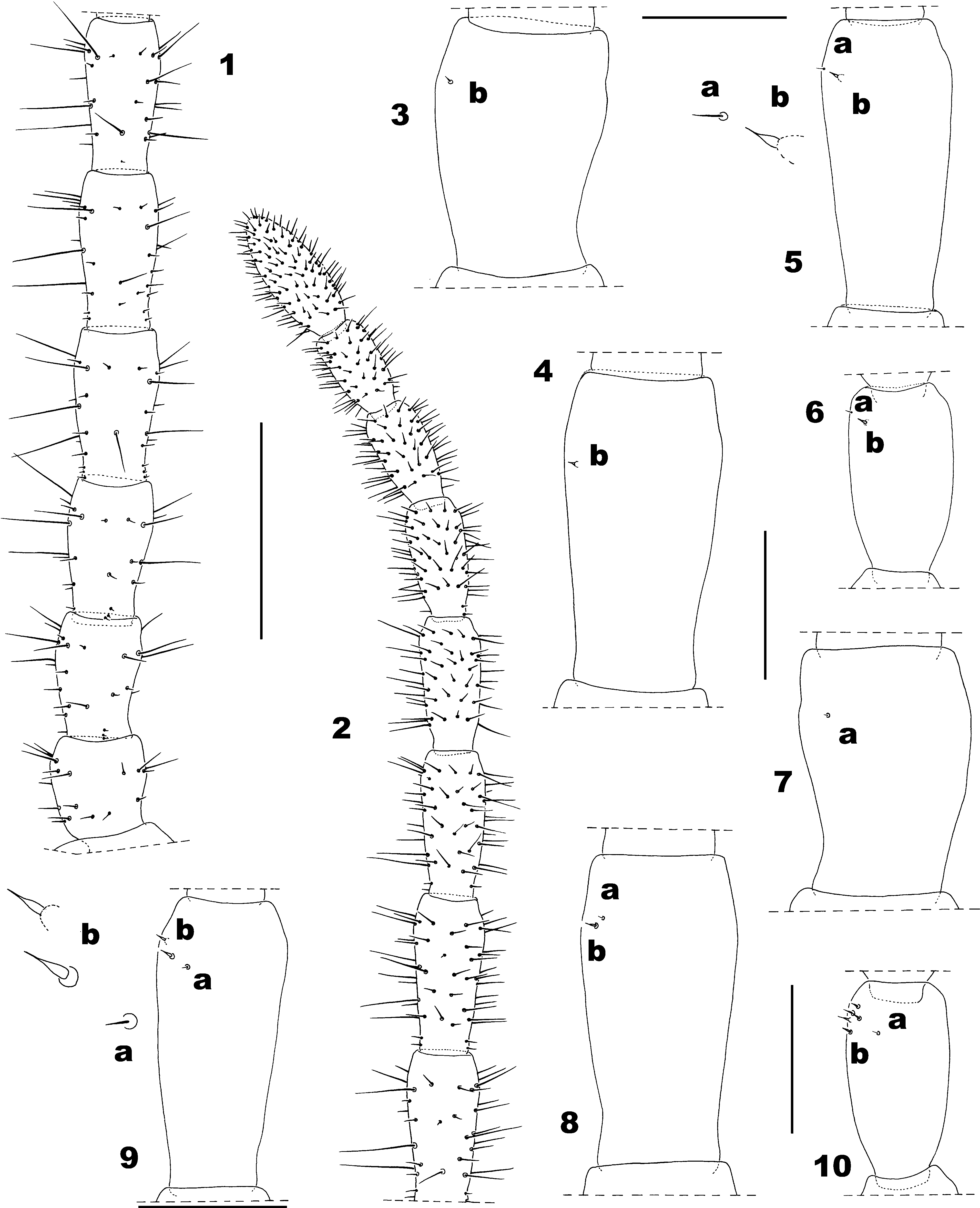
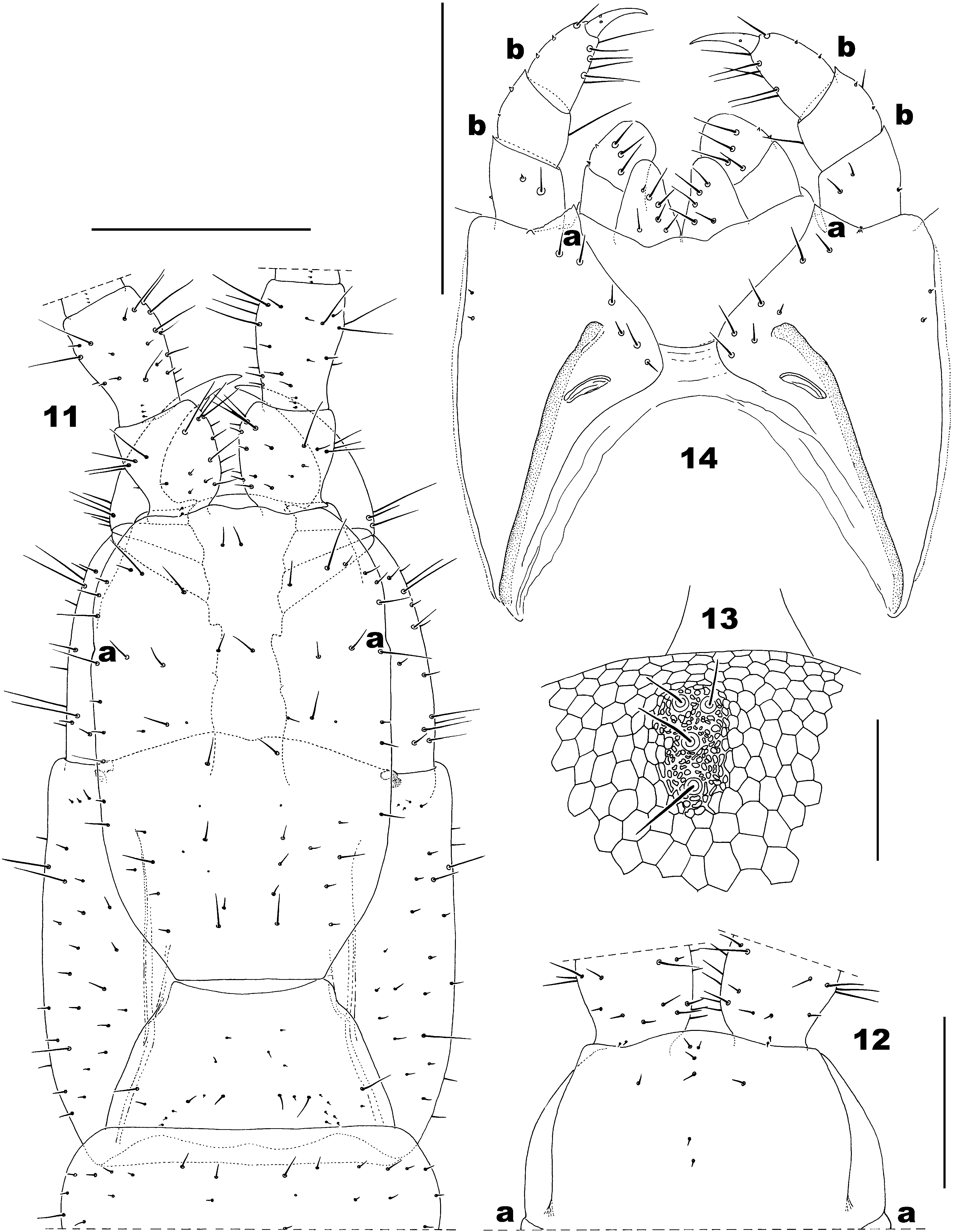

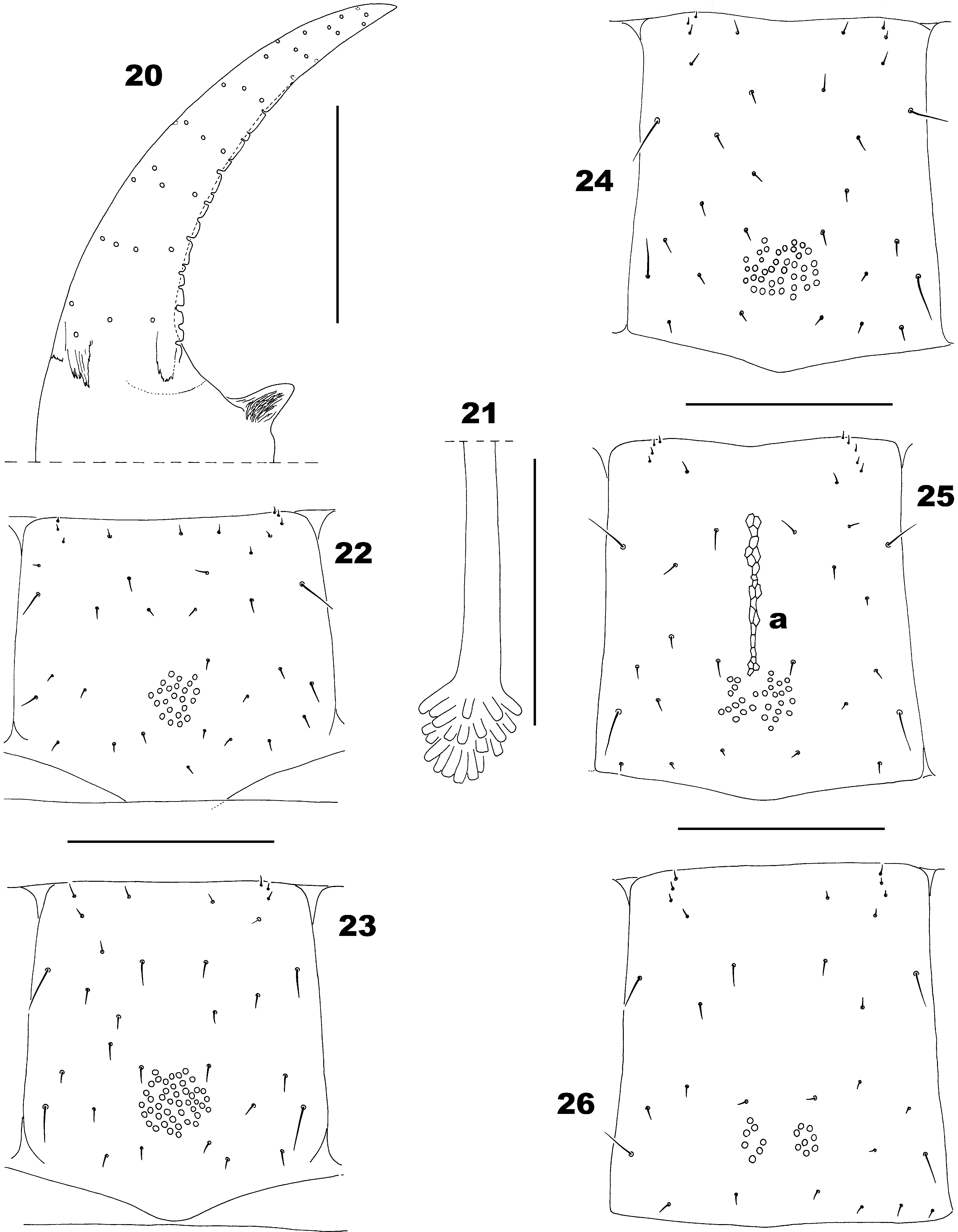
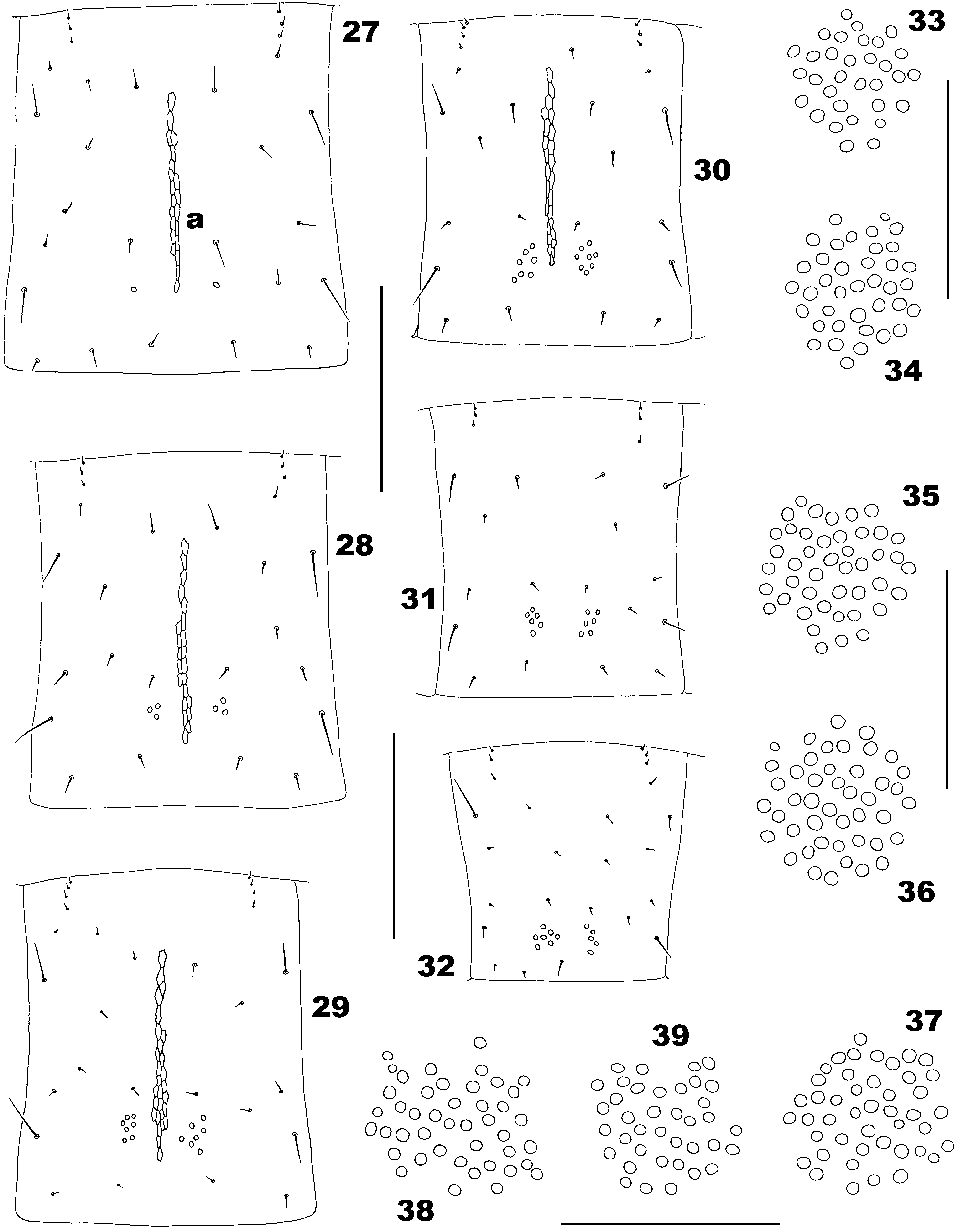
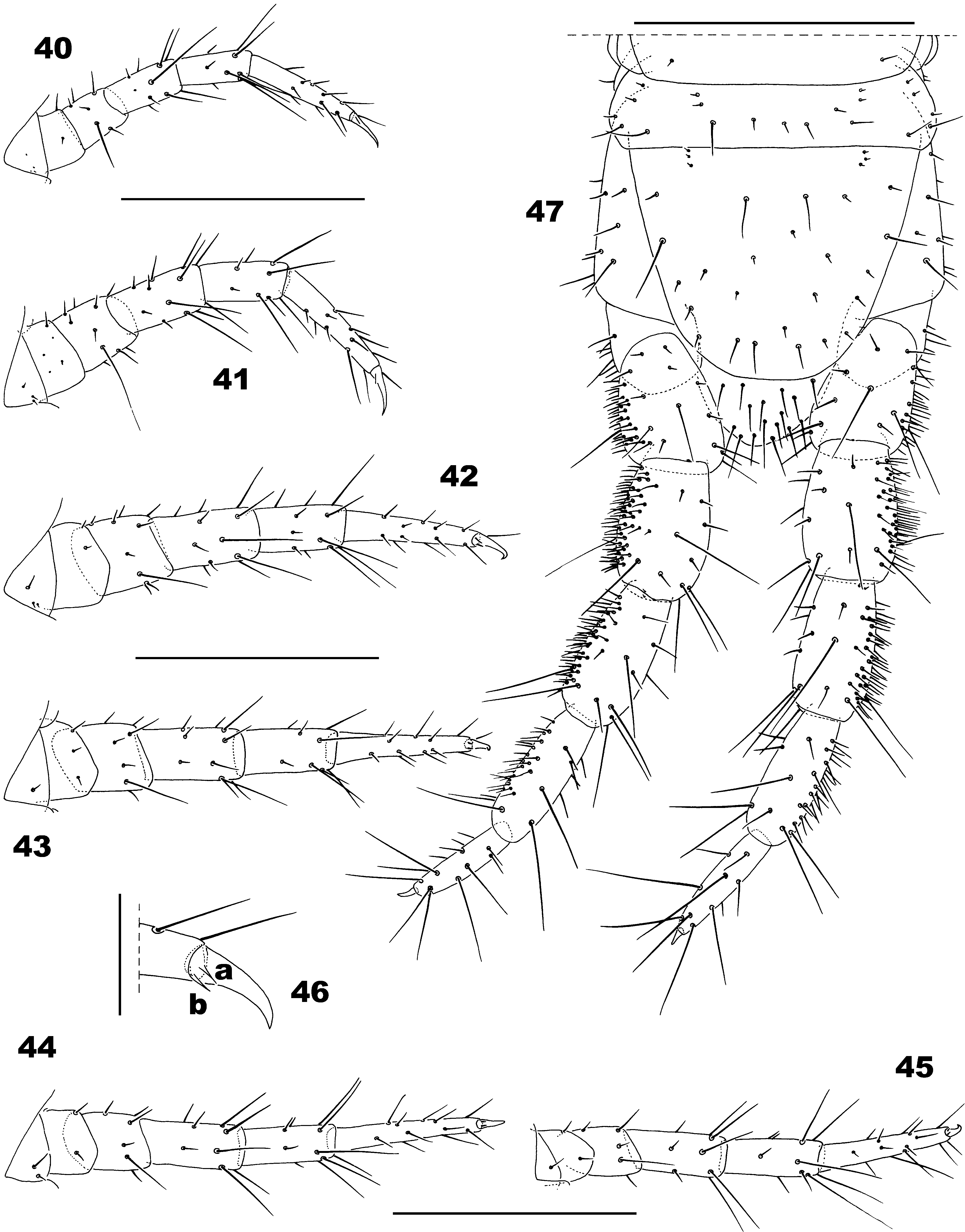
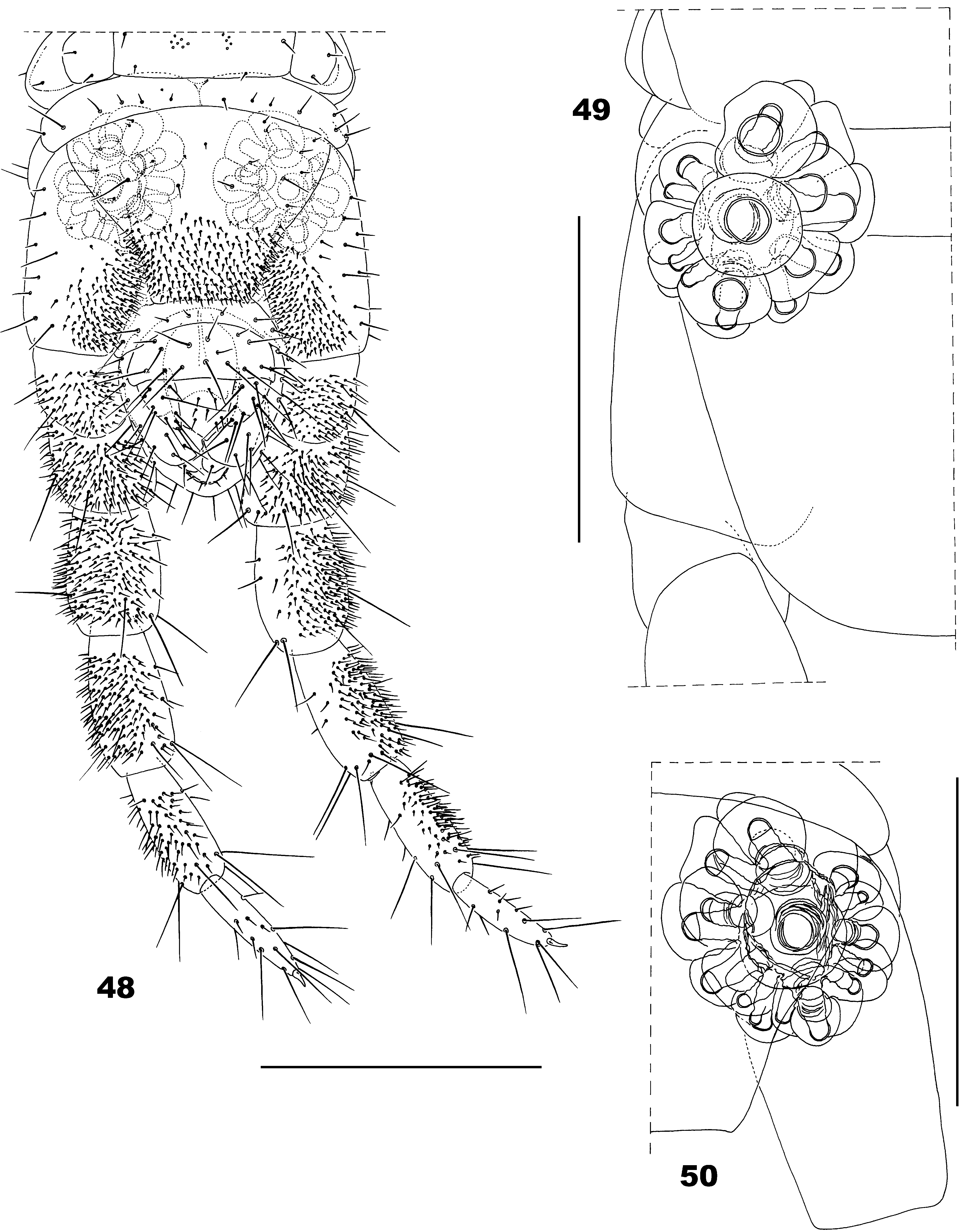
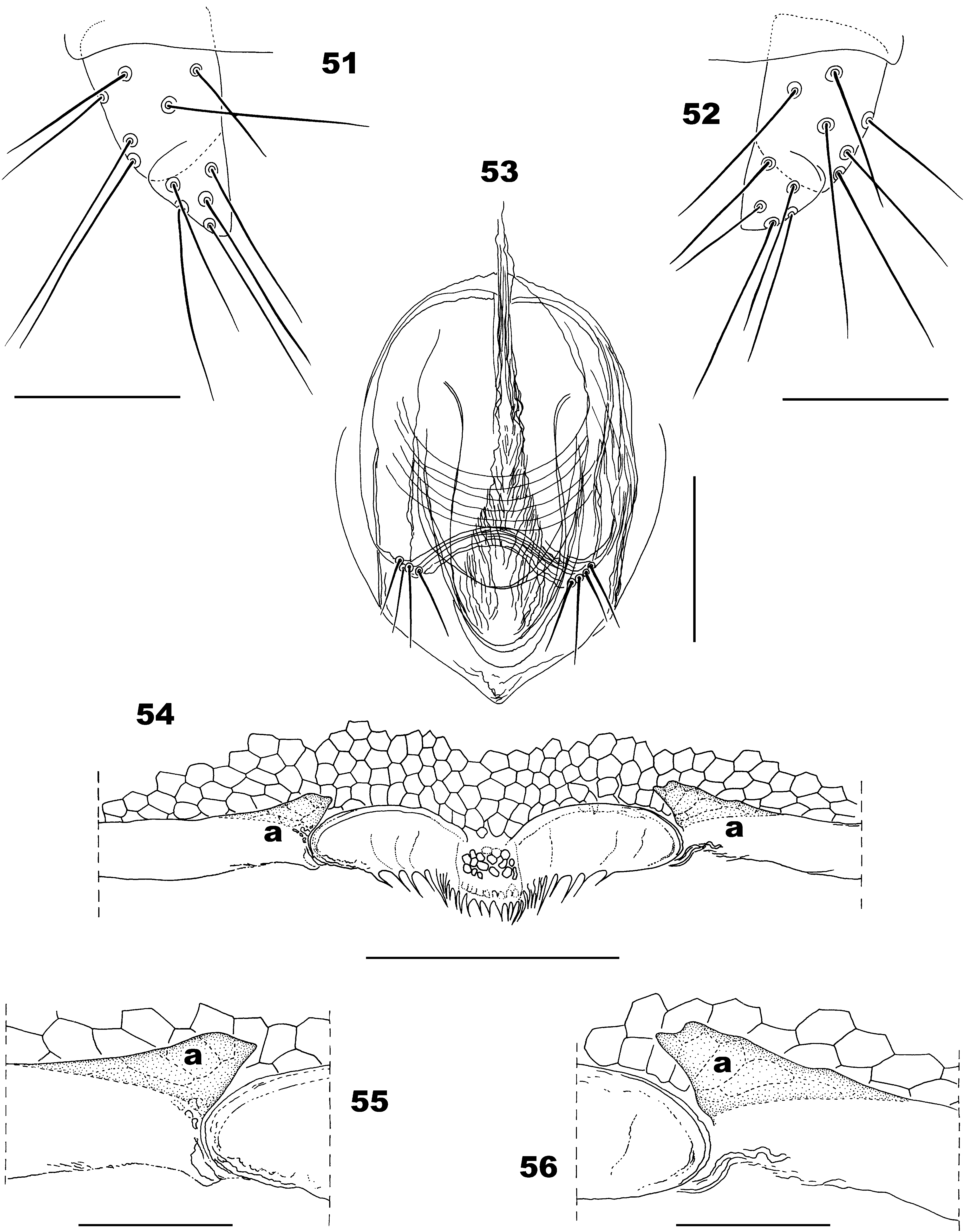
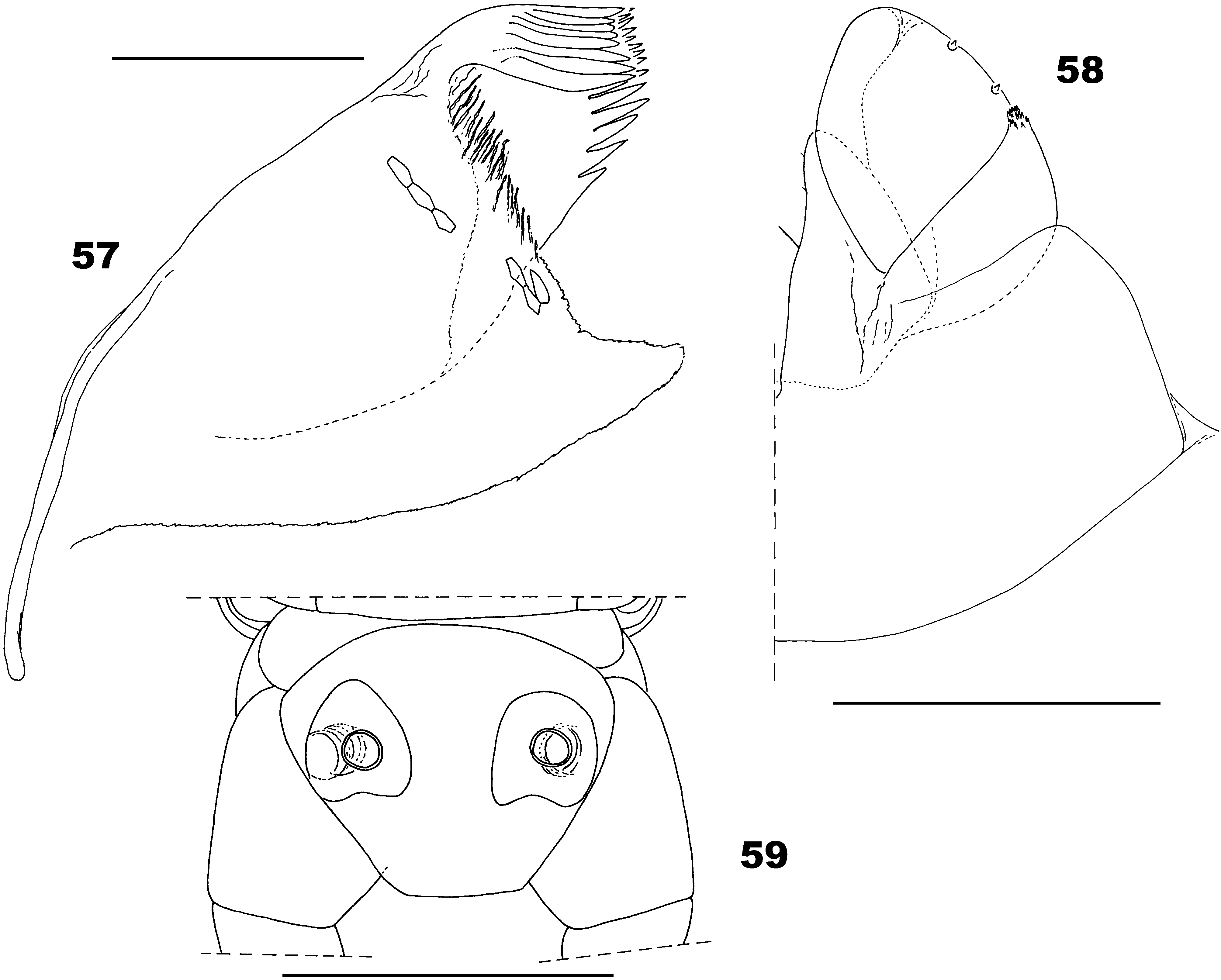
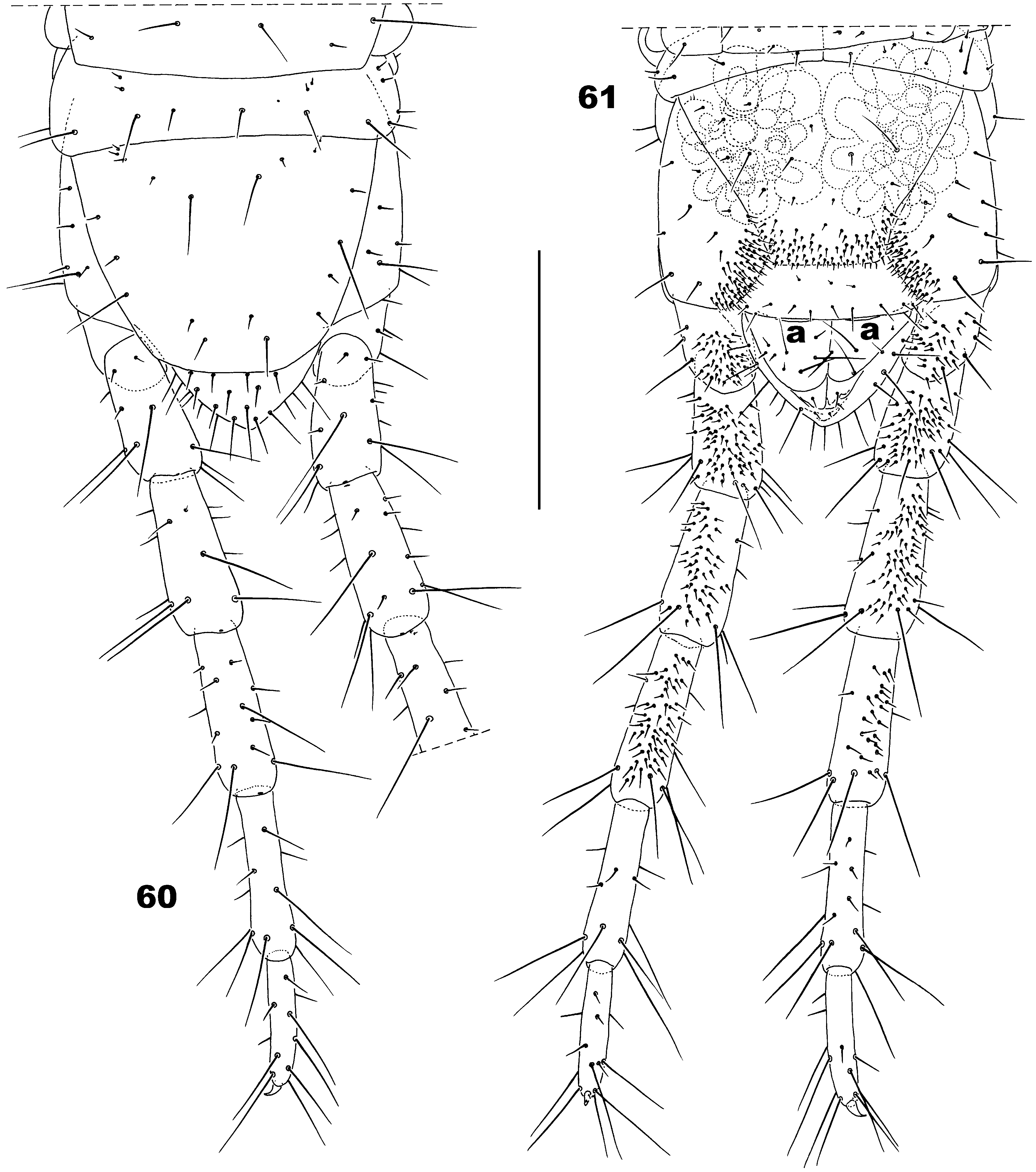
TABLE 2. Number of type a and b sensilla on antennal articles II, V, IX and XIII in the male holotype of Ribautia lewisi sp. nov.
| Ventral | Dorsal | Figs. | |||
|---|---|---|---|---|---|
| a | b | a | b | ||
| II | - | 1 | 1 | - | 3, 7 |
| V | - | 1 | 1 | 1 | 4, 8 |
| IX | 1 | 1 | 1 | 2 | 5, 9 |
| XIII | 1 | 1 | 1 | 5 | 6, 10 |
No known copyright restrictions apply. See Agosti, D., Egloff, W., 2009. Taxonomic information exchange and copyright: the Plazi approach. BMC Research Notes 2009, 2:53 for further explanation.