
Qwaqwaia scolopiae Liljeblad, Nieves-Aldrey & Melika
|
publication ID |
https://doi.org/ 10.5281/zenodo.205510 |
|
DOI |
https://doi.org/10.5281/zenodo.6182688 |
|
persistent identifier |
https://treatment.plazi.org/id/03AE87D4-FFB2-FFBE-FF66-F8F0B27FFB81 |
|
treatment provided by |
Plazi |
|
scientific name |
Qwaqwaia scolopiae Liljeblad, Nieves-Aldrey & Melika |
| status |
sp. nov. |
Qwaqwaia scolopiae Liljeblad, Nieves-Aldrey & Melika sp. n.
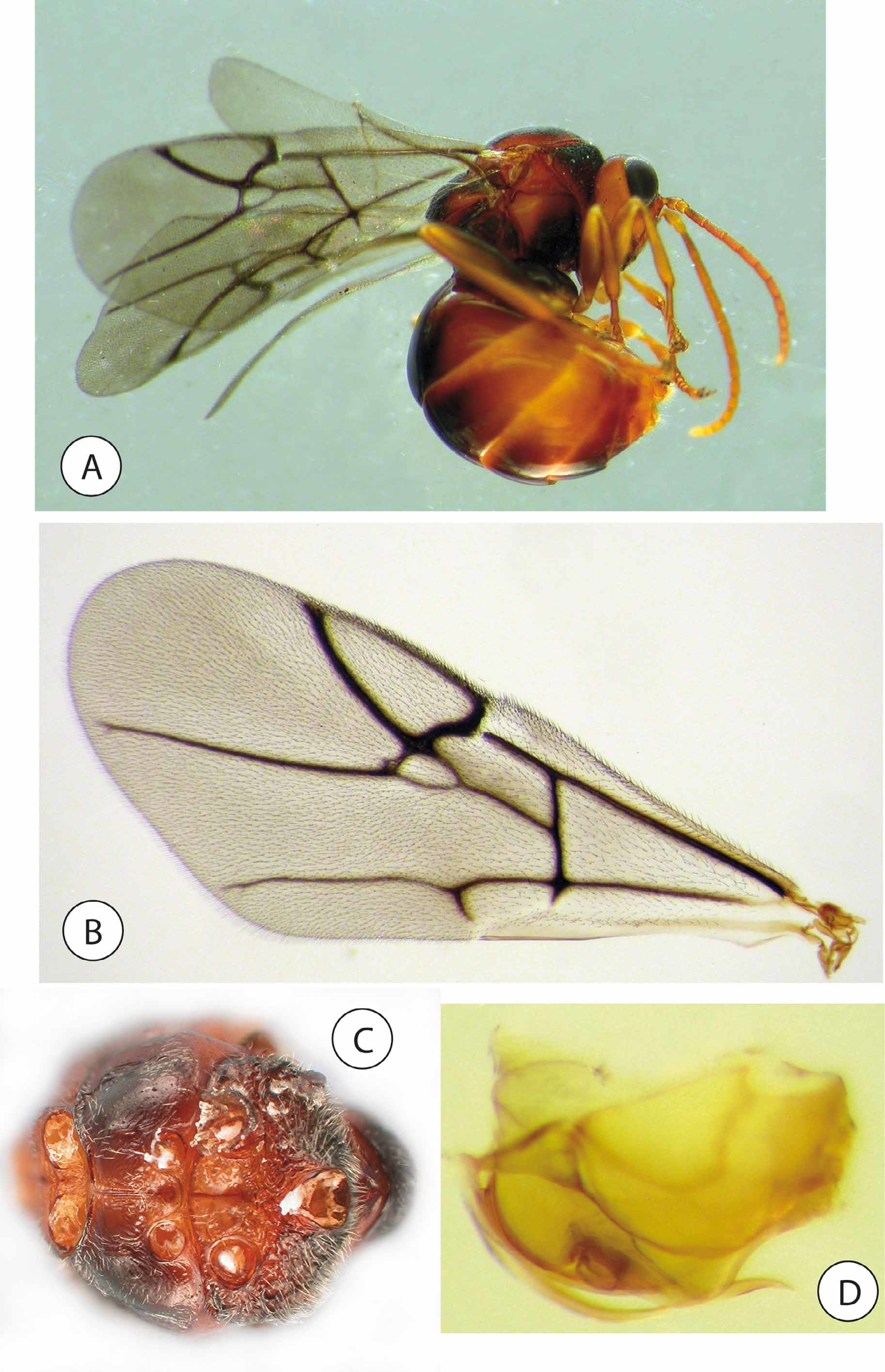
Figs 1–7 View FIGURE 1 View FIGURE 2 View FIGURE 3 View FIGURE 5 View FIGURE 6 View FIGURE 7 .
urn:lsid:zoobank.org:act:BA3F8D2B-FD34-4C93-8E4F-F30869C7670C Type material. Holotype female with type number TYPH01857, labeled “QWAQWA, S of Witsieshoek, Kgotswane R, 28.37S 28.44E, 12.x.1986, S Neser”, “ex galls on Scolopia mundii ”, and “AcSN 1324”. Paratypes: 3 females with the same label data as the holotype. The holotype female and 1 paratype are deposited in SANC (Pretoria, South Africa). One paratype is deposited in SNHM, one in MNCN and one in HPDL.
Material examined. 32 females labeled “ SOUTH AFRICA MPU, Mooihoek Farm nr Wakkerstroom, 27o13’S, 30o31’E, 27.ix.2009 S. Neser”, „Ex galls on Scolopia mundii (Flacourtiaceae) ”. Additional, typical galls collected on S. mundii containing larvae were collected in July 2010 by SN in KZN Province, southern Drakensberg, S 27degr.13’20.95”; E 30degr. 31’31.29”.
Etymology. Named after the host plant Scolopia mundii .
Diagnosis. Occiput with a strong, sharp occipital carina. Pronotum dorsally wide but anteroadmedian pits close and only shallowly separated; dorsal part of pronotal plate with ventral carina, ventral transverse impression almost uniting admedian pits. Mesoscutum distinctly angled in relation to pronotum in lateral view. Mesoscutellum distinctly margined. Radial cell of forewing closed, with veins heavily pigmented. Metatarsal claw simple. Hypopygium short, abrupt, without projecting ventral spine, with dense setae forming apical tuft.
Because of a dorsally wide pronotum, the new species is most readily confused with members of the Synergini and the Aylacini (especially some members of the genus Phanacis ). However, Q. scolopiae can be distinguished from all members of those tribes based on two morphological features: a) it only has two prominent teeth on the right mandible; b) the parascutal carina extends anteriorly all the way to notaulus.
Description. Adult female. Size Body. length 4.2–4.4 mm (n=5) from head to tip of metasoma. Color ( Fig. 3 View FIGURE 3 A). Head brown, with darker parts along inner margins of compound eyes, especially at level of frons; mandibles reddish brown, maxillary and labial palps much lighter; mesosoma reddish brown, with blackish-brown pronotum, internotaular area of mesoscutum, mesopleuron, metanotal troughs and propodeum; coxae and femura darkish brown, remainder of legs reddish brown, lighter; metasoma reddish brown, slightly darker dorsad. Habitus ( Fig. 3 View FIGURE 3 A). Mesosoma predominantly pubescent, but mesopleuron, coxae and metasoma visibly glabrous. All veins of forewing heavily pigmented. Hypopygium abrupt, short, not projected into a ventral spine, habitus resembling that of a typical cynipid male.
Head, anterior view ( Fig. 1 View FIGURE 1 A). Lower face broad, sub-trapezoid, 1.8 times wider than high; relatively densely pubescent, less so dorsally. Upper face smoothly colliculate-coriarious. Ratio of POL/OOL/LOL about 3/3/1, posterior ocellus separated from inner orbit of eye by almost 2 times its diameter. Gena not expanded behind eye. Distance between antennal sockets about same as diameter of socket including rim; sockets situated at mid-height of eye; transfacial distance 1.7 times height of eye; distance between antennal rim and compound eye 1.5 times width of antennal socket including rim. Malar space 0.63 times height of eye. Strong, regular striae radiating from clypeus, laterally reaching margin of eye. Anterior tentorial pits distinct; epistomal sulcus weakly visible, clypeopleurostomal lines obscured by facial striae. Clypeus rectangular, not set off from margin of head, nor protruding over mandibles. Head, posterior view ( Fig. 1 View FIGURE 1 B). Glabrous, pubescence more dense ventrally. Dorsally with sharp occipital carina. Height of occipital foramen equal to height of hypostomal bridge. Posterior tentorial pits deep and high, positioned dorsad ventral margin of occipital foramen. Gular ridges weak but complete, ventrally meeting just after reaching hypostoma; median hairy strip narrow throughout. Hypostomal ridges distinctly margined medially, almost meeting. Antenna ( Fig. 1 View FIGURE 1 C–E). About 0.6 times length of body; consisting of scape, pedicel and 12- segmented flagellum, ratios of their lengths being 30:15:39:38:35:34:28:28:22:20:17:15:14:24; pedicel as long as wide; F1 3.2 times longer than wide; F12 2.1 times longer than wide; all flagellomeres of about equal width. All segments covered with sensilla triochodea. Elongate multiporous plate sensilla (MPS) present on all flagellomeres. Each flagellomere also with one or a few lateral subapical sensilla basiconica. Dorsal pit present, positioned around middle of apical flagellomere, together with sensilla basiconica suggesting a fusion of two flagellomeres. Mouthparts ( Fig. 1 View FIGURE 1 A). Both mandibles with two teeth with corresponding internal rods. Basal region of anterior surface abundantly pubescent. Maxillary palp 5-segmented. First segment short, not longer than wide, second segment long, longer than half length of segments 3–5 combined. Labial palp 3-segmented. Pronotum ( Fig. 1 View FIGURE 1 G & 2A). Pubescent, with rugose-areolate sculpture. Pronotum longitudinally wide medially; ratio of dorsomedian to lateral length 0.36. Anteroadmedian depressions of pronotum superficially separated medially by about ¼ of median length of pronotum; dorsal part of pronotal plate distinctly separated from ventral part by transverse shallow impression connecting admedian depressions; depressions and ventral part of pronotal plate smoothly sculptured, with a single row of setae extending laterad. Prepectus (propleuron and prosternum) ( Fig. 3 View FIGURE 3 C). Propleura meeting medially along all of externally visible surface. Profurcal pit small, rounded; positioned anterior to middle of furcasternum. Mesonotum ( Fig. 1 View FIGURE 1 G–F & 2A). Evenly pubescent with colliculate-coriarious sculpture. Parascutal carina anteriorly extending to notaulus. Notaulus distinct and complete. Anterioadmedian signa visible. Median mesoscutal impression distinct and running for ¾ length of mesoscutum from posterior margin. Mesoscutum and scutellum separated by transscutal fissure. Scutellar foveae distinct, smoothly sculptured, with strong median septum; anterior margin of foveae curved, posteriorly margined by rugose sculpture of scutellum. Axillula closed, heavily pubescent with reduced rugose sculpture. Subaxillular strip without dorsal projection. Mesoscutum posterodorsally margined by a carina/ruga. Mesopectus ( Fig. 2 View FIGURE 2 A & 3C). Glabrous except for dense pubescence in mesopleural triangle and sparse pubescence lateroventrad. Subalar pit large, deep and reaching posterior margin of mesopleuron. Mesopleuron with transverse carinate impressions medially. Acetabular carina well developed; mesodiscrimen distinctly marked by carina. Mesofurcal pit longitudinally situated around centers of mesocoxal foramina. Metanotum ( Fig. 2 View FIGURE 2 B). Metanotal trough relatively densely pubescent, with diffusely rugose sculpture, ventral bar broad and smooth. Metascutellum distinctly constricted medially, as high as height of ventral impressed area of metanotum. Metapectal-propodeal complex ( Fig. 2 View FIGURE 2 A–B). Evenly pubescent, with rugose sculpture, similar to propodeum medially. Metepimeron distinctly impressed, marked by a distinct ledge posteriorly. Metapleural sulcus meeting posterior margin of mesopectus high, removed about 1/3 of height of metapectal-propodeal complex from its dorsal margin. Lateral propodeal carinae straight, diverging ventrad while increasingly obscured by rugose sculpture. Nucha relatively long, distinctly set off from propodeum; dorsally smooth. Petiolar foramen posteriorly positioned, ventral margin far removed from posterior margins of metacoxal foramina. Surface of metasternum anterior to metafurcal pit smooth, at most with some superficial sculpture. Anterior margin of metasternum and metacoxal foramina not contiguous. Metafurcal pit longitudinally situated posterior to centers of metacoxal foramina. Legs ( Fig. 2 View FIGURE 2 A & 2C). Lateral surface of metacoxa more or less evenly pubescent. Tarsal claws simple; pubescence sparse, apically absent. Forewing ( Fig. 3 View FIGURE 3 B). About 1.16 times length of body. Wing margin with distinct cilia. Unusually complete and distinct venation; veins of radial cell especially heavily pigmented, closed along anterior margin, 2.4 times longer than wide. R1 laterad 2r short, anteriorly directed, almost at right angles with anterior wing margin. Areolet present, conspicuously large. Rs+M meeting basalis at middle of the latter. Metasoma ( Fig. 2 View FIGURE 2 C–D). Mostly smooth; short and high, not much laterally compressed; extending more dorsad than ventrad petiole. Dorsal length of 3tg about ¼ that of whole gaster. Petiolar hump absent. Ninth tergum at position of base of third valvula without any distinct point of weakness. Hypopygium ( Fig. 2 View FIGURE 2 E). Ending abruptly, not prolonged into a ventral spine, without apical incision but with dense tuft of setae; with straight lateral flaps. Cercus positioned relatively close to apex of 9tg. Ovipositor ( Fig. 3 View FIGURE 3 D). Terebra short, articulation between second valvifer and second valvula situated anterior to dorsalmost part of second valvifer, basal part of dorsal margin of 9tg oblique.
Final-instar larva. Size. Body length 4.2 mm (range 3.8–5.0); body width 1.2 mm (1.0–1.5) (n=3).
Habitus. Larval type caudate hymenopteriform ( Clausen 1940; Gauld & Bolton 1988; Quicke 1997). Maggotlike, segmented, without appendages, except a caudal appendage arising from last abdominal segment.
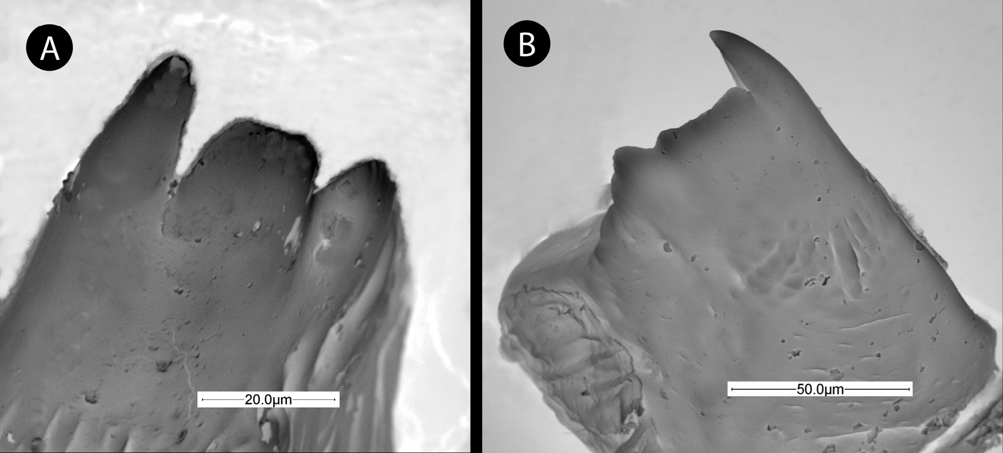
Body (Fig.4A, 4D & 6A–B). Spindle shaped, narrower posteriorly. Integument whitish, smooth and bare, except for a few short setae concentrated to head, thorax and last abdominal segments. Consisting of head and 13 visible segments: 3 thoracic, 9 abdominal and one caudal segment. Nine spiracles visible on TS2– AS 7. TS1, in ventral view, not narrowed medially under ventral part of head; with minute blister-like sculpture and a few short setae laterad. TS1–TS3 of similar length in lateral view; AS 1 similar to TS3, AS 2– AS 4 larger than AS 1; AS 4 prolonged ventrally into two large, stump-like protuberances; last segment elongate, trapezoid, slightly broader than long in ventral and lateral view; caudate segment long and slender, 3.6 times longer than broad, of about same length as AS 8+ AS 9 combined; caudal segment, or “tail,” composed of two parts, a basal cylindrical, smooth part and an apical conical part, with minute blister-like sculpture (Fig. 4C). Head (Fig. 4E). More or less rounded in anterior view. Antennae indistinct, visible only as a pair of small spots on face. A pair of short genal setae present. Mouth parts (Fig. 4F). Clypeus triangular; with straight ventral margin, slightly projecting over mandibles; with a pair of clypeal setae. Labrum trapezoid. Maxillae triangular, well differentiated from labium, with two pairs of maxillary palpi and one pair of maxillary setae well visible. Labium with labial palpi and labial setae also visible. Salivary opening a conspicuous, rounded hole. Mandibles ( Fig. 5 View FIGURE 5 ) asymmetrical, without sculpture or hairs; bases visible beneath labrum. Right mandible ( Fig. 5 View FIGURE 5 B) with three teeth, one larger apical, acute tooth, an intermediate broad, blunt tooth and a basal acute, small tooth. Left mandible ( Fig. 5 View FIGURE 5 A) with three teeth of about similar length; intermediate tooth broader and blunt at apex.
Remarks. The larva of Qwaqwaia scolopiae differs from all other described cynipid larvae by two important diagnostic characters, viz. the conspicuous caudal appendage, and the fourth abdominal segment, ventrally prolonged into two large protuberances. The presence of a conspicuous caudal appendage (caudate larva) has been recorded from many groups of Hymenoptera (e.g. Ichneumonoidea, Proctotrupoidea, Chalcidoidea and also Cynipoidea) ( Clausen 1940; Quicke 1997). However, in the case of the Cynipoidea, a caudate larva has been reported from the Figitidae , and is relatively common in the first larval stages of eucoilines ( Clausen 1940; Nieves-Aldrey et al. 2005; Ovruski 1994). Among cynipids, caudate larvae were not known, nor larvae bearing protuberances on the FIGURE 4. Qwaqwaia scolopiae , final-instar larva: A) body, lateral view. B) 4th abdominal segment, lateral view. C) caudal appendage, lateral view. D) body, ventral view. E) head, anterior view. F) detail of mouth parts.
fourth abdominal segment. This is most similar to larvae of Diastrophus rubi (Bouché) and the Diplolepidini , where the body tapers abruptly posteriorly and the last segment is cylindrical and slightly elongated, but not, however, forming a tail ( Nieves-Aldrey et al. 2005). The mandible asymmetry found in Q. scolopiae is relatively rare among larval cynipids, but is present in the larvae of one genus of aylacine gall inducers ( Diastrophus ) and four genera of inquilines ( Periclistus in cynipid rose galls, and Saphonecrus and Synophrus in cynipid oak galls as well as in Rhoophilus ) ( Nieves-Aldrey et al. 2005; Van Noort et al. 2007). The shape of the teeth of the right mandible, especially the second tooth which is broadly truncate and blunt, resembles that of the larvae of Synergini . The right mandible is strikingly similar to that of the larva of Synophrus politus Hartig. Interestingly , the behaviors of the larvae of S. politus and Q. scolopiae inside the gall are quite similar – both moving freely in a wide, rounded cell with free space ( Nieves-Aldrey et al. 2005; Nieves-Aldrey & Vårdal unpublished data).
Biology. Reared in summer from Scolopia mundii (Eckl. & Zeyh.) Warb. ( Salicaceae , formerly in Flacourtiaceae ). Host determination confirmed by Ms. E. Retief of the South African National Biodiversity Institute. Galls ( Fig. 7 View FIGURE 7 ) are rounded, 6–10 mm in diameter and occur on twigs (growth tip and axillary). A single wasp develops in each gall cavity with space around the mobile larva; larger, paired or otherwise compound galls may have two, three or perhaps more cavities. Older galls become woody and remain attached and exhibit remarkably large emergence holes. New galls are soft, smooth and shiny green, arising in shoot tips or more commonly in leaf axils near shoot tips, sometimes at the base of galls from the previous season(s). They may be green or develop a shiny red surface color before reaching final size and taking on a more greenish grey. They have lenticels on the surface that turn more conspicuous as they mature, giving the gall a roughish surface. Galls arising in shoot tips show remains of nodal bracts and leaf scars, indicating that they are developing in modified twig tissue. The wall width of larger galls is thicker than the diameter of the central cavity, but in small and compound galls the wall may be relatively thinner.
When a gall is carefully cut around the periphery and the halves separated, the larva is usually found in a curved position with the dorsum against the inner wall of the gall cavity, and the mandibles and the tail pressed against the substrate; if disturbed it wriggles very actively within the cell, typically in a gyrating fashion, swinging the tail rapidly with a circular motion. The pupa lies free in the cell with appendages free from the body. The pupa is whitish, later developing a smoky grey coloration in parts. At the stage when the pupa starts showing pigmentation, but before molting into a teneral adult, it is capable of moving the legs, and when disturbed it may flex and move the hind legs alternately in a sweeping movement around the body. Behavior of females after emerging from galls in October and November has not been observed.
Distribution. Galls on the host plant, Scolopia mundii , have so far been encountered at only three sites, all in the Drakensberg escarpment between Lesotho and Swaziland and about 250 km inland. S. mundii has a wide distribution in mainly eastern parts of South Africa from Limpopo Province in the north into Gauteng and NW Province through Mpumalanga to the Eastern Cape and SW Cape as far south-west as Table Mountain on the Cape Peninsula. It is a small to medium-sized tree 3–10 m, occasionally to 20 m at medium to high altitudes in Afromontane forest and on forest margins and rocky outcrops in mountain grassland ( Schmidt et al. 2002). The genus Scolopia has about 45 species in Africa, Asia and Australia, with five occurring in South Africa ( Germishuizen et al. 2006). Two other species co-occurred with S. mundii at the type locality, but no cynipid galls were found on them. Galls were not common on the trees, and only some trees at any of the collection sites had galls, usually sparsely at most. It is worth noting that there are no other cynipids recorded from the Salicaceae , even though there are many common plant species in the temperate region of the northern hemisphere that belong here. This, in spite of the family seeming like a suitable host with several nematine sawflies ( Hymenoptera : Tenthredinidae ) inducing galls on Salix ( Nyman 2000) .


No known copyright restrictions apply. See Agosti, D., Egloff, W., 2009. Taxonomic information exchange and copyright: the Plazi approach. BMC Research Notes 2009, 2:53 for further explanation.