
Semperella jiaolongae, Gong, Lin, Li, Xinzheng & Qiu, Jian-Wen, 2015
|
publication ID |
https://doi.org/ 10.11646/zootaxa.4034.1.9 |
|
publication LSID |
lsid:zoobank.org:pub:7FDC215C-9722-41BB-B752-A9C38942CE82 |
|
DOI |
https://doi.org/10.5281/zenodo.5624231 |
|
persistent identifier |
https://treatment.plazi.org/id/03BB87FC-FFD9-5A28-FF24-FE8612DF37CD |
|
treatment provided by |
Plazi |
|
scientific name |
Semperella jiaolongae |
| status |
sp. nov. |
Semperella jiaolongae View in CoL sp. nov.
( Figures 1–2 View FIGURE 1 View FIGURE 2 )
Material examined. Holotype: MBM179993, South China Sea (22°7.21'N, 119°18.67'E), 19 June 2013, 1120 m depth, muddy bottom.
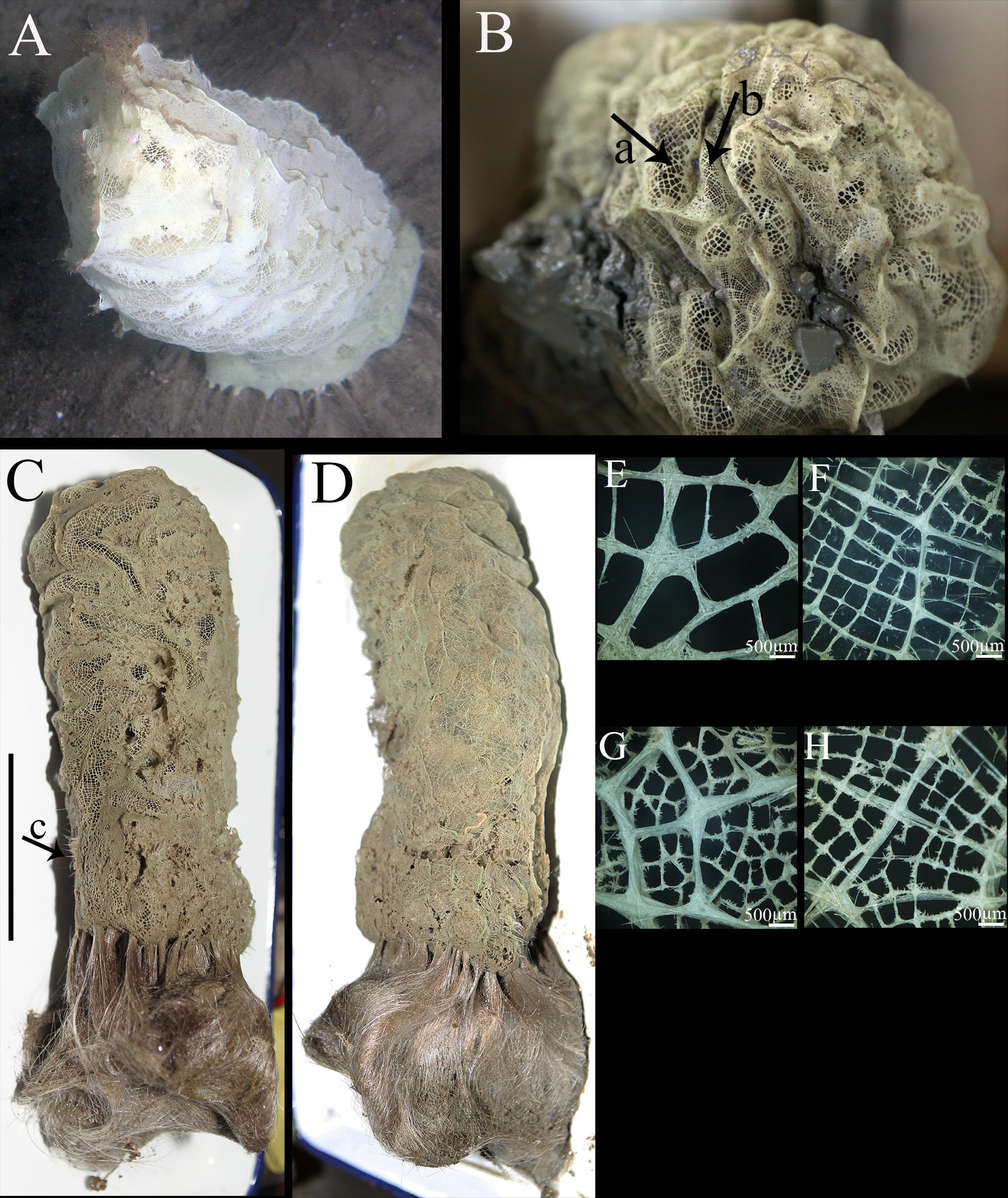
Description. Columnar body is 250 mm long (not including basalia) and 112 mm in maximal diameter. The color is pure white when alive ( Fig. 1 View FIGURE 1 A), but grey after collection ( Fig. 1 View FIGURE 1 B–D) due to contamination by mud during sampling. Some pleuralia rows are present on the bottom of lateral surface ( Fig. 1 View FIGURE 1 C, arrow c). Numerous elongated atrial areas ( Fig. 1 View FIGURE 1 B, arrow a) are scattered on one side of (side 1) the specimen’s surface ( Fig. 1 View FIGURE 1 C), separated by many dermal areas ( Fig. 1 View FIGURE 1 B, arrow b). Atrial surface covered by large-meshed latticework with meshes 0.38 to 2.5 mm in diameter ( Fig. 1 View FIGURE 1 E). The sponge presents two differently organized sides ( Fig. 1 View FIGURE 1 F): (1) side 1 has dermal areas randomly present together with atrial ones, (2) side 2 only contain dermal areas ( Fig. 1 View FIGURE 1 D). The dermal areas on side 1 have thinner main hypodermal beams of the meshed latticework than side 2. Meshes on dermal areas consist of two parts: bigger meshes with thicker edges (main hypodermal beams) forming a framework ( Fig. 1 View FIGURE 1 G), and smaller meshes (minor hypodermal beams) with finer edges dividing bigger meshes into many smaller meshes ( Fig. 1 View FIGURE 1 H). Dermal areas on side 1 have smaller meshes with diameter 0.26 to 0.81 mm, and bigger meshes of 0.80 to 2.1 mm in diameter. Dermal areas on side 2 have smaller meshes ( Fig. 1 View FIGURE 1 G) with diameter 0.19 to 0.93 mm, and bigger meshes ( Fig. 1 View FIGURE 1 H) of 1.5 to 5.0 mm in diameter. Large openings into tissues are present underneath atrial and dermal lattices. The tufts of basalia, more than 110 mm long and 2.6 to 5 mm wide, consist of many small single, basal spicules. Individual shafts of basal spicules are thin, 0.05 to 0.4 mm diameter.
Spicules. Pentactins ( Fig. 2 View FIGURE 2 A) with smooth rays make up choanosomal skeleton, tangential rays 216.6–4217.9 µm long, proximal rays 265.8–4000.1 µm long. Dermal pentactins mainly occur as two types ( Fig. 2 View FIGURE 2 F–G): those with sharply pointed or conical pinular rays with straight tangential rays and those with shorter pinular rays and slightly curved, smooth or slightly spined tangential rays. Pinular rays are 270.1–592.2 µm long; tangential rays are 49.8–198.8 µm long. Atrial pentactins similar to dermal pentactins always present ( Fig. 2 View FIGURE 2 H). Atrial pentactins’ pinular rays 206.7–326.2 µm long, and tangential rays 39.5 –108.5 µm long. Pentactins ( Fig. 2 View FIGURE 2 I) with fewer short spines, and similar lengths for all rays are present mainly in the choanosome, but can also be found in dermal and atrial areas. Basalia ( Fig. 2 View FIGURE 2 B–D) have a spiny shaft and terminal anchor bearing two teeth. Uncinates ( Fig. 2 View FIGURE 2 K) rare, with small short spines, 927.9–3293.7 µm long. Prostalia are sceptres ( Fig. 2 View FIGURE 2 E), common to Semperella , with shafts mostly smooth, except for their distal part with spines; they are very easily broken.
Microscleres consist of amphidiscs, microhexactins, micropentactins and microstauractins. Amphidiscs are micramphidiscs ( Fig. 2 View FIGURE 2 N) only, with shafts covered by numerous spines, total length 15.1–25.5 µm, umbel length 4.2–6.9 µm. Microhexactins ( Fig. 2 View FIGURE 2 L) very rare. Rays straight, 31.9–79.3 µm long, covered by numerous small spines. Micropentactins ( Fig. 2 View FIGURE 2 J) with a very short and minute spiny pinular ray, and four straight, smooth or spiny tangential rays 37.9–87.6 µm long. Microstauractins ( Fig. 2 View FIGURE 2 M) are rare.
Etymology. The species is named after the Chinese manned submersible “Jiaolong”.
Remarks. For Semperella , the shape of the body and the morphology of the spicules are important for species identification. By the external morphology of Semperella species described in Tabachnick & Lévi (2000), S. jiaolongae sp. nov. is most similar to S. crosnieri Tabachnick & Lévi, 2000 . The two species share the same morphological character of having dermal areas present on both sides of the body, and on one side in combination with atrial areas. However, the holotype of S. crosnieri differs from S. jiaolongae sp. nov. in having a tongue-like body and having many microuncinates. In contrast, S. jiaolongae sp. nov. has a cylindrical body shape, and its microuncinates are very rare. In addition, there are other notable differences between the two species: (1) the shape of micropentactins is very different in both species: those in the new species have smooth tangential rays and a very short slightly spined proximal ray, while those in S. crosnieri are always covered with numerous, or rarely with sparse spines; (2) micropentactins and microhexactins of the new species are thinner than those of S. crosnieri ; (3) the umbels of amphidiscs in S. crosnieri have sharper teeth than those of the new species; (4) dermal pinules are a little larger than the atrial pinules in the new species, the contrary of what is seen in S. crosnieri . (5) the new species has a pentactine ( Fig. 2 View FIGURE 2 I) with fewer short spines, and similar lengths for every ray, which is present not only in the choanosome, but also in the dermal and atrial areas, while this kind of pentactin was not reported for S. crosnieri .

No known copyright restrictions apply. See Agosti, D., Egloff, W., 2009. Taxonomic information exchange and copyright: the Plazi approach. BMC Research Notes 2009, 2:53 for further explanation.