
Strieremaeus illibatus Sellnick, 1918
|
publication ID |
https://doi.org/ 10.11646/zootaxa.2993.1.3 |
|
persistent identifier |
https://treatment.plazi.org/id/E067450A-2047-F44F-FF20-FB0BC5ECFC6F |
|
treatment provided by |
Felipe |
|
scientific name |
Strieremaeus illibatus Sellnick, 1918 |
| status |
|
Redescription of Strieremaeus illibatus Sellnick, 1918
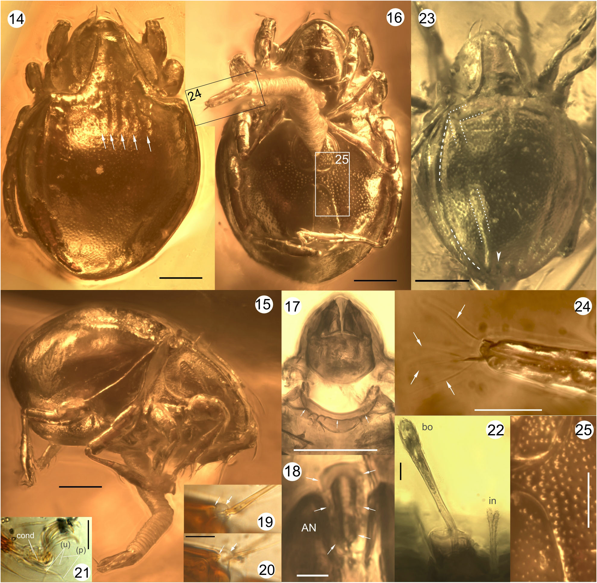
Adult ( Figs. 1–25 View FIGURES 1–4 View FIGURES 5–13 View FIGURES 14–25 ). Measurements: body length 430–650 µm (n=9), width 280–470 µm (n=8), height 310 µm (n=1).
General form and color: Body relatively broad and deep, subglobular or drop-shaped, with hysterosoma little larger than proterosoma ( Figs. 1–3 View FIGURES 1–4 , 14–16, 23 View FIGURES 14–25 ). Brown in transmitted light, with lighter dots evenly distributed over notogaster and ventral plate; color pattern of venter variable [specimen KMA 197-34 shows dark bands bordering epimeres and anal and genital apertures while MGCP Ar 14 shows no distinct bands, but cuticle somewhat darkened around anal and genital apertures].
Integument: Smooth or with fine punctation on prodorsum; uniformly punctate (corresponding to lighter dots in transmitted light) on notogaster, ventral plates, coxisternum and subcapitulum ( Figs. 14–16, 23, 25 View FIGURES 14–25 ). Cerotegument microtuberculate (granules less than 1 µm), apparent on legs, prodorsal ridges and in sejugal region; covering whole body in some specimens.
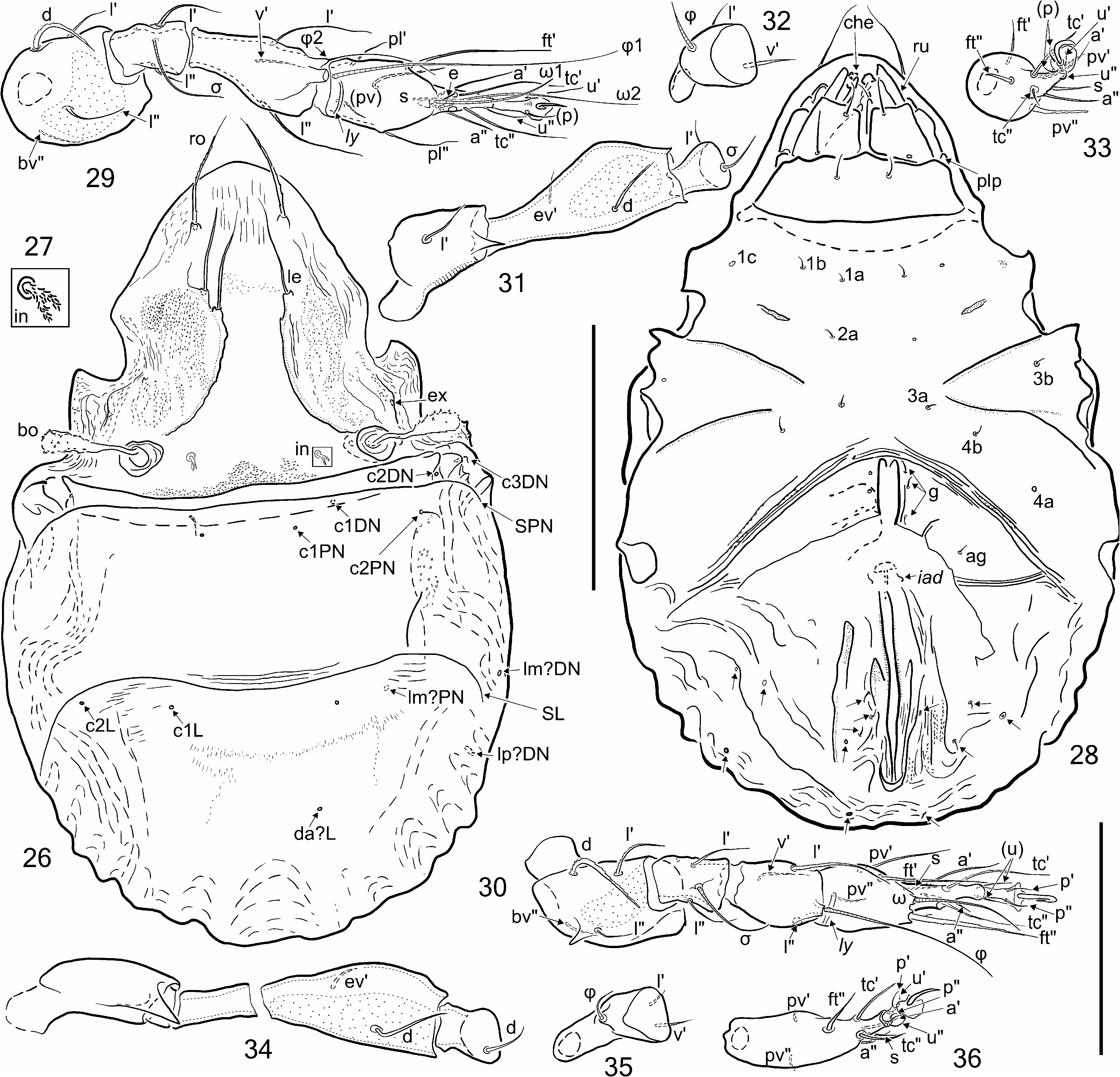
Prodorsum: ( Figs. 1–4 View FIGURES 1–4 , 14–16, 23 View FIGURES 14–25 ). Appears functionally fused to notogaster without intervening groove or articulation, but attachment line (dorsosejugal suture) well marked, relatively straight and uninterrupted. No dorsophragma or pleurophragma apparent. Rostral margin rounded, entire, without genal notch or incision; anteromedially, between setae ro, with bulge of thinner cuticle, appearing like rectangular fenestration in transmitted light. Two pairs of well-developed narrow, carinate, longitudinal ridges present, all four ridges connected by thin fold anteriorly ( Figs. 3 View FIGURES 1–4 , 14, 23 View FIGURES 14–25 ). Medial pair (lamellae) start on medial side of respective bothridium and curve gently anteriad in basal third; middle and distal portions straight, slightly converging to end in short, blunt cusp. Lateral ridge (tutorium) of approximately same size, starting anterior to lateral side of bothridium and diverging anteriorly, ending on lateral face of prodorsum at about same level as lamellar cusp. In oblique lateral projection on dry specimens, prodorsal ridges form isosceles triangle. Seta ro (65 µm, n=5) on small tubercle, acuminate, slightly rough. Seta le (75 µm, n=7) inserted on distal cusp of lamella, finely spinose, acuminate. Setae in (50 µm, n=4) relatively thin, stiff; distal half two-branched (in five specimens) or three-branched (in two specimens, MGCP Ar 33 and UA- 2195a); barbed along whole length, but more densely on branches than on stalk. Bothridium cup-shaped, with internal spiral ridge. Seta bo (65 µm, n=4) with long stalk, gradually widened to elongated, spinose head ( Figs. 4 View FIGURES 1–4 , 22 View FIGURES 14–25 ). Seta ex (ca. 20 µm slightly curved, rough. Mutual distances of setal pairs ro–ro, le–le, in–in, and bo–bo 80 (n=2), 50 (n=3), 60 (n=8), and 140 (n=8) µm, respectively.
Notogaster ( Figs. 1, 3 View FIGURES 1–4 , 14, 15, 23 View FIGURES 14–25 ): Slightly longer than wide; relatively wide and truncate anteriorly but widest just anterior to mid-length. Several short parallel folds extend posteriorly from dorsosejugal suture for about onequarter length of notogaster, resulting in anterocentral region of apparent shallow grooves, usually with five ( Fig. 14 View FIGURES 14–25 , arrows), sometimes fewer ( Fig. 28 View FIGURES 26–36 ). Several other folds converge in each humeral region, resulting in characteristic pattern of apparent grooves and weak complementary ridges. Two closely parallel grooves ( Fig. 23 View FIGURES 14–25 , white dotted lines) extend almost laterally from region of central folds, then turn at right-angle to delineate chevron-like humeral angle between them. These grooves continue obliquely posteriad, converging to delineate wedge-shaped central plateau; innermost groove weaker or absent in this region, but can be prominent in pygidial regon where grooves bend to nearly vertical orientation (white arrowhead). Third, more lateral groove ( Fig. 23 View FIGURES 14–25 , white dashed line) curves posteriorly from humeral region, nearly parallel to notogastral margin, but effaces in pygidial region. Grooves clearly visible only in reflected, oblique illumination ( Figs. 14, 23 View FIGURES 14–25 ); in transmitted light grooves demarcated mainly by change in apparent shape of punctation at their margins. Twelve pairs of setae present: c 1 just lateral to anterocentral folds, c 2 and c 3 close together within humeral angle ( Fig. 1 View FIGURES 1–4 ). Seta la (at least 26 µm) longest notogastral seta, inserted just lateral to humeral angle, near its posterior end. Setae la–h 1 slightly shorter, arranged in longitudinal row in region between medial and lateral grooves, curved posteriorly, roughened along dorsal curvature. Setal row p 1 - p 3 external to grooves, not aligned with other setae ( Figs. 1, 3 View FIGURES 1–4 ). Lyrifissures arranged in row outside lateral groove: ia lateral to humeral angle, seen only in dorsal view; im, ih, ips, and ip longer, surrounded by darker integument in transmitted light. Opisthonotal gland opening (gla) present but small, indistinct.
Podosomal region ( Figs. 2, 3 View FIGURES 1–4 , 15, 17 View FIGURES 14–25 ): Pedotecta I and II well developed, of similar size. Laterally, sejugal groove deep and well-defined by carinae; anterior carina curving anteroventrally from behind bothridium and connecting with pedotectum I. Posterior carina nearly vertical, effacing behind acetabulum III ( Figs. 3 View FIGURES 1–4 , 15 View FIGURES 14–25 ). Discidium absent.
Venter ( Figs. 2 View FIGURES 1–4 , 15, 16–18, 24, 25 View FIGURES 14–25 ): Apodeme II and sejugal apodeme discernible, other apodemes not seen. Without well-marked epimeral borders, but sejugal line indicated externally by irregular ridges and depressions ( Fig. 16 View FIGURES 14–25 ); epimere I with similar transverse, irregular surface ridge that nearly connects pedotecta I ( Fig. 17 View FIGURES 14–25 , arrows).
Epimeral setae visible only on one specimen: 1a, 1b, 1c, and 2b ( Fig. 2 View FIGURES 1–4 ); none discernible on epimeres III or IV. Genital setae poorly visible, at least four pairs present. One pair of aggenital, two pairs of anal, and three pairs of adanal setae. Lyrifissure iad far removed from anal opening, lateral to seta ad 3 ( Fig. 2 View FIGURES 1–4 ). Preanal organ hollow, appearing caecum-like in ventral view ( Fig. 18 View FIGURES 14–25 ). Ovipositor ( Figs. 15, 16, 24 View FIGURES 14–25 ) protruded in one specimen; almost as long as body, with typical fine, longitudinal striae but also broad ringlike folds (perhaps preservation artifact); four long, thin terminal setae observed ( Fig. 24 View FIGURES 14–25 , arrows), others not discernible.
Gnathosoma: Subcapitulum diarthric, rutellum pantelobasic, hypostomal setae thin, smooth. Chelicera chelatedentate; palp five-segmented, setation unknown.
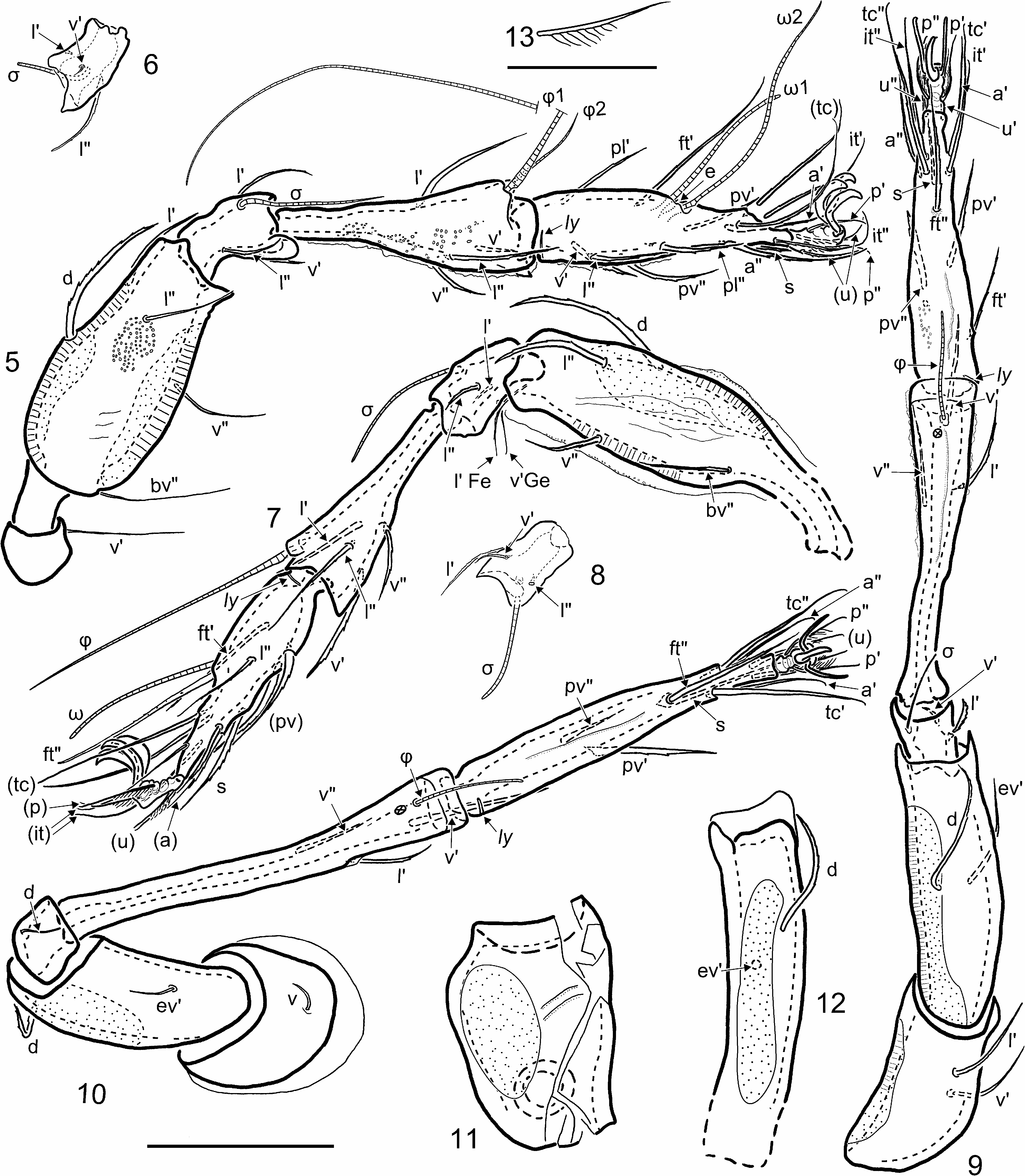
Legs, general aspects ( Figs. 5–13 View FIGURES 5–13 , 15, 16, 19–21 View FIGURES 14–25 ): Long (leg IV longest, about equal to body length), slender, covered with granular cerotegument. Tarsal lyrifissures (ly) laterally displaced to antiaxial position. Porose areas on all femora and on trochanters III–IV. Tarsi with noticeable sclerotization ending abruptly, just distal to setae (u); extended by narrow, hyaline and apparently flexible ambulacral stalk, broadening distally as pad into which setae (p) and claws insert ( Figs. 5, 9 View FIGURES 5–13 , 21 View FIGURES 14–25 ); pulvillus apparently absent (see Remark 1, below). Apotele tridactylous, with lateral claws slightly thinner than empodial claw. Leg setation presented in Table 1 and on respective figures. Distal setae—(p) on legs II–IV and (u) on all legs—with comb of fine cilia along ventral side ( Fig. 13 View FIGURES 5–13 ); other setae more or less barbed, no setae strongly curved except for curled tip. Each leg segment except tarsi with some type of ventrodistal projection encompassing base of subsequent segment, but forming tectum only on femora and genua.
1 Based on information from nymphs (see text), the following can be inferred: the iteral setae (it) on tarsi I–III first appear as a pair in the tritonymph; the accessory proximal setae l” and v’ on tarsus I, and l” on tarsus II, appear in the adult.
Leg I ( Figs. 5, 6 View FIGURES 5–13 , 19–21 View FIGURES 14–25 ): Tarsus without apophyses. Solenidion ω 1 nearly isodiametric, ω 2 much longer, attenuate, at least ω 2 having oddly shaped, keel-like base, unilaterally broadened on distal side, then narrowed to insertion; setae (it), (tc), ft' long, with ends slightly curved backwards and mostly smooth; (a), s, (pl), (pv), l ”, v ' more robust, pointed, barbed; seta ft” absent. Famulus baculiform, blunt, slightly inclined proximad (barely visible and probably partly destroyed on illustrated specimen, but seen intact on specimen UA-2195). Tibia: φ 1 very long, flagellate, curved, with conspicuous basal keel, inserted on blunt apophysis ( Fig. 19 View FIGURES 14–25 , arrows); φ 2 much shorter, attenuate, inserted proximolateral to φ 1, on tapered apophysis ( Fig. 20 View FIGURES 14–25 , arrows); setae (l), (v) similar in size, almost smooth. Genu: σ slightly widened basally, relatively long, attenuate; setae (l), v' setiform, slightly barbed, v' shorter than laterals and inserted almost in midline. Femur: with separate ventral and dorsal porose areas; seta d large, barbed; (l), v”, bv ” thinner, slightly roughened. Trochanter: small, poorly visible, bearing one seta (presumably v ’). Setal formula: 1–5–3(1)–4(2)–19(2).
Leg II ( Figs. 7, 8 View FIGURES 5–13 ): Tarsus: ωrather long, little tapered, without well-visible basal keel; ω 2 absent. Setae (it), (tc), (ft) thin, almost smooth, ft” inserted near ω; (a), s, (pv) stronger, clearly barbed. Tibia: φ long, straight, with basal keel, borne on small tubercle; (l), (v) setiform, slightly barbed. Genu: σ relatively long, slightly tapered; (l) slightly barbed, longer than v ', latter almost smooth, inserted near ventral midline as on genu I. Femur: with separate ventral and dorsal porose areas and longitudinal folds. Setae d and l" long, robust; l', v ”, bv ” thinner, all barbed. Trochanter bearing one long, straight barbed seta, presumably v’ (discerned only on MGCP 14). Setal formula: 1– 5–3(1)–4(1)–16(1).
Leg III ( Fig. 9 View FIGURES 5–13 ): Tarsus: setae (a), s, (it), (tc), (pv) similar to those on tarsus II; ft ” more robust, with short barbs; ft ' thin, positioned relatively low and proximal on segment. Tibia: solenidion φ slightly tapered, similar in length to setae; l' and (v) barbed. Genu: σ relatively long, thin; setae l ', v ' thick, barbed, half length of σ. Femur: with two conspicuous lateral lobes embracing genu; single porose area on paraxial side; setae d, ev ' barbed. Trochanter large, robust, with paraxial porose area; setae l ', v ' thin, barbed. Setal formula: 2–2–2(1)–3(1)–15.
Leg IV ( Figs. 10–12 View FIGURES 5–13 ): Tarsus very long, slightly curved ventrad, with longitudinal dorsal fold; setae (it), ft ”, s robust, (a) thinner, all almost smooth; setae (pv) robust and roughened. Tibia: very long, slightly curved ventrad, with unusually thick walls; φ thin, slightly tapered; v' robust, as long as φ; l' and v ” thinner, all setae barbed. Genu: only thin seta d visible. Femur very long and robust, with single, oval dorsal porose area; setae d and ev ' barbed. Trochanter large, with dorsal porose area and one thin seta. Setal formula: 1–2–1–3(1)–12.

No known copyright restrictions apply. See Agosti, D., Egloff, W., 2009. Taxonomic information exchange and copyright: the Plazi approach. BMC Research Notes 2009, 2:53 for further explanation.
|
Kingdom |
|
|
Phylum |
|
|
Class |
|
|
Order |
|
|
Family |
|
|
Genus |