
Stylochus alexandrinus Steinböck, 1937
|
publication ID |
https://doi.org/ 10.1080/00222930500081997 |
|
publication LSID |
lsid:zoobank.org:pub:E6427790-6DB4-4633-AD85-7A5FB041FF2D |
|
persistent identifier |
https://treatment.plazi.org/id/03EB1969-0306-FFEA-B6B7-E0F3CE26FB6D |
|
treatment provided by |
Felipe |
|
scientific name |
Stylochus alexandrinus Steinböck, 1937 |
| status |
|
Stylochus alexandrinus Steinböck, 1937 View in CoL
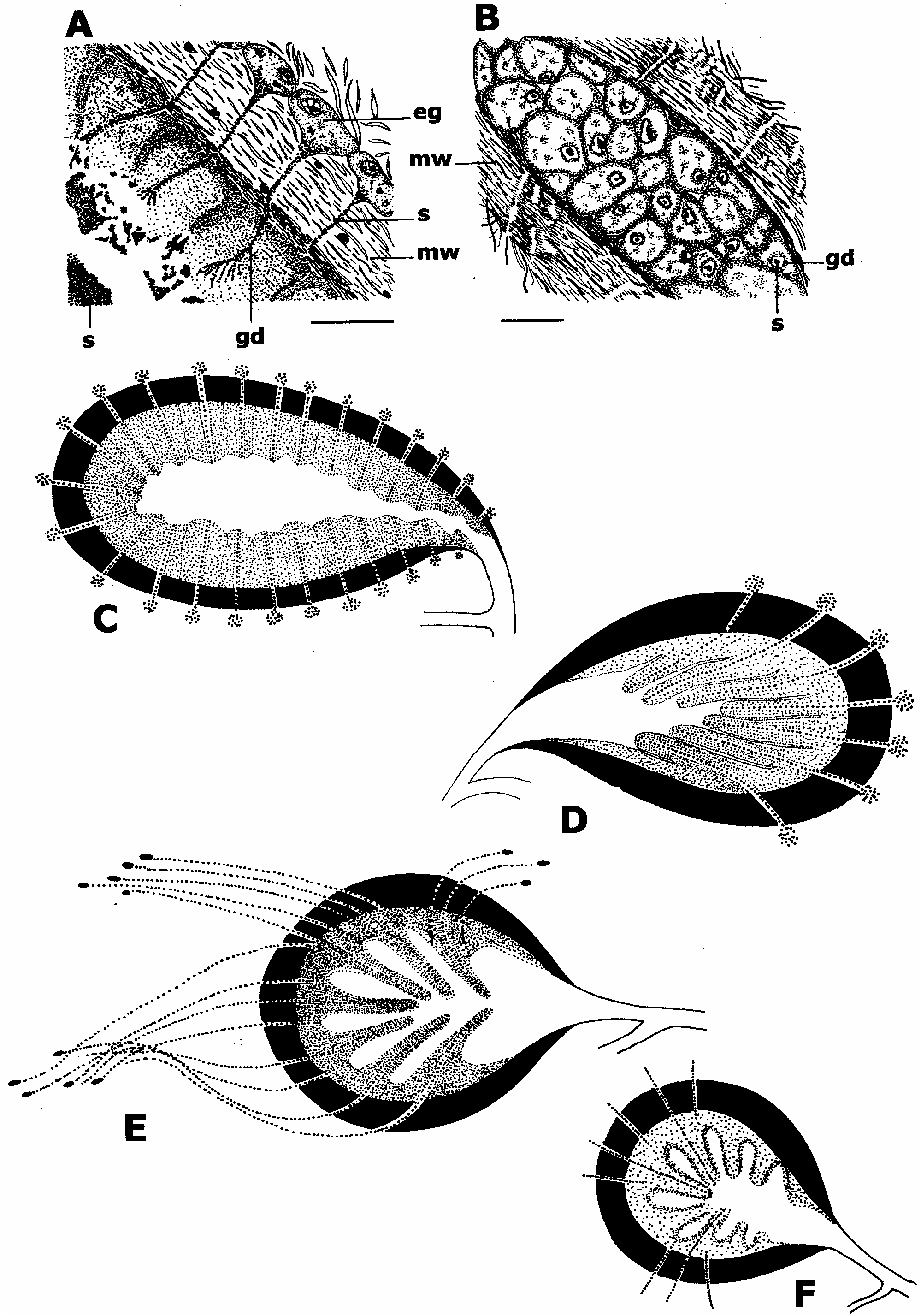
( Figure 2D View Figure 2 )
Material examined
Holotype: No. 3624, one sagittally sectioned specimen, locality: Alexandria, Egypt, leg. 8 August 1933, deposited in MNHV.
Morphological notes
In the following, only morphological characters which deviate from the description of Steinböck (1937, p 1–5) are added or re-described.
Reproductive system. Male copulatory apparatus directed backwards with elongate seminal vesicle, oval and muscular prostatic vesicle, and distal penis papilla. A common vas deferens joins the seminal vesicle from anterior. The prostatic duct joins the ejaculatory duct at the base of the penis papilla. Interior lining of prostatic vesicle with few finger-like extensions attached to the proximal half; finger-like extensions directed distad. Along the glandular epithelium of the prostatic vesicle, the ducts of the extra-vesicular glands open through multiple outlets into the lumen of the prostatic vesicle. Extra-vesicular glands numerous, positioned dorsally and ventrally in the parenchyma, surrounding the anterior part of the male complex ( Figure 2D View Figure 2 ). Male and female gonopores separate. Oviducts entering vagina from dorsal. From this junction the vagina continues frontad, then turns ventrad and opens via a female atrium to the exterior. Female tract non-ciliated; cement glands open into the distal part of the vagina.
Stylochus frontalis Verrill, 1892 View in CoL
( Figure 2E View Figure 2 )
Stylochus inimicus Palombi 1931, p 218 View in CoL –222, Figures 1–4 View Figure 1 View Figure 2 View Figure 3 View Figure 4 , Plate 4.
Stylochus tenax Palombi 1936, p 4 View in CoL –10, Figures 1 View Figure 1 –7, Plate I Figures 1 View Figure 1 , 2 View Figure 2 .
Material examined
Voucher specimen. No. 67505, one specimen sagittally sectioned, locality: Piscadera Baai, Curacao, Caribbean Sea, leg. D. D. Correa, 18 December 1965 ; Nos 67506–07, two specimens sagittally sectioned, locality: Virginia Key, Florida, North America , leg. D. D. Correa; January 1959 ; deposited in SMNH.
Morphological notes
In the following, only morphological characters which deviate from the description of Palombi 1931 (p 218–222) and Hyman 1940 (p 461–462) are added or re-described.
Reproductive system. Male genital system with ventral testes follicles, seminal vesicle, prostatic vesicle of polyglandular type, and distal penis papilla. The prostatic duct joins the ejaculatory duct at the base of the penis papilla. Prostatic vesicle roundish oval, with few long finger-like extensions directed distad, attached to proximal half of vesicle. Extravesicular glands at some distance from prostatic vesicle, with very long glandular ducts which cross the muscular wall of the prostatic vesicle and open along the glandular lining of the prostatic vesicle ( Figure 2E View Figure 2 ). Male atrium lined with glandular epithelium being completely ciliated. Female system as in Palombi (1936).


| SMNH |
Department of Paleozoology, Swedish Museum of Natural History |
No known copyright restrictions apply. See Agosti, D., Egloff, W., 2009. Taxonomic information exchange and copyright: the Plazi approach. BMC Research Notes 2009, 2:53 for further explanation.
|
Kingdom |
|
|
Phylum |
|
|
Order |
|
|
Family |
|
|
Genus |
Stylochus alexandrinus Steinböck, 1937
| Bulnes, V. N., Faubel, A. & Park, J. - K. 2005 |
Stylochus tenax
| Palombi A 1936: 4 |
Stylochus inimicus
| Palombi A 1931: 218 |