
Symmachia uirassu Dolibaina, Dias & Casagrande, 2020
|
publication ID |
https://doi.org/ 10.11646/zootaxa.4780.3.3 |
|
publication LSID |
lsid:zoobank.org:pub:CA5DA614-D448-4D95-B271-DDC8D56A37F60 |
|
DOI |
https://doi.org/10.5281/zenodo.3857488 |
|
persistent identifier |
https://treatment.plazi.org/id/A0AC5F0F-D580-4484-BE4F-C1314694EFA6 |
|
taxon LSID |
lsid:zoobank.org:act:A0AC5F0F-D580-4484-BE4F-C1314694EFA6 |
|
treatment provided by |
Plazi |
|
scientific name |
Symmachia uirassu Dolibaina, Dias & Casagrande |
| status |
sp. nov. |
Symmachia uirassu Dolibaina, Dias & Casagrande , sp. nov.
( Figs 25–26 View FIGURES 21–38 , 45 View FIGURES 39–47 , 54 View FIGURES 48–56 , 63 View FIGURES 57–66 , 97–98 View FIGURES 95–102 , 115–116 View FIGURES 113–122 , 129 View FIGURES 128–132 , 144 View FIGURES 138–146 , 153 View FIGURE 153 , 155 View FIGURES 154–156 ) http:// urn:lsid:zoobank.org:act:A0AC5F0F-D580-4484-BE4F-C1314694EFA6
Symmachia ? sp.; D’Abrera, 1994. Butt. Neot. Reg. IV Riod., p. 1044, figs [1–2] (male dorsal and ventral).
Symmachia praxila [misidentification]; Hall & Harvey, 2007. Trop. Lep. Res. 16(1–2), p. 7 [in part].
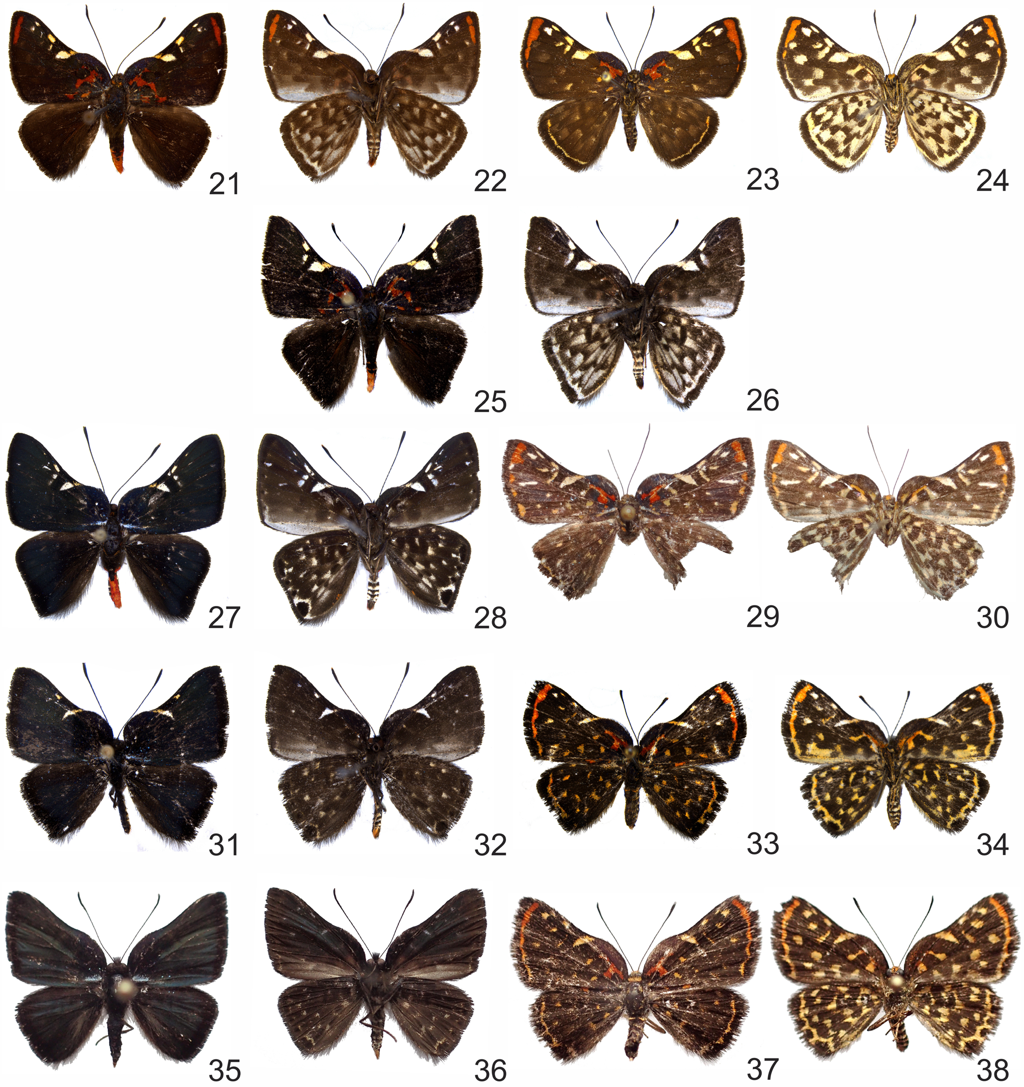
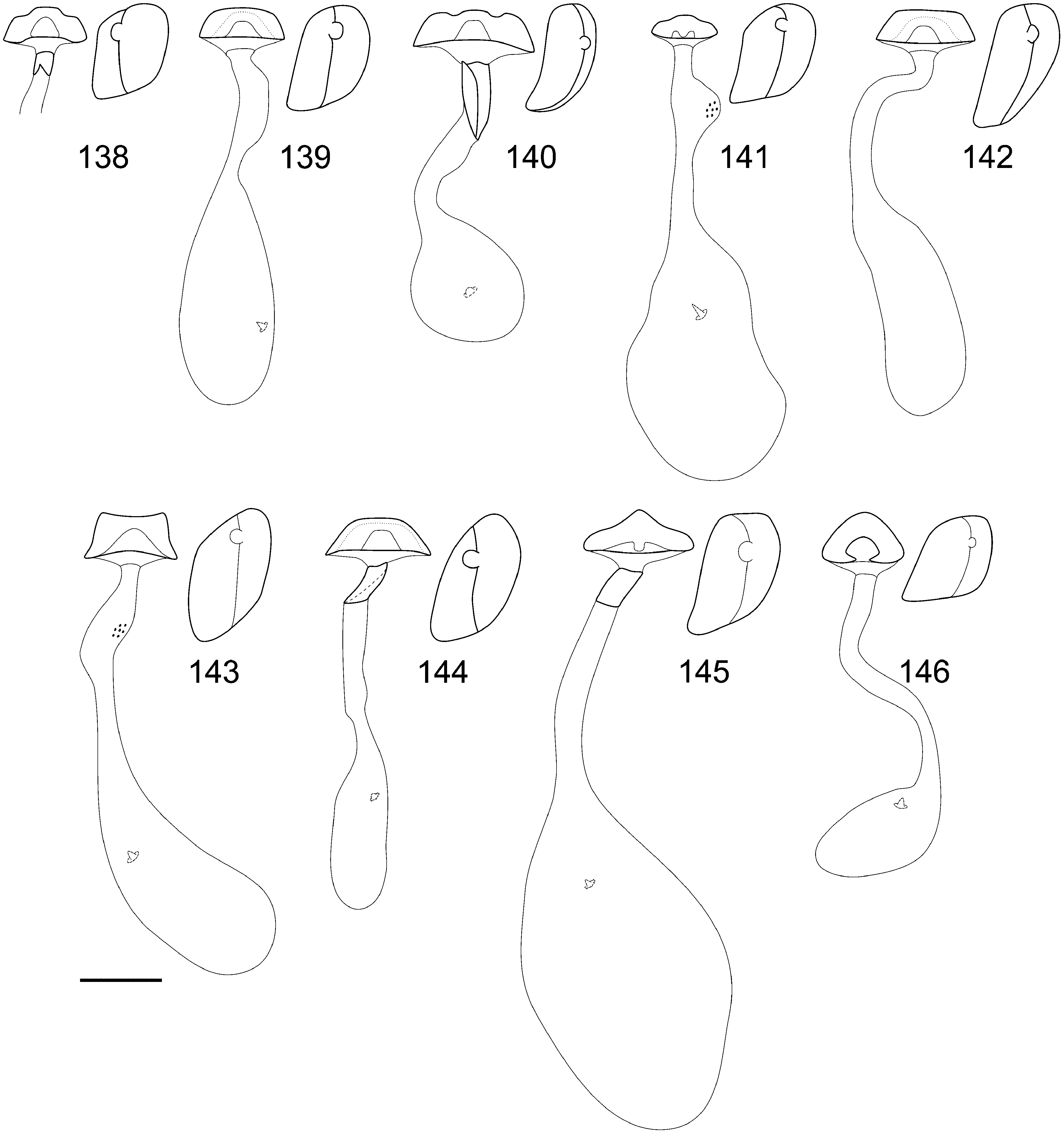
Diagnosis. Symmachia uirassu is sympatric with S. atlantica ( Figs 152–153 View FIGURE 152 View FIGURE 153 ). Males are most similar to S. praxila and S. divisora ( Figs 25–26 View FIGURES 21–38 ) and females although unknown likely to be similar to most species of the “Probetoriformes” species group. Males with frons, patagia, dorsal thorax and tegulae as in S. praxila ; FW strongly bulged; bulged area about 38% of the FW length; end of bulge aligned with the origin of M 3 ( Figs 25 View FIGURES 21–38 , 63 View FIGURES 57–66 ); FWD mostly black, with reddish orange spots on the basal area and a dark green sheen on the discal and postdiscal areas, and some light bluish green scaling on 1A ( Figs 25 View FIGURES 21–38 , 63 View FIGURES 57–66 ); FWD with a translucent white and creamy sickle-shaped spot between the discal cell and the costal margin, a small round whitish spots on the postdiscal area in R 2 –R 3 and M 1 –M 2 near the discal cell ( Figs 25 View FIGURES 21–38 , 63 View FIGURES 57–66 ); HWD homogeneously dark brown, with some reddish orange scales along the inner margin ( Fig. 25 View FIGURES 21–38 ); abdomen color pattern as in S. praxila ( Figs 97–98 View FIGURES 95–102 ); CAS size and color as in S. praxila ( Figs 115–116 View FIGURES 113–122 ); valva larger than in S. probetor ; aedeagus thick and long; vesica with a rounded patch of cornuti ( Fig. 129 View FIGURES 128–132 ).
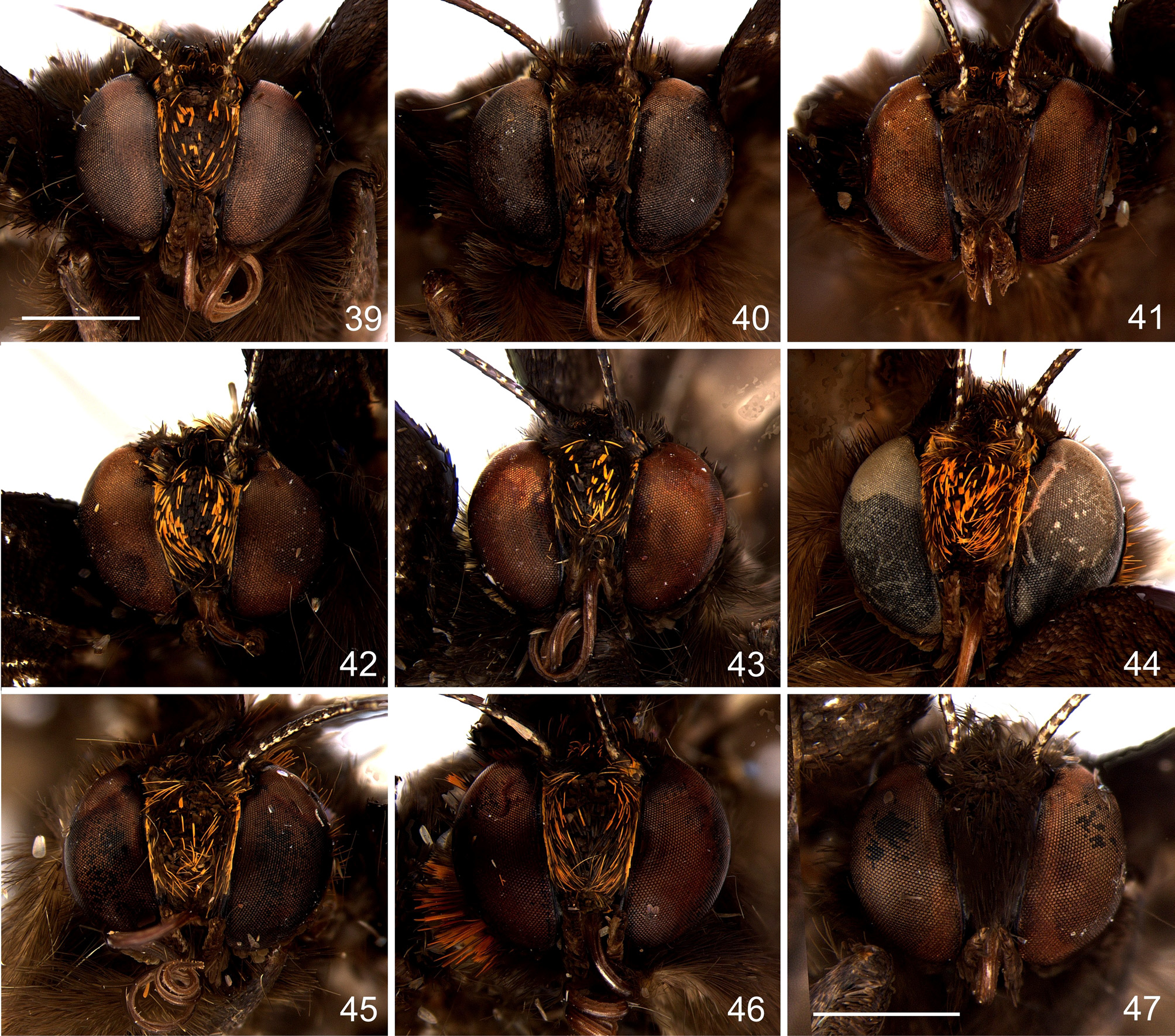
Description. Male. Head ( Figs 45 View FIGURES 39–47 , 54 View FIGURES 48–56 ): mostly dark brown and black, with some scattered reddish orange scales on the frons and bordering the compound eyes (some of the orange may have been lost); antenna black, each segment with proximal white scales, antennal club formed by the last 10–12 segments tip yellowish; labial palpus dark brown, short, not extending beyond middle of frons, first segment as long as half of the second segment, third segment about 1/6 as long as second.
Thorax ( Figs 25–26 View FIGURES 21–38 ): ventrally brown and dark brown; prothorax dorsally with reddish orange scales; mesothorax dorsally black, with a dark bluish sheen and some reddish orange scales; metathorax dorsally with long, dark brown and some reddish orange scales; legs mostly dark brown, each segment ventrally with creamy and white scales.
FW, shape and size: average size 1.16 cm (1.1–1.2 cm; n=3); roughly triangular; costal margin sinuous, strongly bulged in the basal area; apex pointed, angled; outer margin almost straight; tornus angled; inner margin almost straight.
FWD ( Figs 25 View FIGURES 21–38 , 63 View FIGURES 57–66 ): mostly black with iridescent dark blue and dark green scales, and translucent white, reddish orange and creamy spots; dark blue sheen on the costal margin bulge and on the basal area; dark green sheen roughly on the postbasal, discal and postdiscal areas; irregular reddish orange spots in the basal area of the discal cell, Sc–R, CuA–1A, and 1A and the inner margin; sickle-shaped spot formed by a translucent white spot in the discal cell, and creamy spots between the costal margin and Sc, Sc–R 1, R 1 –R 2 and most of the discal cell width; and further postdiscal spots in R 2 –R 3, M 1 –M 2 and M 2 –M 3, nearer to the discal cell, and in R 3 –R 4 and R 4 –M 1, nearer to the apex; fringe black.
FWV ( Fig. 26 View FIGURES 21–38 ): mostly brown to dark brown in a more or less checkered pattern, with a white band along the inner margin below 1A and on the postdiscal and submarginal areas of CuA 1 –1A; sickle-shaped spot and postdiscal spots as on the upper side, and further three fainter postdiscal spots in M 2 –M 3, M 3 –CuA 1, and CuA 1 –CuA 2.
HW, shape: roughly triangular; costal margin slightly convex; apex rounded; outer margin straight; tornus pronounced, angled; inner margin slightly convex.
HWD ( Fig. 25 View FIGURES 21–38 ): mostly black, with long, reddish orange scales in the basal area of CuA 2 –1A and along inner margin; fringe black.
HWV ( Fig. 26 View FIGURES 21–38 ): mostly dark brown, with whitish spots and a white submarginal band; whitish spots in a more or less checkered pattern from the basal to the postdiscal area; submarginal band straight and more or less regular from the apex to the tornus; some reddish orange scales along the inner margin.
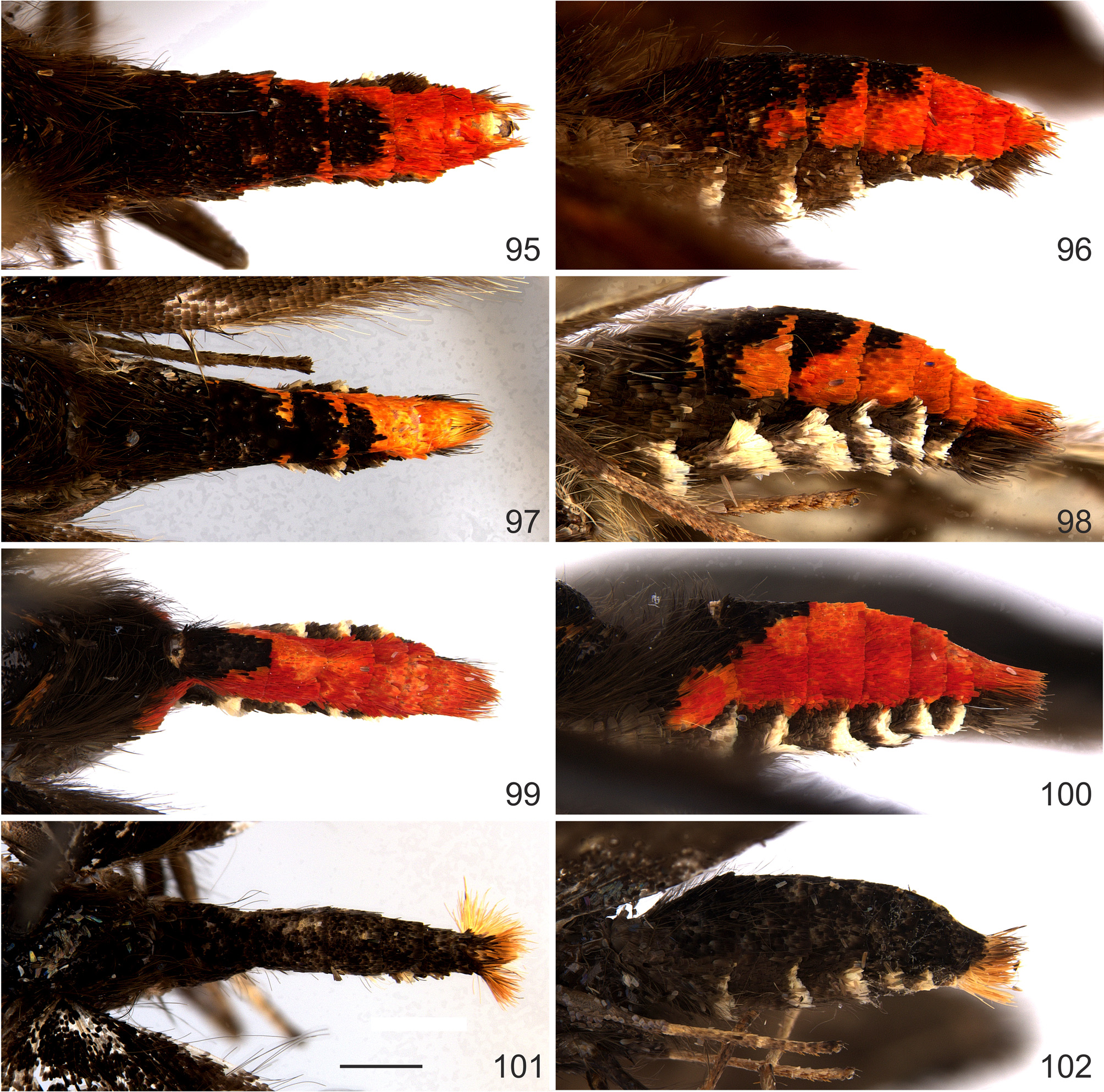
Abdomen ( Figs 97–98 View FIGURES 95–102 , 115–116 View FIGURES 113–122 ): terga mostly, dorsally and laterally, black, with a dark blue sheen, except segments 5–8 completely reddish orange, segments 3–4 with a narrow reddish orange stripe dorsally but almost completely laterally, and only few reddish orange scales laterally in segment 2; pleura black; sternites ventrally dark brown, with large distal white scales; genitalia surrounded by long scales, dorsally reddish orange, laterally and ventrally black; tergites 4 and 5 with an undivided band of CAS on the posterior margin, as long as 1/9 of the tergites’ length and as wide as half the tergites’ width, not projected anteriorly; CAS scales pale yellow.
Genitalia ( Fig. 129 View FIGURES 128–132 ): tegumen rectangular; membranous area between the uncus and the tegumen more or less triangular; uncus rectangular, with a medial anterior projection; gnathos C-shaped, angled, ventral part about one third larger than the dorsal, connected to the tegumen by a small dorsal projection; ventral arm of the tegumen thin and obliquely projected, connected ventrally to the wider dorsal arm of the saccus; anterior projection of the saccus wider than long and bilobed anteriorly; valva small, roughly triangular in lateral view, connected ventrally to the saccus and the pedicel, and dorsally to the opposite valva; pedicel posteriorly sclerotized and keeled, connected to the middle of the aedeagus; aedeagus roughly cylindrical, anterior opening wide and on the left side; vesica with an basal patch of very small pointed cornuti of similar size and a apical band of fewer and larger cornuti; subscaphium sclerotized.
Female. Unknown.
Etymology. This species is dedicated to the Instituto Uiraçu, a non-profit association established in 2001 by the eminent Brazilian lepidopterist Dr. Victor O. Becker to protect the Serra Bonita mountain range in southern Bahia, Brazil, and type locality of this species. The specific epithet means “Harpy Eagle” in the Brazilian indigenous Tupi language, and is composed of two elements: “uirá” (bird) and “açu” (big). The name is treated as a noun in apposition.
Distribution. This species is apparently restricted to coastal Atlantic Forest of Brazil (Bahia and Espírito Santo ( D’Abrera 1994)) ( Fig. 153 View FIGURE 153 ). It is not known if this species occurs further south along the Brazilian coast, in sympatry with S. praxila .
Comments. Symmachia uirassu is described based on three males specimens collected in RPPN Serra Bonita, Camacan, Bahia, Brazil. The specimen identified as “S ymmachia sp.?” illustrated by D’Abrera (1994: 1044 [1,2]) from Espírito Santo, Brazil corresponds to S. uirassu , and is recognized as an “undescribed taxon (…) probably probably related to S. accusatrix ”, and is identified as a male of S. praxila by Hall & Harvey (2007). However, the wings size and shape, the dark ground color, and the absence of the FWD reddish orange submarginal band and postdiscal costal spots clearly indentify this specimen as S. uirassu . Females still are unknown although it is possible they are misidentified with other species of the group in museum collections.
The holotype and paratypes were all collected in a hilltop at 920 m covered by a dense understory with bamboo and liana where they were flying and resting on leaves of trees up to five meters high ( Figs 154–155 View FIGURES 154–156 ) at about 11:00 h. Two males were collected in the second and one in the fourth day of collecting at this site occurring simultaneously with other species of Symmachia , including S. atlantica .
Type material. Male holotype of Symmachia uirassu with the following labels: / HOLOTYPUS / HOLOTY- PUS Symmachia uirassu Dolibaina & Dias det. 2019/ DZ 43.449 / BRASIL, BAHIA, CAMACAN, RESERVA SERRA BONITA, TRILHA DAS BROMÉLIAS 15º23’ S, 39º34’ W, 3–10.XII.2016 920m GoogleMaps , MIELKE, CARNEIRO, DIAS, DOLIBAINA & SANTOS LEG /. ( DZUP).
Paratypes. 2 males, same data as the holotype, DZ 36.622*, DZ 43.459* ( DZUP)


| DZUP |
Universidade Federal do Parana, Colecao de Entomologia Pe. Jesus Santiago Moure |
No known copyright restrictions apply. See Agosti, D., Egloff, W., 2009. Taxonomic information exchange and copyright: the Plazi approach. BMC Research Notes 2009, 2:53 for further explanation.
|
Kingdom |
|
|
Phylum |
|
|
Class |
|
|
Order |
|
|
Family |
|
|
Genus |