
Thaumatocranaus magnificus, Hara, Marcos R., Bragagnolo, Cibele & Pinto-Da-Rocha, Ricardo, 2017
|
publication ID |
https://doi.org/ 10.11646/zootaxa.4254.4.3 |
|
publication LSID |
lsid:zoobank.org:pub:29A83502-70C0-4A8F-82D5-CD692FC3219E |
|
DOI |
https://doi.org/10.5281/zenodo.6010184 |
|
persistent identifier |
https://treatment.plazi.org/id/F179007A-FF9B-FFC1-6CC5-CC28FC41FE1F |
|
treatment provided by |
Plazi |
|
scientific name |
Thaumatocranaus magnificus |
| status |
sp. nov. |
Thaumatocranaus magnificus sp. nov.
( Figs 3 View FIGURE 3 , 4 View FIGURE 4 , 7 View FIGURE 7 D, E)
Type material. COLOMBIA. Amazonas: Araracuara , ix.1988, M. Torres leg., male holotype ( ICN AO 352).
Diagnosis. Thaumatocranaus magnificus sp. nov. resembles T. splendidus sp. nov. because of the median longitudinal groove of scutal area I inconspicuous, lateral margin of dorsal scutum with an external row of tubercles increasing in size posteriorly, becoming large, conical and slightly acuminated, and the robust, unbranched apophysis on free tergite III. It can be distinguished from T. splendidus sp. nov. by the femur III unarmed on ventral face and retrodorsal apical spine on femur IV curved backwards and slightly retrolaterally.
Etymology. From the Latin magnificus , meaning eminent, distinguished, in reference to the distinguished and amazing effect of the depigmented spot on the dorsal scutum.
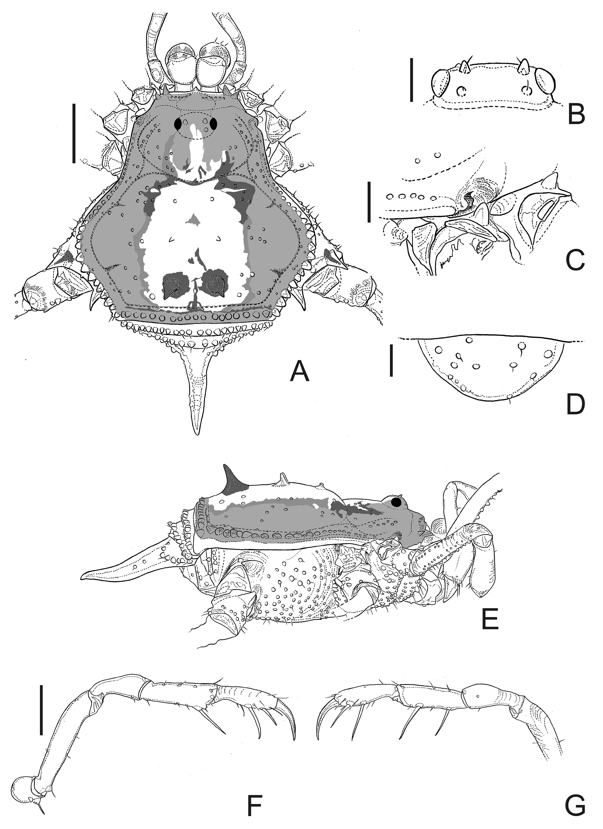
Description. Male (holotype): Dorsum ( Fig. 3 View FIGURE 3 A–E): Measurements: DSL 3.55; DSW 3.75; LI 8.95; LII 16.25; LIII 11.4; LIV 15.75. Carapace and scutal areas I–III with tubercles sparsely distributed. Median longitudinal groove of scutal area I inconspicuous. Lateral margin of dorsal scutum with an external row of tubercles increasing in size posteriorly, becoming large, conical and slightly acuminated. Free tergite III with a robust, unbranched apophysis.
Venter: Coxae I–IV tuberculate; coxae I–II with median and apical enlarged tubercles; coxa III with apical enlarged tubercles.
Chelicerae ( Fig. 3 View FIGURE 3 A): Segment I dorsal face smooth; movable finger with 10–11 teeth; fixed finger with 7–8 teeth.
Pedipalps ( Fig. 3 View FIGURE 3 F, G): Trochanter dorsal face smooth, unarmed, ventral face with 1 pair of enlarged setiferous tubercles (prolateral largest). Femur dorsal face smooth, with 2 ventral tubercles. Patella–tarsus with few dorsolateral scattered setiferous tubercles. Tibial setation: prolateral II, retrolateral I. Tarsal setation: prolateral and retrolateral IIi placed on distal half.
Legs ( Fig. 4 View FIGURE 4 ): Coxa IV with 1 prolateral and 1 retrolateral conical, apical apophyses (similar size). Trochanters I–IV with ventral slightly enlarged tubercles and 1 basoventral median enlarged tubercle; trochanters II–III with retrolateral enlarged tubercles. Trochanter IV with 1 prodorsal, enlarged, blunt subapical tubercle; 2 retrodorsal enlarged median tubercles (basalmost largest); retrolateral face with few slightly enlarged tubercles, 2 subbasal enlarged tubercles, and 1 apical, acuminated enlarged tubercle (this one largest). Femora I–II dorsal face unarmed; ventral face with 2 rows of tubercles; ventroapical face unarmed. Femur III with 1 pair of dorsoapical spines (retrolateral largest); ventral face with 2 rows of tubercles; ventroapical face unarmed. Femur IV slightly curved inwards (in dorsal view), with 1 pair of dorsoapical spines (similar size); ventral face with 2 rows of tubercles; ventroapical face unarmed. Patellae I–III unarmed; patella IV with 1 retrodorsal, acuminated, enlarged apical tubercle and few slightly enlarged tubercles on retrolateral face. Tibiae I–III unarmed. Tibia IV dorso- and ventroapical faces unarmed; ventral face with 2 rows of tubercles, retrolateral one with 4–5 acuminated, large tubercles, far from each other. Metatarsi I, IV and tarsi III–IV normally built (not inflated); metatarsus IV almost straight. Tarsal segmentation: 6(3), 10(3), 7, 7.
Penis ( Fig. 7 View FIGURE 7 D, E): Glans stylus unknown (stylus broken and lost during dissection). Ventral plate with 3 pairs of long MS C (distal most pair longest and strongly curved inwards), 1 pair of MS D laterally placed, 1 pair of MS A, 2 pairs of MS B, without MS E.
Coloration: Depigmented (pale beige) large spot on the median longitudinal third of dorsal scutum (from scutal grooves I–IV and as narrow stripes on carapace). Pair of spines on scutal area III dark brown. Body, trochanter IV, apexes of coxae I–IV, femora I–IV, tibiae I–IV, metatarsi I–IV and entire patellae I–IV brown. Trochanters I–III, remaining parts of the legs and body pale brown.
Female: Unknown.
Type locality. Colombia, Amazonas, Araracuara.
Geographical distribution. Known only from the type locality.


| ICN |
Instituto de Ciencias Naturales, Museo de Historia Natural |
No known copyright restrictions apply. See Agosti, D., Egloff, W., 2009. Taxonomic information exchange and copyright: the Plazi approach. BMC Research Notes 2009, 2:53 for further explanation.