
Trachelas brachialis, Zhang, 2017
|
publication ID |
https://doi.org/ 10.11646/zootaxa.4324.1.2 |
|
publication LSID |
lsid:zoobank.org:pub:09489Dcd-Afed-403C-8Aa2-D3E40A9A314F |
|
DOI |
https://doi.org/10.5281/zenodo.6010977 |
|
persistent identifier |
https://treatment.plazi.org/id/03A6772D-614F-1670-FF73-C992FCBD5273 |
|
treatment provided by |
Plazi |
|
scientific name |
Trachelas brachialis |
| status |
sp. nov. |
Trachelas brachialis sp. n.
Figs 9C–D View FIGURE 9 , 10–12 View FIGURE 10 View FIGURE 11 View FIGURE 12
Type material. Holotype: ♂, CHINA: Hunan Province: Dongan County, Damiaokou Township, Shunhuangshan Mountain National Forest Park , Nvying Creek (26°24′08.042″N, 111°02′03.646″E), 679m a.s.l., 9 October 2015, leg. Chi Jin GoogleMaps . Paratypes: 5♀, same data as holotype GoogleMaps ; 4♀, Hunan Province: Dongan County, Damiaokou Township, Shunhuangshan Mountain National Forest Park , Ehuang Creek (26°24′58.684″N, 111°02′08.473″E), 694m a.s.l., 8 October 2015, leg. Chi Jin, Jingchao He and Xiangbo Guo. All specimens are deposited in MHBU GoogleMaps .
Etymology. The specific name is a Latin adjective meaning "brachial". It refers to the arm-like embolus.
Diagnosis. The male of this species can be easily distinguished from all the other Old World Trachelas species by 1) the palp with one large, long and one small, spine-shaped DTA; 2) embolus arm-like and very thick, with two apophyses apically (considering the spiraling ridges preceding the copulatory openings, these apophyses might both be serving as functional conductors, hooking into the ridges when the embolus enters the CO). The female resembles T. tanasevitchi in having a small upwardly opening hood on the epigynal plate, but can be distinguished from it by: 1) the hood situated posteriorly, whereas hood situated medially in T. tanasevitchi ; 2) connecting ducts long, forming two coiled circles anteriorly before proceeding posteriorly along midline, whereas short and almost straight in the latter species; 3) ST2 smaller than ST1, whereas ST2 larger than ST 1 in T. tanasevitchi .
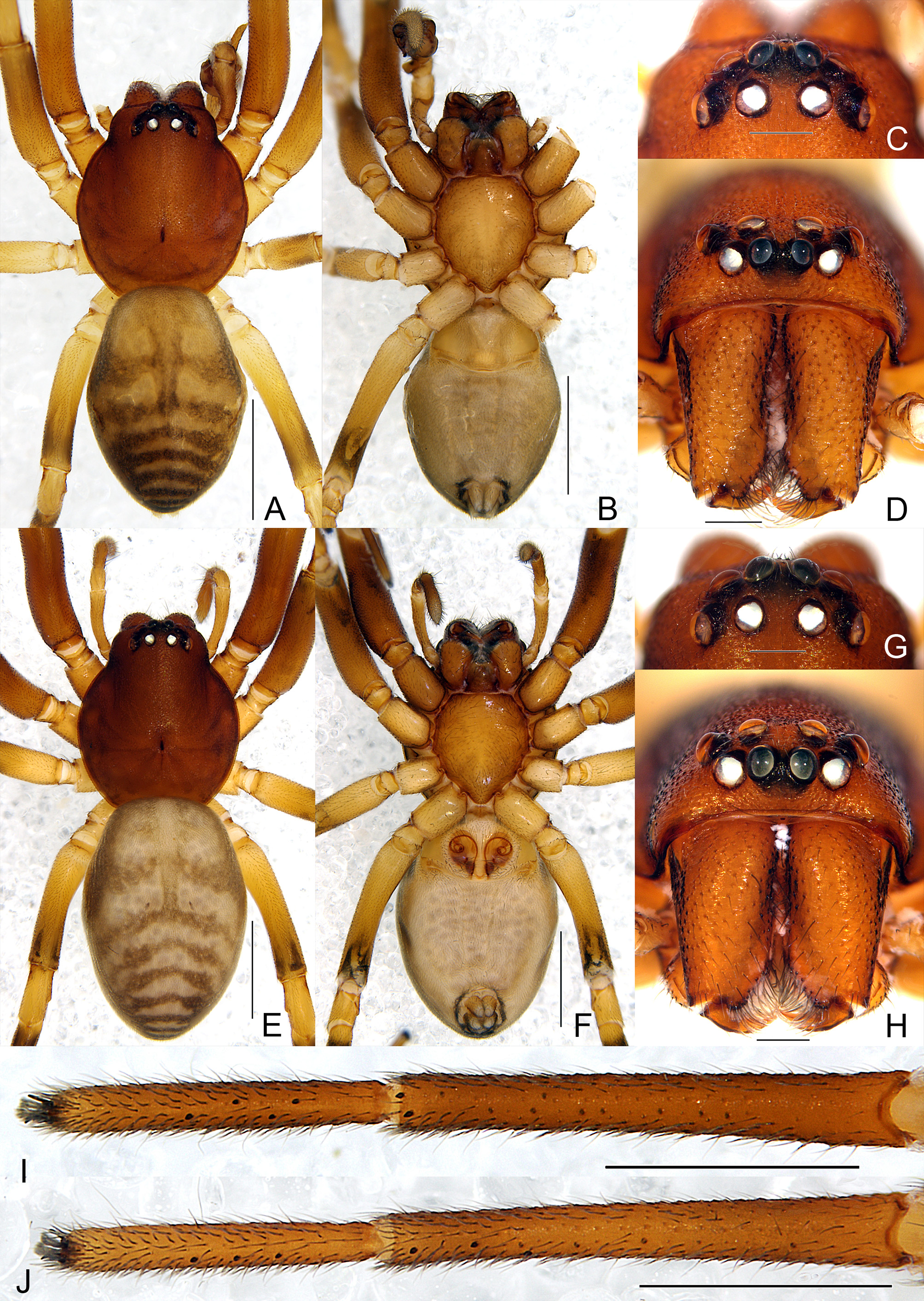
Description. Male ( Figs 10A–D, I–J View FIGURE 10 , 11A–D View FIGURE 11 , 12 View FIGURE 12 ). Holotype: body 3.56 long; carapace 1.66 long, 1.44 wide; abdomen 1.90 long, 1.35 wide. Carapace ( Fig. 10A View FIGURE 10 ) brown, ovoid in dorsal view, highest between fovea and PER, densely covered with tiny granulations. CRW 0.90, 0.63 times carapace width. Fovea dark brown, distinct. Eyes ringed with black. AER and PER recurved in dorsal view ( Fig. 10C View FIGURE 10 ). Eye diameters: AME 0.10, ALE 0.11, PME 0.12, PLE 0.12. Eye interdistances: AME–AME 0.12, AME–ALE 0.07, PME–PME 0.10, PME–PLE 0.09, ALE– PLE 0.07. MOA 0.25 long, anterior width 0.24, posterior width 0.31. PERW 0.59, 0.66 times CRW. Clypeus height 0.14, wider than diameter of AME.
Chilum ( Fig. 10D View FIGURE 10 ) triangular, sclerotized and brown, with posterior median indentation. Chelicerae yellowbrown, granulated as carapace, cheliceral boss pronounced, with three promarginal and two retromarginal teeth. Endites and labium ( Fig. 10B View FIGURE 10 ) light yellow-brown; endites without oblique depression; labium as wide as long. Sternum light yellow-brown, shield-shaped, with sharp precoxal triangles; intercoxal sclerites distinctly present between coxae I and II, II and III, and III and IV.
Legs light yellowish-brown; short, black ventral leg cusps present on tarsi and metatarsi I–II, arranged sparsely in two lines on leg I, in single line on leg II ( Figs 10I –J View FIGURE 10 ). Measurements of legs: leg I 5.10 (1.63, 0.68, 1.26, 0.94, 0.59), II 5.18 (1.57, 0.63, 1.20, 1.12, 0.66), III 3.56 (1.04, 0.48, 0.68, 0.91, 0.45), IV 5.06 (1.47, 0.56, 1.15, 1.38, 0.50). Leg formula: 2143. Abdomen oval, light yellowish-brown, grey laterally and medially, posterior part with several grey chevrons; dorsal scutum absent ( Fig. 10A View FIGURE 10 ). Venter pale grey, with four narrow lines of sclerotized spots, barely visible.
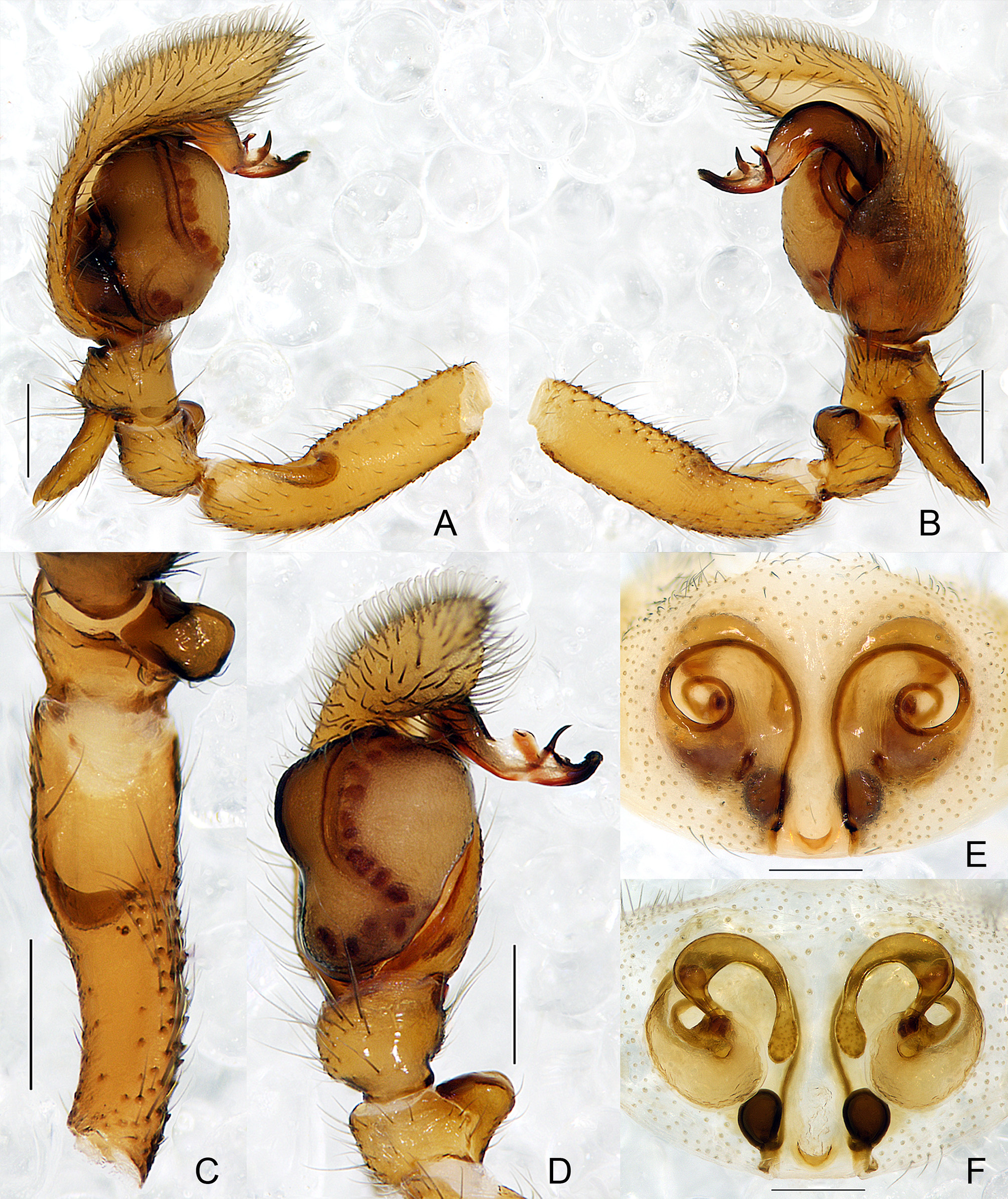
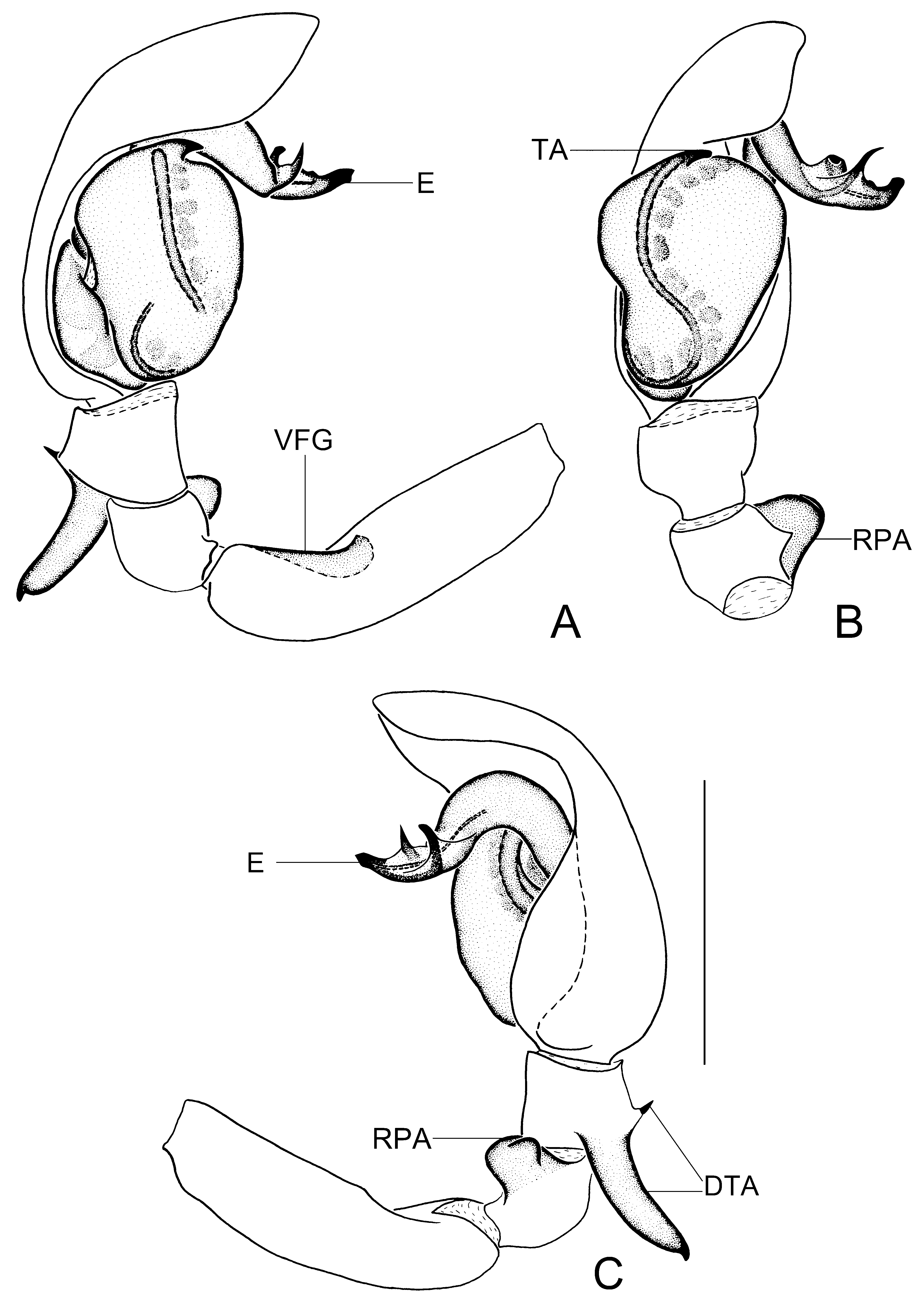
Palp as illustrated ( Figs 11A–D View FIGURE 11 , 12 View FIGURE 12 ). Femur with large, shallow ventral terminal groove; patella with large subrectangular apophysis, almost as long as wide, retrolaterally with small blunt projection. Tibia with one large and one small DTA; large one long and thick, with sharp projection pointed posteriorly; small one spine-shaped, pointed anteriorly. Tegulum inverted pear-shaped, distally rounded, S-shaped sperm duct discernable through translucent cuticle; subtegulum nearly invisible in dorsal view. Embolus well-developed, arm-like, extremely thick, originating retrolaterally, with one finger-shaped and one spine-shaped functional conductors apically. Tegular apophysis short, apically inserted, pointed retrolaterally. Cymbium somewhat bent apically towards retrolateral side, along with embolus and over the tegulum.
Female ( Figs 9C–D View FIGURE 9 , 10E–H View FIGURE 10 , 11E–F View FIGURE 11 ). Total length 3.76–4.46 (n = 9). One paratype: body 4.46 long; carapace 1.95 long, 1.67 wide; abdomen 2.51 long, 1.70 wide. CRW 1.02, 0.61 times carapace width. Eye diameters: AME 0.12, ALE 0.13, PME 0.13, PLE 0.13. Eye interdistances: AME–AME 0.06, AME–ALE 0.01, PME–PME 0.12, PME–PLE 0.12, ALE–PLE 0.07. MOA 0.28 long, anterior width 0.28, posterior width 0.34. PERW 0.65, 0.64 times CRW. Clypeus height 0.14, slightly wider than diameter of AME. Leg measurements: I 6.03 (1.86, 0.76, 1.42, 1.22, 0.77); II 5.88 (1.78, 0.71, 1.38, 1.29, 0.72); III 4.05 (1.20, 0.56, 0.81, 1.03, 0.45); IV 5.85 (1.68, 0.62, 1.42, 1.61, 0.52). Leg formula: 1243. Legs without cusps. Sternum with sharp precoxal triangles; intercoxal sclerites distinctly present between coxae I and II, and II and III, but not between III and IV. Other characters as in male.
Epigyne ( Figs 9C View FIGURE 9 , 11E View FIGURE 11 ): poorly sclerotized, with small hood posteriorly, opening upward; spiraling ridges present, preceding copulatory openings; copulatory openings small, pore-like, situated medially, far away from each other, above pair of oval invaginated luminas. Vulva ( Figs 9D View FIGURE 9 , 11F View FIGURE 11 ): ST2 small and oval, with gland pores, connecting with curved inflated copulatory ducts; connecting ducts long and slender, visible through translucent cuticle, coiled twice anteriorly before entering ST1; ST1 oval, connected to basally weakly sclerotized FD.
Distribution. Known only from the type locality ( Fig. 19 View FIGURE 19 ).


No known copyright restrictions apply. See Agosti, D., Egloff, W., 2009. Taxonomic information exchange and copyright: the Plazi approach. BMC Research Notes 2009, 2:53 for further explanation.
|
Kingdom |
|
|
Phylum |
|
|
Class |
|
|
Order |
|
|
Family |
|
|
Genus |