
Trichomyia pintoi Santos & Leite
|
publication ID |
https://doi.org/ 10.5281/zenodo.213986 |
|
DOI |
https://doi.org/10.5281/zenodo.6180789 |
|
persistent identifier |
https://treatment.plazi.org/id/03B1027A-2C65-9B51-62AC-FE6AFA80FB82 |
|
treatment provided by |
Plazi |
|
scientific name |
Trichomyia pintoi Santos & Leite |
| status |
sp. nov. |
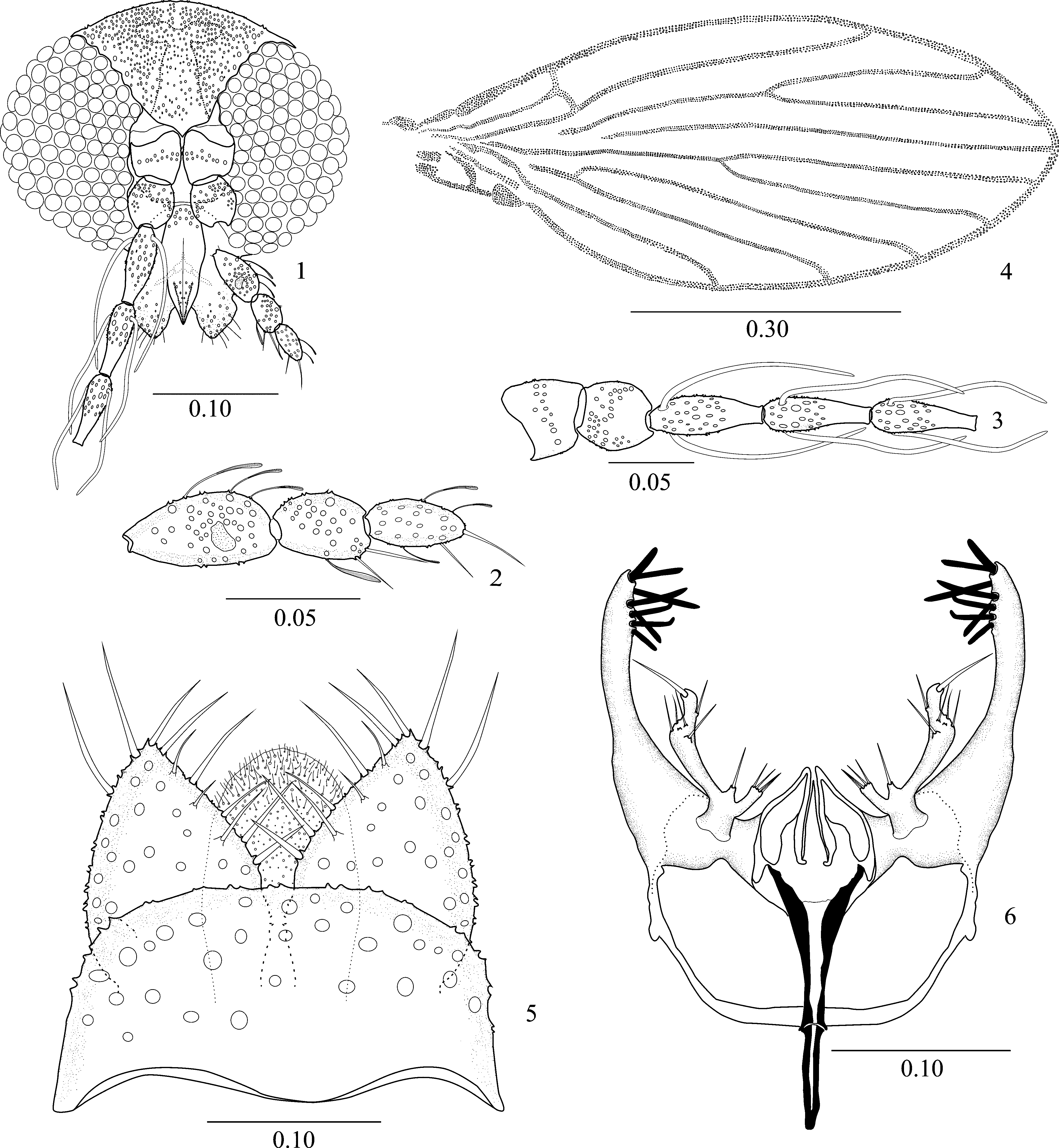
Trichomyia pintoi Santos & Leite sp. nov.
( Figs. 1–6 View FIGURES 1 – 6 )
Diagnosis. Head subcircular. Maxillary palpus with 3 segments, sensory pit present on the first palpomere. Ascoids paired in flagellomeres. Wing with rounded apex. Gonocoxite with 8 strong spines deployed in the distal third. Gonostylus deployed at the base of gonocoxite.
Description. Male holotype. Total length 1.1 mm, pale brown. Head subcircular in frontal view, slightly flattened dorsoventrally. Antenna incomplete, only 7 segments present ( Fig. 1 View FIGURES 1 – 6 ). Scape irregular; spherical pedicels; flagellomeres cylindrical and tapered with a pair of transparent ascoids ( Fig. 3 View FIGURES 1 – 6 ). Palpus with 3 palpomeres, sensory pit on internal surface of the first palpomere; palpomere proportions: 1.0:0.6:0.5 ( Fig. 2 View FIGURES 1 – 6 ). Labrum triangular; clypeus rectangular. Wing 2.4× longer than wide; Sc-R fused distally with R1 and C; r-m and m-cu missing; radial fork in the distal half of wing ( Fig. 4 View FIGURES 1 – 6 ). Terminalia: cercus longer than broad in ventral view, with bristles inserted on posterior extremity; sternite 10 shorter than cercus, extremity truncated micropilose ( Fig. 5 View FIGURES 1 – 6 ). Gonocoxite long, fused basally with hypandrium, wide base and with 8 sclerotized spines deployed in the distal third. Gonostylus, originating at the base of gonocoxite, with 3 tubers. At its base 2 smaller tubes originate: the more robust with 4 apical spines and the less robust with a single apical spine. The long tube is ornamented by a subapical spine followed by an apical spine. Aedeagal apodeme simple, short, and sclerotized. Aedeagus symmetric, long in relation to the apodeme and sclerotized. Parameres bifid in dorsal view ( Fig. 6 View FIGURES 1 – 6 ).
Type material. Brazil, Espírito Santo State, Santa Teresa municipality, Reserva Biológia Augusto Ruschi, (19°54ʹS 40°33ʹW; 820 m. a.s.l.), holotype male, 28.IX.2011, Santos, CB, 3 paratype males with the same data as that of the holotype.
Type locality. The specimens of Trichomyia were collected in Vale do Canaã, Reserva Biológica Augusto Ruschi, Santa Teresa, located in the Atlantic Rain Forest Biome, Brazil.
Etymology. The specific name is in honor of the researcher of Psychodidae Dr. Israel de Souza Pinto.
Taxonomic comments. Few previous studies have examined the taxonomy of Trichomyiinae . Bravo (1999, 2001) classified some Neotropical species of Trichomyia in 2 subgenera: Opisthotrichomyia, including species with the first 2 palpus segments fused and Septemtrichomyia, including species with bristles present on the seventh segment of the abdomen. However, the species with 3 palpus segments were not separated into other subgenera. Bejarano et al. (2009) discussed an evolutionary line for Western Hemisphere species with 4 separate palpus segments. Future studies on phylogeny and phylogeography might clarify the taxonomy of Trichomyiinae .
No known copyright restrictions apply. See Agosti, D., Egloff, W., 2009. Taxonomic information exchange and copyright: the Plazi approach. BMC Research Notes 2009, 2:53 for further explanation.