
Tylopus spinisterna, Nguyen, 2012
|
publication ID |
https://doi.org/ 10.5281/zenodo.5347271 |
|
DOI |
https://doi.org/10.5281/zenodo.5449263 |
|
persistent identifier |
https://treatment.plazi.org/id/191D7C72-CD26-E81F-FE97-9779FCEEF9D0 |
|
treatment provided by |
Tatiana |
|
scientific name |
Tylopus spinisterna |
| status |
sp. nov. |
Tylopus spinisterna View in CoL , new species
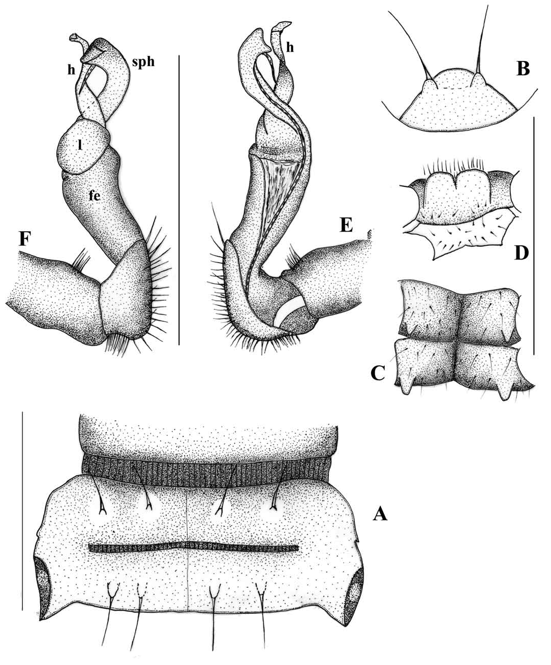
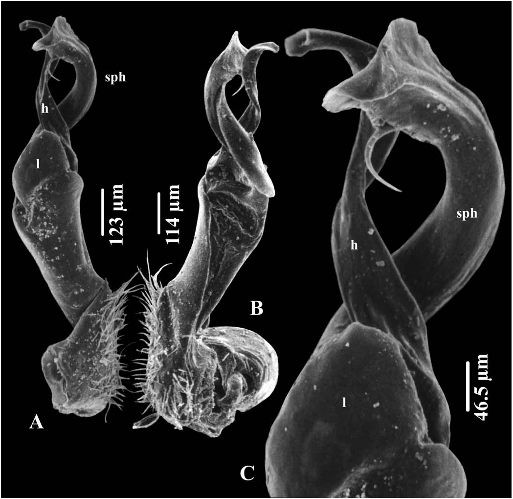
( Figs. 3 View Fig , 4 View Fig , 18 View Fig )
Material examined. — Holotype: Male in the bottle IEBR-142H, with a label “ Lam Dong Province, Bi Doup-Nui Ba National Park (12°00'– 12°19'N, 108°21'– 108°44'E), corn field, 1400 m a.s.l., pitfall traps, coll. Nguyen Duc Anh, 2–9 Apr.2008 ”. GoogleMaps
Paratypes: 7 males, 5 females (IEBR-142P), same data as holotype GoogleMaps .
Diagnosis. — The species can be distinguished from its congeners by the combination of the following characters: all sterna except for sternum 5 with four projections; metaterga strongly rugose, with two rows of 2+2 and 2+2 setiferous knobs/tubercles; solenophore and process h of gonopod subequal; gonopod lamina l present, suboval-shaped, but lobe m and spine z absent.
Etymology. — “ spinisterna ” is a noun in apposition and emphasizes the presence of spiniform projections on the sterna.
Description. — Size: Body length 16–[19]– 20 mm (male), 23–24 mm (female). Width of midbody prozona 1.4–[1.5] mm (male), 2.0– 2.2 mm (female) and metazona [1.8]– 1.9 mm (male), 2.6–2.7 mm (female).
Colouration: Head, pleura, prozona, and anterior half of metaterga brown; posterior half of metaterga divided into three regions: a black one in middle surrounded by two paler regions. Paraterga, legs, sterna and antenna brownish yellow except for distal part of antennomere 6 and whole antennomere 7 blackish brown.
Head: Slightly broader than collum; labrum densely setose. Epicranial suture deep, dividing frons into two parts; each part of frons with 2+2 long setae along epicranial suture. Antenna claviform, long, reaching body ring 4 laterally. Antennomere 3=4=5>6=2>1& 7 in length.
Collum: Slightly narrower than body ring 2, subtrapeziform; surface considerably rugose, with row of 3+3 setae in anterior half, and two additional rows of 1+1 knobs and 2+2 setiferous tubercles in posterior half. Paratergum small, with a setiferous incision laterally.
Body ring 3<4<2= 5 in width, body parallel-sided on rings 5–16, thereafter gradually tapering towards telson. Prozona dull. Metaterga strongly rugose, with rows of 2+2 and 2+2 setiferous knobs in front of and behind transverse sulcus, respectively. Metatergal transverse sulcus broad, finely striate, and present on body rings 4–19, but fully developed (reaching base of paraterga) on body rings 5–19 ( Fig. 3A View Fig ). Pleura with dense microgranules. Pleurosternal keels well developed on body rings 2–7, reduced to small acute spines on body rings 8–11, and obliterated on body ring 12 and subsequent body rings. Axial line evident. Waist between pro- and metazona broad, finely striate.
Paraterga: Well developed, with two lateral setiferous incisions more obvious on poreless paraterga. Caudal corner acute, exceeding posterior contour of metaterga. Calluses largely reduced ( Fig. 3A View Fig ). Ozopores located on lateral side of paraterga of body rings 5, 7, 9–10, 12–13, 15–19.
Telson: Epiproct long, broadly truncated with two small terminal tubercles. Tip with four spinnerets. Hypoproct subtriangular, with two small distolateral setiferous knobs ( Figs. 3B View Fig ).
Sterna: Densely setose; transverse and longitudinal sutures poorly developed. Each sternum with four small projections near coxae ( Fig. 3C View Fig ); anterior pairs of projections slightly stronger and longer than posterior pairs. Male sternum 5 with a bifid lamina between coxae 4 ( Fig. 3D View Fig ).
Legs: Thin and slender, about 1.3 (male), 1.2 (female) times as long as midbody height. Tarsal brushes present on leg pairs 1–15, missing on subsequent leg pairs. Prefemora not swollen. Neither large nor micro tubercles present ventrally.
Gonopod ( Figs. 3E,F View Fig , 4A,C View Fig ): Coxite stout, slightly shorter than femorite; distoventral part densely setose. Prefemur densely setose, set off from femorite by an oblique sulcus laterally. Femorite slender, somewhat expanded distally and grooved mesally. Postfemoral region demarcated from femorite by an oblique suture laterally. Suboval lamina l and process h present, but spine z and both lobe m, n absent. Process h weakly twisted, as high as solenophore. Solenomere flagelliform, completely sheathed by modestly coiled solenomere.
Distribution. — Known only from the type locality ( Fig. 18 View Fig )

No known copyright restrictions apply. See Agosti, D., Egloff, W., 2009. Taxonomic information exchange and copyright: the Plazi approach. BMC Research Notes 2009, 2:53 for further explanation.
|
Kingdom |
|
|
Phylum |
|
|
Class |
|
|
Order |
|
|
Family |
|
|
Genus |