
Veigaia leruthi Willmann, 1935
|
publication ID |
https://doi.org/ 10.1080/00222933.2010.535913 |
|
DOI |
https://doi.org/10.5281/zenodo.10529077 |
|
persistent identifier |
https://treatment.plazi.org/id/03B8D10A-FF86-FF90-FDDD-FA52FB86FB95 |
|
treatment provided by |
Carolina |
|
scientific name |
Veigaia leruthi Willmann |
| status |
|
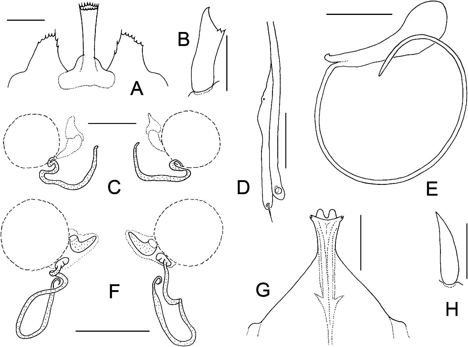
( Figures 2 View Figure 2 E–H, 3)
Veigaia leruthi Willmann, 1935: 12–18 , figs 5–12.
Veigaia leruthi Willmann 1936: 255–256 , fig. 12; Sellnick 1940: 26; Farrier 1957: 47–49, pl. 17, figs 1–9; Błaszak et al. 2004: 169; Błaszak et al. 2006: 7–8.
Material examined
One female – Belgium, Namur Province , Jemelle Village, Grotte du Fayt Cave, soil, 2 December 2000, leg. G. Rochez ; one male – Belgium, Liège Province , Engis Village , Grotte de Rosée Cave , Crystal Palace Dome (“ Palais de Cristal ”), soil, 28 August 2002, leg. G. Rochez and M. Dethier.
Description (female)
Dorsal idiosoma ( Figure 3A View Figure 3 )
Idiosoma 842 µm long and 481 µm wide. Dorsal shield 738 µm long and 451 µm wide, with deep lateral incisions reaching almost to setae J1; those incisions terminally slightly curved and directed posteromedially. Podonotal region longer than wide (longer than opisthonotal region), subtrapezoid, widely rounded anteriorly, with ill-defined lateral shoulders, almost parallel posterolaterally, smooth, bearing 21 pairs of simple, smooth and spine-like setae, excluding setae r4 situated on soft integument off the shield. Anterior margin of podonotum connected with peritremes, peritremes with anterior ends reaching close to bases of setae j1. Opisthonotal region wider than long, semicircle-like, widely and regularly rounded on posterior margin, smooth and without sculptural reticulation on surface, bearing 12 pairs of simple, smooth and spine-like setae. Most of dorsal setae with minute basal mamelons and generally subequal in size; but length of podonotal shield setae slightly decreasing posteriorly, especially in setae of dorsocentral j row: j1 77–81 µm, j2 50–53 µm, j3 45–48 µm, j4 40–44 µm, j5 and j6 37–40 µm. Dorsocentral J setae on opisthonotum subequal in length, 41–46 µm long. Among all dorsal shield setae only s1 and s2 conspicuously shorter (s1 13 –14 µm, s2 15 –17 µm), r5 and r6 moderately shorter (r5 23–26 µm, r6 31–32 µm), j2, z1 and r2 moderately longer (50–53 µm), vertical setae j1 distinctly longer (77–81 µm). Longest opisthonotal setae between 44 and 48 µm long, J5 off the shield and 40–42 µm long. Posterior soft integument with a pair of small suboval sclerites close to posterior margin of idiosoma .
Ventral idiosoma ( Figure 3B View Figure 3 )
A pair of poorly defined presternal platelets present. Sternal shield oblong, 214 µm in length, weakly sclerotized, anterior and posterior margins not clearly defined, surface smooth except for ill-defined short sculptural lines in lateral areas; anterolateral corners well-developed, wide; lateral sternal constriction between coxae II normal and 133 µm wide. Sternal shield with three pairs of setae and two pairs of lyrifissures, setae st1 moderately longer than st2 and st3 (st1 55–58 µm, st2 and st3 37–42 µm). Each metasternal plate free, abutting to endopodal platelets III–IV, bearing seta st4 (42–45 µm) and an associate lyrifissure, suboval to subtriangular in shape, not connected with sternal shield. Genital shield tongue-shaped, sinuous laterally, weakly sclerotized and hyaline in anterior part, smooth, bearing three pairs of unequal setae (st5 41–44 µm, Jv1 45–47 µm, Zv1 13 µm), fused with anterolateral corners of ventral shield in metapodal regions. Spherical endogynial structure with robust and well-sclerotized spine directed posteriorly present and positioned between endopodal platelets III–IV, under anterior surface of genital shield. Ventral shield wider than long, 315 µm in width, subrectangular, with widely rounded posterior corners, bowllike, posteriorly almost straight, only weakly reticulate to transversely striate on lateral surfaces, with four pairs of subequal setae (Jv2 and Jv3 45–48 µm, Zv2 31–32 µm, Zv3 52–53 µm); anterolateral corners free from elements of parapodal or peritrematal shielding, each punctiform organ in metapodal regions with two pores. Peritrematal shields reduced to very narrow posterior strips, seemingly each with a minute poststigmatic seta on posteriormost surface. Anal shield subtriangular, slightly wider than long (111 µm wide and 103 µm long), smooth, with three circum-anal setae and a pair of preanal setae; preanal setae markedly longer than circum-anal setae: Jv4 54 µm, postanal and adanal setae 32–33 µm. All setae on ventral shields simple, needlelike and mostly smooth. Dorsolateral and ventrolateral membranous integument with 12 pairs of simple, smooth, spine-like setae.
Gnathosomal structures
Epistome partly obscure, with a long central process and much shorter lateral cusps; central process barbed distally, lateral cusps serrate on anterior margin. Chelicerae
normal for genus, 683–690 µm long; distal segment 450–467 µm long. Corniculi normal, horn-like, without additional denticles ( Figure 2H View Figure 2 ).
Spermathecal structures ( Figure 2F View Figure 2 )
Coxae IV associated with well-developed and sclerotized tubular structures. Tubes well-observable, evenly sclerotized, thin, strongly elongated, with several bends and short terminal part angustate, hooked and pointed on tip. Each tube connected with a sacculus and an associated horn-like structure, both abutting inner surface of coxa IV.
Legs
All legs with a well-developed pretarsus and ambulacral apparatus including a pulvillus and two claws. All leg segments without projections. Tibia IV with a long and upright dorso-distal sensory macroseta (97 µm). Tarsus I 190 µm, tibia I 162 µm and tarsus IV 238 µm long.
Male
Dorsum with a compact holodorsal shield. Venter with separate sternogenital and ventri-anal shields; ventri-anal shield completely fused to holodorsal shield. Chaetotaxy similar to that of females. Cheliceral spermatodactyl strongly elongated, 318 µm in length, thin and curved ( Figure 2E View Figure 2 ). Epistome bilobed apically, smooth laterally, distinct from that of females, as in Figure 2G View Figure 2 .
Notes
Because of the adequate viewing conditions enabling us to see most of the important morphological features of Veigaia leruthi , we have been able to discern more detail than is reported in the original descriptions of Willmann (1935, 1936), or in their English version translated by Farrier (1957). There is only one more description of this species, that of Błaszak et al. (2004), based on a very brief enumeration of the main diagnostic characters. The components of the sperm induction system and endogynium are described here for the first time, including chaetotaxy of the schizodorsal shield, various metric data, and character of peritrematal regions.
Veigaia leruthi can be easily distinguished from the other known species of the V. exigua group, especially by the length of idiosoma (the largest species in this species group), form of tubular structures of the insemination apparatus (which is associated with coxae IV), and form of spherical structure as a component of the endogynium. For main distinguishing characters see Tables 1 and 2, and the key to species identification.
Key to the females of European species of the Veigaia exigua group
1. Dorsum with a single laterally incised schizodorsal shield; opisthonotal region of dorsal shield with 12 pairs of setae, setae J5 on soft integument off the shield; anal shield with five setae; ventral and genital shields fused in metapodal regions (species of Veigaia exigua group)...................... 2 Dorsum with separate podonotal and opisthonotal shields; opisthonotal region of dorsal shield with more than 12 pairs of setae, setae J5 on the shield; anal shield with three circum-anal setae; ventral shield free, completely separated from genital shield.............................................
................................................... other species of Veigaia
2. Metapodal punctiform organs each with at most two pores; vertical and paravertical setae subequal in length: verticals j1 never more than 1.5 times longer than paraverticals z1; central projection of epistome lanceolate: long, slender, barbed distally, slightly expanded subapically and pointed apically; hypostomal corniculus normal, regularly tapered distally and smooth................................................................. 3
Metapodal punctiform organs each with at least seven pores; vertical and paravertical setae widely varying in length: verticals j1 at least 2.5 times longer than paraverticals z1; central projection of epistome columniform (truncate and serrate apically) or calycoideous (conspicuously expanded distally); hypostomal corniculus usually partly modified, slightly broadened distally, and with two small denticles or tubercles on subapical surface............. 6
3. Lateral projections of epistome narrowed, tapering apically and serrate on lateral margins; spermathecal structures associated with coxae IV welldeveloped, with long tubular components................................ 4
Lateral projections of epistome widened and with well-differentiated anterior margin widely rounded and densely serrate; spermathecal structures associated with coxae IV rudimentary, without tubular components........
............................................. Veigaia exigua (Berlese, 1916)
4. Small species, idiosoma never more than 600 µm long; spherical part of endogynium absent or weakly sclerotized, without any spine-like projection; terminal parts of spermathecal tubes formed otherwise: widely rounded or connected to sacculus................................................... 5
Large species, idiosoma more than 775 µm long; endogynium with wellsclerotized spherical structure bearing robust spine directed posteriorly; tube of insemination apparatus with short terminal part angustate, hooked, and apically pointed..........................................................
............................................ Veigaia leruthi Willmann, 1935
5. Tube of insemination apparatus simple, very elongated, ribbon-like, and with numerous bends, each coxa IV associated with one tube....................
.............................................. Veigaia agilis (Berlese, 1916)
Insemination apparatus with a pair of tubes mutually connected in central sacculus, each coxa IV associated with two tubes..........................
............................................. Veigaia pusilla (Berlese, 1916)
6. Central process of epistome strongly expanded distally, bowl-like to calyxlike, with well-differentiated and serrate anterior margin.................. 7 Central process of epistome narrow, columniform, with almost parallel lateral margins, truncate and serrate apically.................................... 8
7. Metapodal punctiform organ with about 17 pores; podonotal shield with 22 pairs of setae, marginal setae r4 situated on the shield; spermathecal structures associated with coxae IV poorly developed, tubular components absent; hypostomal corniculus normal, regularly tapered distally and smooth; lateral projections of epistome narrowed and pointed distally.....................
................................. Veigaia garraldensis Athias-Henriot, 1961
Metapodal punctiform organ usually with seven pores; podonotal shield with 21 pairs of setae, setae r4 off the shield; spermathecal structures and tubular components well-developed, terminal part of tubes more sclerotized and broadened, almost club-like or rostrum-like; hypostomal corniculus slightly broadened distally and with two small obtuse denticles on subapical surface; lateral projections of epistome with well-developed anterior margin widely rounded and densely serrate..............................................
.............................................. Veigaia slovaca Mašán, 2008
8. Podonotal shield with 21 pairs of setae, setae r4 off the shield; spermathecal tubes relatively thick; hypostomal corniculus with two subapical tubercles; central process of epistome shorter, not exceeding lateral projections, and with parallel lateral margins; post-stigmatic setae placed on soft integument, off the peritrematal shields...................................................
................................... Veigaia perinsolita Athias-Henriot, 1961
Podonotal shield with 22 pairs of setae, marginal setae r4 on the shield; spermathecal tubes relatively thin; hypostomal corniculus with two subapical denticles; central process of epistome longer, markedly reaching beyond lateral projections, and slightly expanded distally; post-stigmatic setae positioned on peritrematal shields......................................................
..................................................... Veigaia hubarti sp. n.

No known copyright restrictions apply. See Agosti, D., Egloff, W., 2009. Taxonomic information exchange and copyright: the Plazi approach. BMC Research Notes 2009, 2:53 for further explanation.
|
Kingdom |
|
|
Phylum |
|
|
Class |
|
|
Order |
|
|
Family |
|
|
Genus |
Veigaia leruthi Willmann
| Mašán, Peter, Grazyna & Madej 2011 |
Veigaia leruthi
| Blaszak C & Ehrnsberger R & Skorupski M 2006: 7 |
| Blaszak C & Skorupski M & Ehrnsberger R 2004: 169 |
| Farrier MH 1957: 47 |
| Sellnick M 1940: 26 |
| Willmann C 1936: 256 |
Veigaia leruthi
| Willmann C 1935: 18 |