
Xenogaster pilosula Seevers, 1957: 103
|
publication ID |
https://doi.org/ 10.1016/j.jcz.2023.07.005 |
|
publication LSID |
lsid:zoobank.org:pub:5FDAD7BE-908C-448A-872E-42B72E38145E |
|
DOI |
https://doi.org/10.5281/zenodo.10374979 |
|
persistent identifier |
https://treatment.plazi.org/id/0390EB6A-FFE4-D735-7138-6F8AFE5FA07A |
|
treatment provided by |
Felipe |
|
scientific name |
Xenogaster pilosula Seevers, 1957: 103 |
| status |
|
Xenogaster pilosula Seevers, 1957: 103 View in CoL (original description).
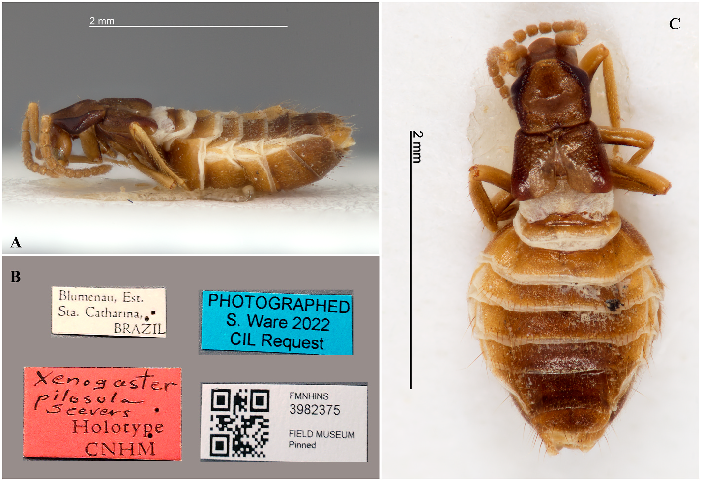
Material Examined: HOLOTYPE ( Fig. 3A–C View Fig ). BRAZIL. Santa Catarina: Blumenau, with Nasutitermes sp. PARATYPES (3 specimens, undetermined sex, pinned): BRAZIL. Santa Catarina: Blumenau, with Nasutitermes sp. , FMNH (ex Reichensperger collection). 1 specimen (undetermined sex, pinned): BRAZIL. Minas Gerais: Passa Quatro (ex Reichensperger collection), with Nasutitermes ehrhardti, FMNH. Additional material: 5 specimens (3 #m, 2 #f; in EtOH 70%): BRAZIL, Passa Quatro – Minas Gerais, FMNH 4128765 (= Paratypes of X. subnuda ). 1 #M, BRAZIL, S˜ao Paulo: Ubatuba (L. R. Fontes col.) last segments dissected on slides, with Nasutitermes ehrhardti, MZSP 21432. 1 #F, BRAZIL, Santa Catarina: Blumenau ( R. L. Araújo col.), dissected on slides, with Nasutitermes sp. , MZSP 21154.
Measurements: Female: 3–3.2 mm.
Male: 2.9–3.1 mm.
Diagnosis: Body light-brown, with margins of pronotum and elytra; and posterior region of head and tergites, darker. Abdomen physogastric, with membranous areas between tergites and sternites. X. pilosula differs from the other species in Xenogaster by the shape of the frons, which is broadly arcuate.
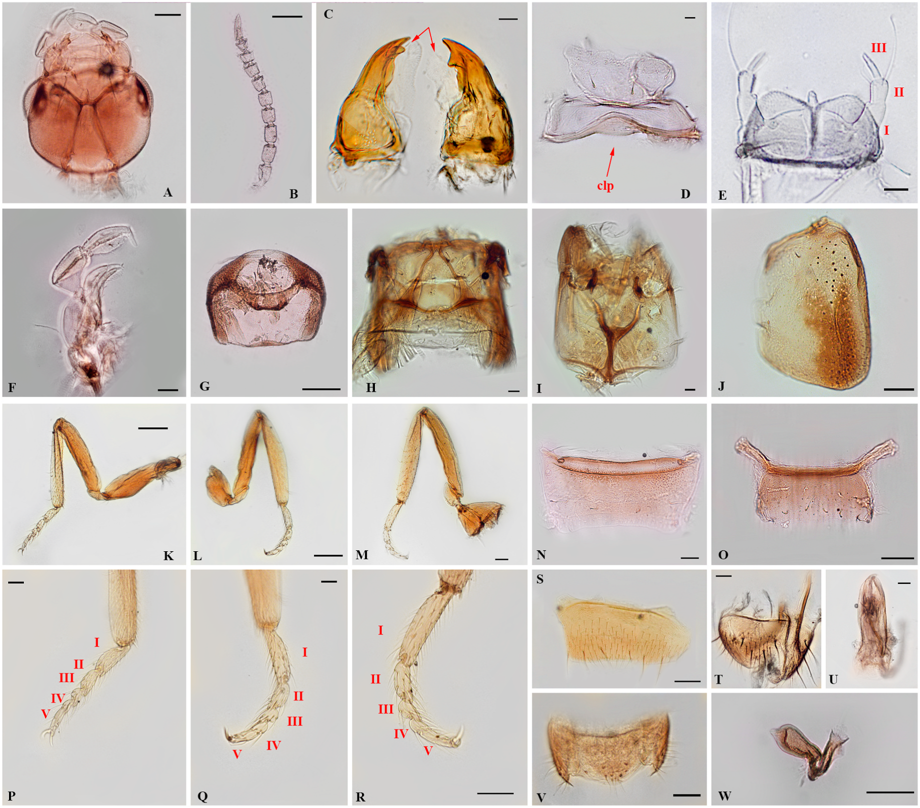
Redescription. Head ( Fig. 4A View Fig ) slightly longer than wide, gradually forming rounded posterior angles; dorsal surface of head finely punctuate, slightly depressed between antennal insertions. Frons distinctly and broadly arcuate, bearing row of bristles on margin. Clypeus ( Fig. 4D View Fig , indicated by arrow) transverse and membranous, broadly and deeply emarginate on anterior margin. Labrum ( Fig. 4D View Fig ) membranous, trapezoidal and transverse, covered with sparse and short bristles. Gula fused with submentum with almost 2/3 of head length, diverging and wider posteriorly. Antennae ( Fig. 4C View Fig ) with 11 antennomeres, scape elongate and antennomere 2 with sparse and short bristles; 2–9 longer than wide, densely covered with short and long bristles; 2–4 subequal in length; 5–10 slightly decreasing in length; 10 distinctively transverse; antennomere 11 conical, bearing a pair of coeloconic sensilla. Mandibles ( Fig. 4C View Fig ) assymetrical; most differences occurring on subapical tooth, closer to apical region on left mandible; broader at base and gradually constricting at apex; subapical region well projected on both mandibles, but more conspicuously on left mandible. Mandibular p prostheca occupying almost entire mesal length (In Fig. 4C View Fig indicated by arrows). Small sensitive pores at base. Maxilla ( Fig. 4F View Fig ) with cardo subelliptical and transverse; stipes rectangular, longer than wide, bearing long bristles near basal region; lacinia and galea arcuate, similar in length; lacinia narrow, with a distal comb of bristles across inner margin; distal lobe of galea with a comb of thick bristles; sparse and moderately long bristles across internal margin of galea. Maxillay palpi 4-articulated, arcuated; palpomere 1 reduced, subquadrate; 2 subcylindrical, dilated at apex; 3 oval, covered with long bristles, shorter than previous; 4 narrow, subulate. Labium. Labial palpi ( Fig. 4E View Fig ) 3-articulated; palpomere 1 broader, longer than wide; 2 the shorter, with two long bristles on each side of anterior margin; 3 longer and narrow, longer than previous, with almost same length of palpomere 1. Ligula ( Fig. 4E View Fig ) broader, bilobed; emarginate at middle, with rounded digitiform projection at center of emargination. Prementum rounded in general; anterior margin slightly curved, with a pair of bristles symmetrically distributed near edges; two longitudinal rows of three bristles centered; small pores on whole surface.
Prothorax ( Fig. 4G View Fig ). Pronotum slightly elongate and pentagonal; light-brown with sides darker; pronotum surface irregularly grooved and with finely punctures and short bristles; pronotal disc broad and deeply impressed; anterior region slightly elevated, with straight angles diverging posteriorly; lateral margins parallel on posterior 2/3, convergent on third; posterior margin weakly sinuose, with rounded angles. Each lateral margin with a longitudinal row of three long bristles; two bristles near anterior margin and one on posterior region. Meso- and metathorax ( Fig. 4I View Fig ) partially membranous, with one pair of bristles on posterior region. Endosternite Y-shaped; basal stalk being gradually broader anteriorly; furcal arms almost reaching level of mesocoxal cavities, without any apparent anterior arms; bilobed at base, lobes slightly longer; distally bifurcate. Scutellum transverse, somewhat trapezoidal. Metanotum ( Fig. 4H View Fig ) partially membranous; one pair of long bristles on anterior region. Elytra ( Fig. 4J View Fig ) elongate, wider on posterior region; clear ferruginous, outer margin darker, giving impression of distinct longitudinal stripe; coarsely punctate, with grainy appearance; lateral margins of elytra parallel with posterior angles slightly rounded; anterior margin declivous; inner margins of elytron completely separated from each other behind scutellum; distal margins slightly declivous and convergent; angles slighly rounded; elytra surface covered by sparse and short bristles, but bearing one long bristle near anterior margin and two bristles centered on medial region. Legs ( Fig. 4K – M View Fig ) with procoxa transverse, somewhat flattened; mesocoxa subspherical, mesocoxal cavities widely separated; metacoxa triangular, narrowly separated by basal process of metendesternite. Tibia slightly longer than femur, coarsely covered with long and thick bristles along entire length; inner and outer margin of protibia straight; outer margin of meso- and metatibia slightly convex and inner margin straight; meso-, and metatrochanter almost same length, protrochanter shorter, each bearing a long bristle. Pro-, meso-, and metatarsi each with two unciform claws, and a dentiform projection between claws; tarsomeres ( Fig. 4P – R View Fig ) bearing sparse bristles; tarsal formula 5-5-5, with 4th and 5th tarsomeres almost fused, present a clear line of division between them. Pro-, meso-, and metatarsi increasing in length; metatarsi with almost same length of tarsomeres 2–5 combined.
Abdomen. physogastric. Tergites and sternites separated by 2 pairs of paratergites in abdominal segments III – VI; inner paratergites well sclerotized, subquadrate, bearing a transversal row of bristles on posterior margin; paratergites of segment VII narrow and triangular; inner paratergites of segments V – VII with a single erect bristle on surface; outer paratergites narrow, with two long bristles on surface. Surface of abdominal tergites and sternites coarsely porose; with row of long bristles on posterior margin. Segment I membranous, with tergite fused to metanotum, glabrous; segment II with reduced tergite and sternite; tergite II shaped as transverse plate with anterior margin broadly emarginate at middle; sternite II transverse, sclerotized, present only in fully-grown physogastric specimens. Tergites III – VI transverse; III with anterior margin weakly emarginate at middle; IV – VI with anterior margin heavily sclerotized; tergites III – VI with two transversal rows of long bristles: one row of erect bristles near posterior margin, and one row on posterior edge. Tergite VII ( Fig. 4N View Fig ) subtrapezoidal, wider than long, anterior margin straight with a pair of glandular reservoirs, each widely separated; posterior margin broadly and weak emarginate, forming rounded angles; tergite with transversal row of four long setae in medial region, densely punctate on whole surface, with short and sparse bristles; posterior margin bearing fringe of bristles. Tergite VIII wider than long, anterior margin slightly emarginate at middle, with prominent angles on female, and broadly rounded on male; lateral margins rounded; one row of four long bristles near posterior margin, symmetrically distributed. Sternite VIII subquadrate, wider than long, with long lateral extensions on anterior margin of female ( Fig. 4O View Fig ), and with narrow and shorter angles on male ( Fig. 4S View Fig ); transversal row of six bristles near posterior margin; symmetrically distributed small bristles and pores on surface near posterior region. Sternite IX represented by a pair of hemisternites laterally attached to tergite IX on females. Tergite IX subtriangular, covered with long bristles and two long bristles in a longitudinal row parallel to lateral margin; male tergite IX with long thick apodemes ( Fig. 4T View Fig ). Tergite X ( Fig. 4T View Fig male, 4V female) in unique piece, wider at base, slightly tapering through apex, which is rounded; surface covered with short bristles, and two transversal rows of long bristles: more distantly separated proximal row, and a closely arranged distal row. Aedeagus ( Fig. 4U View Fig ). Median lobe piriform, curved dilated at base. Lateral lobes with three segments; distal segments longer; proximal segment broader, with line of division from medial segment. Spermatheca ( Fig. 4W View Fig ) sclerotized, stem elongated; capsule dilated at apex.
Remarks: X. pilosula is easily distinguished in the genus by the shape of the frons, which is broadly arcuate, contrasting with the straight to emarginate in the other species. Moreover, some X. pilosula specimens studied were intermediate stages of the development of physogastry. Some studied specimens display a distinctive physogastric abdomen, but the lack of the sternite II, the absence of post-sternite areas, and further areas of sclerotization on the paratergites led to conclude that they are not full-grown specimens; and some conditions related to post-imaginal growth have been underestimated in literature, herein detailed discussed in the next section.
It is noteworthy that we analyzed five out of nine paratypes of X. subnuda Seevers (FMNH 4128765) from Passa Quatro – Minas Gerais, and in fact, they are misidentified specimens and belong to X. pilosula . The paratypes from Blumenau - Santa Catarina, and Ilha Grande - Rio de Janeiro are definitely X. subnuda .
No known copyright restrictions apply. See Agosti, D., Egloff, W., 2009. Taxonomic information exchange and copyright: the Plazi approach. BMC Research Notes 2009, 2:53 for further explanation.
|
Kingdom |
|
|
Phylum |
|
|
Class |
|
|
Order |
|
|
Family |
|
|
Genus |
Xenogaster pilosula Seevers, 1957: 103
| Pires-Silva, Carlos M. & Zilberman, Bruno 2023 |
Xenogaster pilosula
| Seevers, C. H. 1957: 103 |