
Yorknia aprostatica, Schockaert & Curini-Galletti & Ridder & Volonterio & Artois, 2009
|
publication ID |
https://doi.org/ 10.1111/j.1096-3642.2008.00463.x |
|
persistent identifier |
https://treatment.plazi.org/id/BE459720-FFCB-A849-E6F7-D905FC3F6CBE |
|
treatment provided by |
Felipe |
|
scientific name |
Yorknia aprostatica |
| status |
sp. nov. |
YORKNIA APROSTATICA SP. NOV.
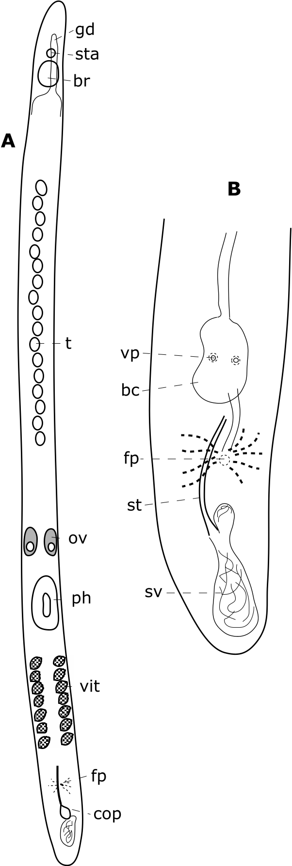
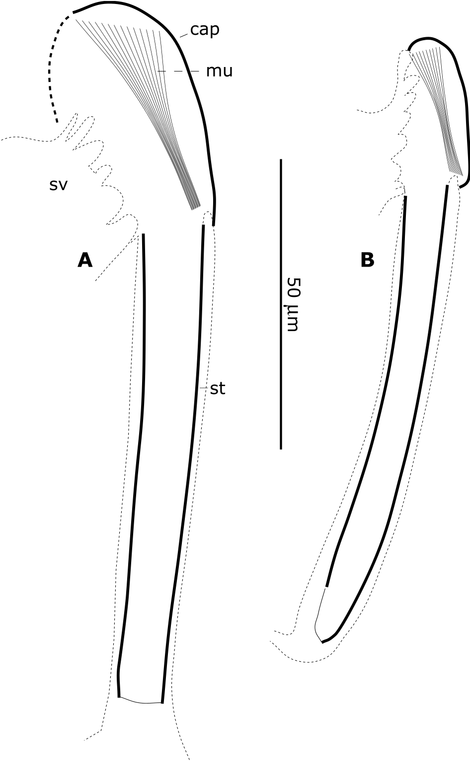
Diagnosis: Species of Yorknia with 15 testes in a row, without either prostate glands or vesicle, and with an almost straight stylet of about 80-Mm long, with a proximal ‘cap’ of 30–40 Mm in diameter, and with a single large seminal vesicle. Female pore only slightly behind the male pore; prepenial copulatory bursa with two vaginas.
Occurrence: Cairns, Australia (Queensland), at Yorkney’s Knob ; intertidal in fine to medium-fine sand (type locality) (October 1993) .
Material studied: A whole mount, designated as the holotype, ( QM G 230107 ). A second whole mount and six sectioned animals (UH 386 and UH 387–392) .
Etymology: The genus name is derived from the locality where the species was found; the species epithet is derived from the most striking character, namely the absence of prostate glands.
Description: The animals are long and slender, 3–4-mm long, with some anterior sensory bristles. Adhesive papillae were not seen in the whole mounts, nor were they seen in the sections. The brain is encapsulated, and there is a clear extension of the gut over the brain. The pharynx is directed ventrally, is slightly lobate, and is located at between two-thirds and three-quarters of the body ( Fig. 10A View Figure 10 ). The epidermis is 1.5-Mm thick, with 3-Mm-long cilia.
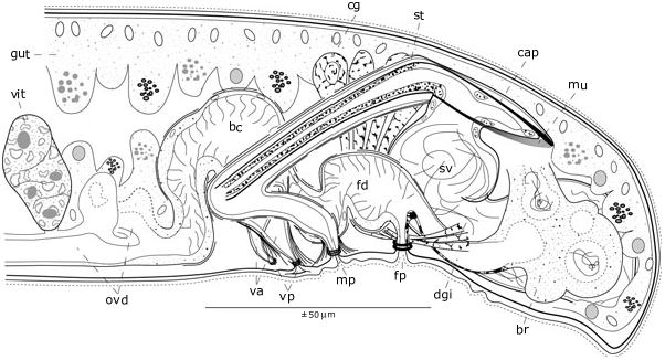
The ovaries are just in front of the pharynx, and all vitellarian follicles are behind the pharynx, extending to the middle of the post-pharyngeal body part. Behind the last follicle the two ovovitelloducts join into the common female duct, which is swollen in the living animal, and clearly functions as a copulatory bursa. At about its middle, two vaginal pores can be seen ( Fig. 10B View Figure 10 ). In the sections ( Fig. 11 View Figure 11 ), the two vaginas depart as a wide funnel from the dorsallysituated copulatory bursa. The female duct narrows towards the female pore, where the duct is enlarged again, and continues backwards as a genito-intestinal duct communicating with a large resorbing bursa. The epithelium covering the female duct is ciliated. This epithelium is very low in the copulatory bursa, in the vaginas, and at the dorsal side of the swollen part at the pore, and is extremely thin in the genitointestinal duct, where it is devoid of cilia. At their very end, the ovovitelloducts show aspects that suggest some resorbing function, but no degenerating sperm was observed. The cement glands, which open at the female pore, are very large and are found in the whole posterior part of the animal.
The copulatory organ is provided with an almost straight stylet, lying in a tubiform, narrow male atrium that enlarges proximally to a narrow space, which receives at its ventral side the wide opening of the seminal vesicle. Dorsally, this proximal space is covered by a sclerotized ‘cap’, which has a fan-like muscle, with its wider side attached to the cap, and its narrower side attached to the stylet (see also Fig. 12 View Figure 12 ). The seminal vesicle is extremely large, occupying almost the whole postpenial part of the animal. One of the most striking characters of this species is the complete absence of any prostate glands (and vesicle). The stylet ( Fig. 12 View Figure 12 ) is about 80-Mm long, with an almost constant diameter of 8–10 Mm; the ‘cap’ is 45-Mm long in the holotype, and is 30-Mm long in the paratype. The fan-like muscle is clearly visible in the whole mount.
| QM |
Queensland Museum |
No known copyright restrictions apply. See Agosti, D., Egloff, W., 2009. Taxonomic information exchange and copyright: the Plazi approach. BMC Research Notes 2009, 2:53 for further explanation.
|
Kingdom |
|
|
Phylum |
|
|
Order |
|
|
Family |
|
|
Genus |