
Asterocheres complexus Stock, 1960
|
publication ID |
https://doi.org/ 10.11646/zootaxa.3827.4.6 |
|
publication LSID |
lsid:zoobank.org:pub:2C480B30-8005-4F66-8FB3-5C54A44F68F3 |
|
DOI |
https://doi.org/10.5281/zenodo.5613647 |
|
persistent identifier |
https://treatment.plazi.org/id/03938795-FFD4-1C56-37C2-9405C21E93D4 |
|
treatment provided by |
Plazi |
|
scientific name |
Asterocheres complexus Stock, 1960 |
| status |
|
Asterocheres complexus Stock, 1960
( Fig. 1 View FIGURE 1 )
Asterocheres boecki Giesbrecht, 1899 (non Brady 1872)
Material examined. holotype female (preserved in ethanol, deposited in ZMA under registration number ZMA- Co.100.571b) and 1 female plus 1 copepodid paratypes (ZMA-Co. 100.571) associated with Spongelia fragilis (Schmidt) var. ramose; collected in Cap Béar ( France), 30 m depth, June 16 1959, coll. by Dr. J.H. Stock.
Description of adult female. Body cyclopiform, slender with cephalothorax oval and cylindrical urosome (see Fig. 2 View FIGURE 2 of Taf. 1 in Giesbrecht 1899). Total length from anterior margin of rostrum to posterior margin of caudal rami 680 µm and maximun width 360 µm. Prosome comprising cephalothorax fully incorporating first pedigerous somite and three free pedigerous somites. Cephalothorax (see Fig. 28 of Taf. 2 in Giesbrecht 1899) with posterolateral angles straight and slightly produced into backwardly directed processes.
Urosome 4-segmented, comprising leg 5-bearing somite, genital double-somite and 2 free abdominal somites (see Fig. 3 View FIGURE 3 B in Stock 1960). Leg 5-bearing somite wider than long, with serrate dorsal margin. Posterior hyaline frills of urosomites with serrate free margins. Urosomites ornamented with numerous integumental pores and sensilla and apparently devoid of epicuticular scales. Genital double-somite slightly wider than long; paired genital apertures bipartite, each comprising lateroventral copulatory pore and dorsolateral gonopore (oviduct opening); lateral margins with row of long spinules in middle third, close to gonopore area (see Fig. 3 View FIGURE 3 B in Stock 1960). Seta of genital area not observed.
Caudal rami about as long as wide (measured along outer margin); armed with 6 terminal setae (see Fig. 3 View FIGURE 3 B in Stock 1960). Seta I absent and setae II and VII slightly displaced onto dorsal surface.
Antennule ( Fig. 1 View FIGURE 1 D) 21-segmented, about 310 µm long. Segmental fusion pattern as follows (Roman numerals indicating ancestral segments): 1(I)-2, 2(II)-2, 3(III)-2, 4(IV)-2, 5(V)-2, 6(VI)-2, 7(VII)-2, 8(VIII)-2, 9(IX-XII)-7, 10(XIII)-2, 11(XIV)-1+1espina, 12(XV)-2, 13(XVI)-2, 14(XVII)-2, 15(XVIII)-2, 16(XIX)-2, 17(XX)-2, 18(XXI)- 2+1 aesthetasc, 19(XXII)-2, 20(XXIII-XXIV)-4 and 21(XXV-XXVIII)-7. Segment 10(XIII) reduced and partly overlapped by distal expansion of compound segment 9(IX-XII).
Antenna (see Fig. 3 View FIGURE 3 E in Stock 1960) biramous, about 220 µm long (including terminal claw). Coxa small, with a tuft of minute spinules on inner margin. Basis elongated with a row of fine spinules on inner margin. Exopod small, one-segmented with one short subterminal seta and one long terminal seta, both of them smooth. Endopod 3- segmented; proximal segment elongated, ornamented with a row of long spinules on inner margin; middle segment produced distally on medial side but articulating with distal segment proximally on lateral side, bearing one distal seta longer than entire segment; distal segment with 2 subterminal setae, one of them pinnate, and a terminal claw with a row of fine spinules on inner margin. Distal claw as long as proximal segment of endopod.
Siphon about 180 µm long, conical, reaching the insertion of maxillipeds. Mandible ( Fig. 1 View FIGURE 1 A) comprising slender two-segmented palp and stylet-like gnathobase with 5 large subapical teeth. Proximal segment of palp longest, ornamented with spinules on distal outer margin; distal segment with spinules apically, armed with 2 terminal setae.
Maxillule bilobed (see Fig. 3 View FIGURE 3 D in Stock 1960); praecoxal gnathobase (inner lobe) 2.5 times longer than palp (outer lobe). Praecoxal endite ornamented with a tuft of long spinules proximally, a row of short spinules apically on outer margin and a row of long setules medially; armed with 5 distal setae, one of them smooth and short. Palp with 4 barbed terminal setae (illustrated as naked by Stock).
Maxilla ( Fig. 1 View FIGURE 1 B) 2-segmented but with partial transverse surface suture on syncoxa (proximal segment) possibly marking plane of praecoxa-coxa fusion; praecoxal portion bearing flaccid aesthetasc-like element medially, representing tubular extension of external opening of maxillary gland; coxal portion unarmed. Basis claw-like with a minute seta at middle length and a row of spinules along medial distal part.
Maxilliped ( Fig. 1 View FIGURE 1 C) 5-segmented, comprising short syncoxa, long basis and 3-segmented endopod. Syncoxa with one short seta distally and a row of spinules along inner proximal margin. Basis elongated, with rows of spinules on both margins. First endopodal segment short, bearing 2 smooth short setae; second endopodal segment with a smooth seta subapically; third endopodal segment bearing recurved terminal claw (65 µm long) plus additional plumose apical seta. Distal margin of claw provided with a row of minute spinules.
Swimming legs 1–4 (see Fig. 3 View FIGURE 3 A,C in Stock, 1960) biramous, with 3-segmented rami. Intercoxal sclerite present in legs 1–4. Spine and seta formula as Table 1 View TABLE 1 .
Lateral margins of exopodal segments with minute serrations or spinular rows; those of endopodal segments with rows of setules.
Fifth leg (see Fig. 3 View FIGURE 3 B in Stock, 1960) with protopod incorporated into somite; outer basal seta displaced to laterodorsal surface (not longer than entire free segment). Free segment (exopod) elongate, with 2 smooth terminal setae and one short subterminal seta; outer and inner margins with spinules.
Sixth leg (see Fig. 3 View FIGURE 3 B in Stock, 1960) usually represented by paired opercular plates closing off gonopores on genital double somite; none seta neither spiniform element observed.
Adult male: only known from the habitus, antennule and exopod of leg 2 illustrated by Giesbrecht in 1899. Antennule 18-segmented, with aesthetascs on segments 13 and 17.
Distribution. Italy ( Giesbrecht 1899), France ( Stock 1960), India ( Ummerkutty 1966; under the name of A. latum ).
Remarks. This species was described by Stock (1960) from two females collected in Cap Béar (Mediterranean coast of France). As Stock pointed out, this species was originally described by Giesbrecht (1899) under the incorrect name A. boecki . Following the detailed description of A. boecki provided by Sars (1918), Stock stated that Giesbrecht´s specimens could belong to another Nordic species, A. latum (Brady) . Comparisons among the material collected in Banyuls by Stock and the figures of A. boecki and A. latum illustrated by Sars revealed that Stock´s specimens belonged to a distinct, undescribed species. This new species was not properly described by Stock but was based on descriptions of A. boecki by Giesbrecht (habitus; Pl. 2, II; Giesbrecht 1899), A. latum by Sars (antennules, maxilla, maxilliped, and the exopods of leg 1-4; Sars 1918) and Stock added some illustrations (antenna, maxillule, urosome, exopod of leg 1 and endopod of leg 4 for female and leg 5 for male; Fig. 3 View FIGURE 3 in Stock 1960).
The re-examination of the holotype resulted in some discrepancies with respect to previous descriptions: (1) the antennule is 21-segmented in the female, in contrast Sars described this antennule as very slender and composed of 20 segments; (2) the mandible was illustrated by Giesbrecht and Sars, but only the palp which is twosegmented, because the stylet is located inside the oral cone. The stylet with 5 large subapical teeth is illustrated and described here for the first time; (3) the maxilla possesses a flaccid element similar to an aesthetasc which was overlooked by Sars; (4) the maxilliped illustrated by Sars has some elements missing.
This species belongs to a group whose females have 21-segmented antennules and a 2-segmented mandibular palp; it contains 19 species: A. astroidicola Conradi, Bandera & López-González, 2006 , A. ellisi Hamond, 1968 , A. espinosai Varela, Ortiz & Lalana, 2007 ; A. flustrae Ivanenko & Smurov, 1997 , A. genodon Stock, 1966 , A. hirsutus Bandera, Conradi & López-González, 2005 , A. hoi Bandera & Conradi, 2013 , A. jeanyeatmanae Yeatman, 1970 , A. kervillei Canu, 1898 , A. latus ( Brady, 1872) , A. lilljeborgi Boeck, 1859 , A. peniculatus Kim, 2010 , A. reginae Boxshall & Huys, 1994 , A. simulans (Scott, 1898) , A. suberitis Giesbrecht, 1897 , A. tarifensis Conradi & Bandera, 2011 , A. tenerus (Hansen, 1923) , A. tenuicornis Brady, 1910 , A. tubiporae Kim, 2004 , and A. urabensis Kim, 2004 .
Considering the shape of the body, A. complexus can be separated from a few of its congeners. While this species has the usual cyclopiform body, with an oval cephalothorax and a cylindrical urosome, A. ellisi , A. espinosai , A. jeanyeatmanae , A. lilljeborgi , A. reginae , and A. tubiporae have a dorsoventrally flattened prosome ( Bandera & Conradi 2009b; Varela et al., 2007; Yeatman 1970; Ivanenko & Ferrari 2003; Boxshall & Huys 1994; Kim 2004b). Also, A. espinosai is here treated as an incompletely described species due to the lack of accurate information of the oral appendages and the confusion between legs 2 and 3 in the original description. Therefore, the comparison of this species with its congeners is difficult.
The length of the siphon is a good feature to distinguish one species from another. Asterocheres complexus is characterized by its possession of an oral cone reaching the insertion of the maxillipeds, thus differing from A. peniculatus , A. hirsutus , A. urabensis , and A. hoi in which the siphon reaches the intercoxal plate of leg 1 and from A. genodon , A. astroidicola and A. tenerus whose the siphon overtakes the intercoxal plate of leg 2 ( Kim 2010; Bandera et al. 2005; Kim 2004a; Bandera & Conradi 2013; Conradi et al. 2006; Bandera & Conradi 2009a).
Asterocheres complexus possesses a subquadrate caudal rami. In contrast, in this group there are species with a much longer caudal rami; in A. simulans and A. kervillei they are twice longer than wide, 1.5 times longer than wide in A. suberitis , 2.6 times longer than wide in A. latus , and 6 times longer than wide in A. tenuicornis (Ivanenko 1997; Bandera & Conradi 2009c, 2009a; Eiselt 1965).
The remaining species of the group, A. flustrae and A. tarifensis , are the most closely related species to A. complexus . However, these two species can be easily separated from A. complexus by the shape of the posterolateral angles of the cephalothorax. A. complexus presents the posterolateral angles of the cephalothorax straight and slightly produced into backwardly directed processes. In contrast, A. flustrae and A. tarifensis possess rounded posterior corners ( Ivanenko & Smurov 1997; Conradi & Bandera 2011).
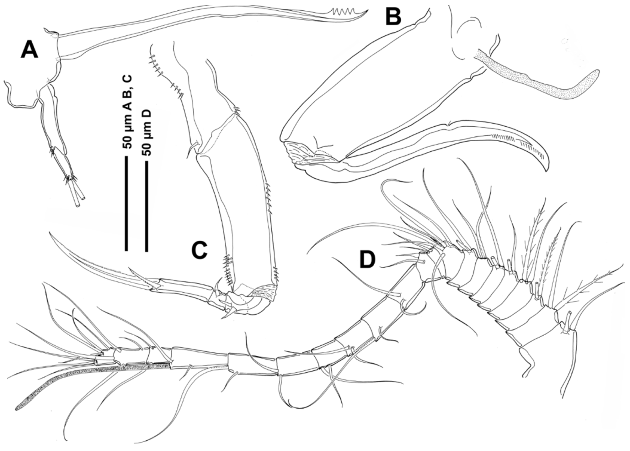
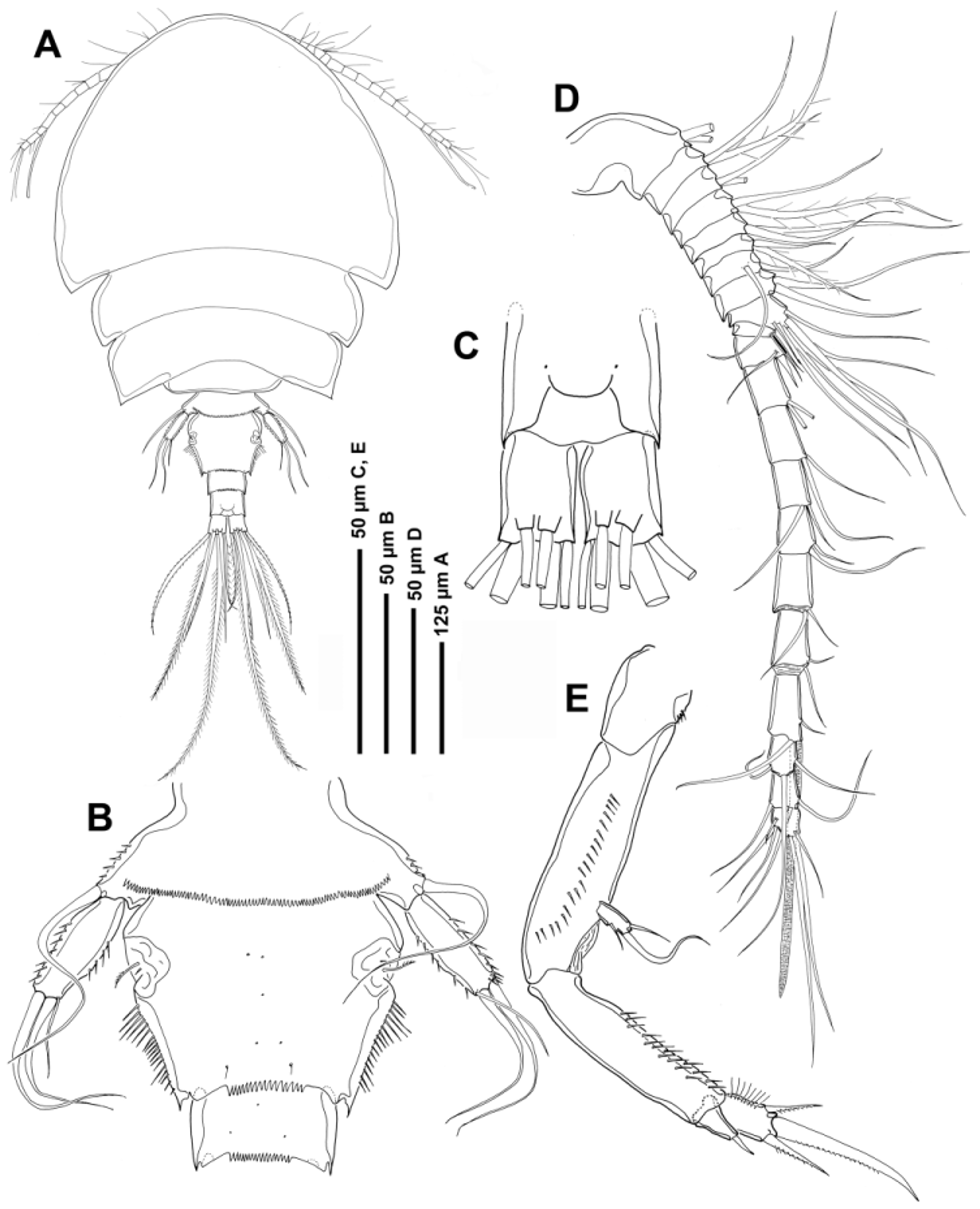
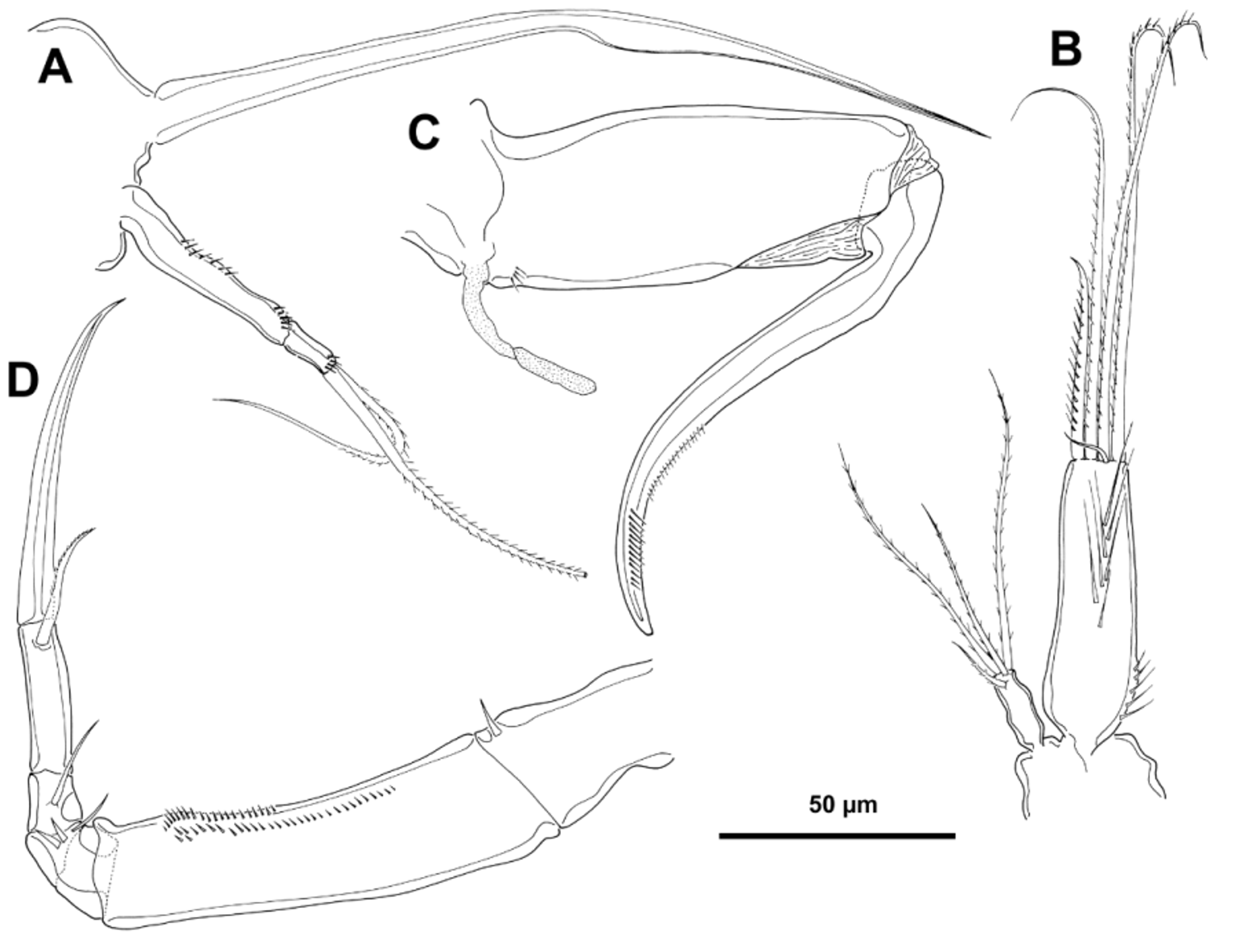
TABLE 1. Spine and seta formula of swiming legs for Asterocheres comple xus Stock 1960.
| Coxa | Basis | Exopod | Endopod | |
|---|---|---|---|---|
| Leg 1 | 0-1 | 1-1 | I-1;I-1;III,2,2 | 0-1;0-2;1,2,3 |
| Leg 2 | 0-1 | 1-0 | I-1;I-1;III,I+1,3 | 0-1;0-2;1,2,3 |
| Leg 3 | 0-1 | 1-0 | I-1;I-1;III,I+1,3 | 0-1;0-2;1,1+I,3 |
| Leg 4 | 0-1 | 1-0 | I-1;I-1;III,I+1,3 | 0-1;0-2;1,1+I,2 |
| ZMA |
Universiteit van Amsterdam, Zoologisch Museum |
No known copyright restrictions apply. See Agosti, D., Egloff, W., 2009. Taxonomic information exchange and copyright: the Plazi approach. BMC Research Notes 2009, 2:53 for further explanation.
|
Kingdom |
|
|
Phylum |
|
|
Class |
|
|
Order |
|
|
Family |
|
|
Genus |
Asterocheres complexus Stock, 1960
| Bandera, Eugenia & Conradi, Mercedes 2014 |
Asterocheres boecki
| Giesbrecht 1899 |