
Asterocheres tenerus ( Hansen, 1923 )
|
publication ID |
https://doi.org/ 10.5281/zenodo.185273 |
|
DOI |
https://doi.org/10.5281/zenodo.6221578 |
|
persistent identifier |
https://treatment.plazi.org/id/03AEF308-FFEE-6110-FF4D-19B06E588204 |
|
treatment provided by |
Plazi |
|
scientific name |
Asterocheres tenerus ( Hansen, 1923 ) |
| status |
|
Asterocheres tenerus ( Hansen, 1923)
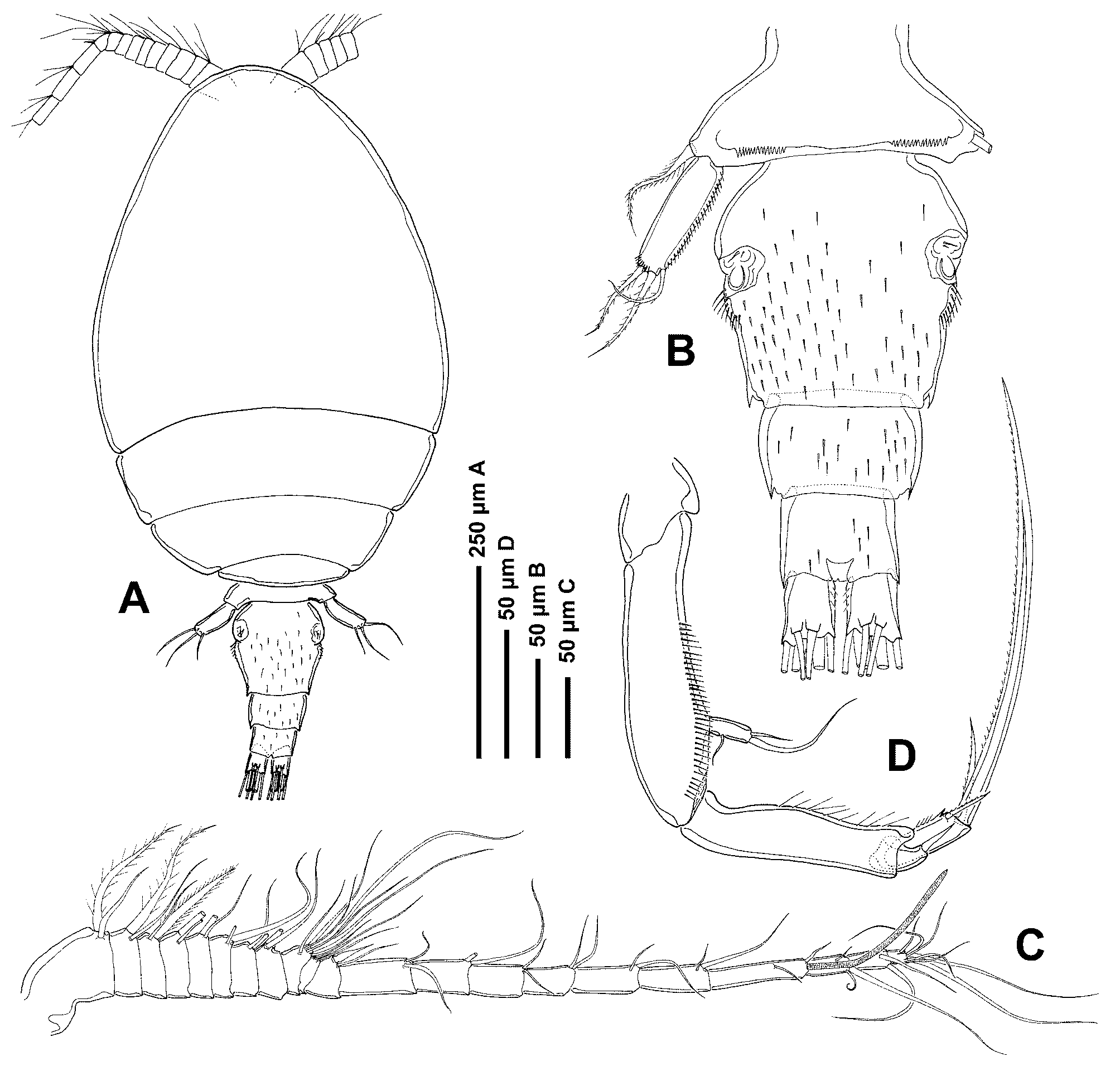
( Figs 2–4 View FIGURE 2 View FIGURE 3 View FIGURE 4 )
Ascomyzon tenerum Hansen 1923: 7 .
Asterocheres tenerus: Stock 1966b: 152 ; Johnsson 1998: 92.
Material examined. Holotype Ψ and 8 paratypes (5 Ψ and 3 ɗ) ( ZMUC. CRU-8357) collected during the “Danish Ingolf Expedition” in Davis Strait, Station 25 (63º30΄N, 54º25΄W), at 582 fathoms depth (ca. 1.06 km).
Redescription of adult female: Body ( Fig. 2 View FIGURE 2 A) cyclopiform, with oval cephalothorax and cylindrical urosome. Total length 904 µm (n = 1); maximum width 452 µm. Ratio of length to width of prosome 1.5:1. Ratio of length of prosome to urosome 2.6:1. Prosome comprising cephalothorax fully incorporating first pedigerous somite and 3 free pedigerous somites. Pedigerous somite 4 much smaller and narrower than preceding somites. Dorsal cephalothoracic shield and free pedigerous somites ornamented with integumental pores and sensillae (these features not shown in Fig. 2 View FIGURE 2 A). Urosome ( Fig. 2 View FIGURE 2 B) 4-segmented, comprising pedigerous somite 5, genital double-somite and 2 free abdominal somites. Genital double-somite and following somites furnished with large spinules. Pedigerous somite 5 narrow, largely concealed under tergite of pedigerous somite 4, with spinular row on each side of dorsal midline. Genital double-somite about as long as wide, with separate genital apertures, each comprising ventrolateral copulatory pore and dorsolateral gonopore (oviduct opening); lateral margins with rows of setules posterior to genital apertures. Caudal rami longer than wide, armed with 6 setae (seta I absent); setae III–VI arranged along posterior margin; setae II and VII inserted subapically on dorsal surface.
Antennule ( Fig. 2 View FIGURE 2 C) 21-segmented, 544 µm long. Segmental homologies (expressed segment given first followed by ancestral segment(s) in brackets) and setation pattern as follows: 1(I)-2, 2(II)-2, 3(III)-2, 4(IV)-2, 5(V)-2, 6(VI)-2, 7(VII)-2, 8(VIII)-2, 9(IX–XII)-7, 10(XIII)-2, 11(XIV)-1+spine, 12(XV)-2, 13(XVI)-2, 14(XVII)-2, 15(XVIII)-2, 16(XIX)-2, 17(XX)-2, 18(XXI)-2+ae, 19(XXII–XXIV)-3, 20(XXV)-2, 21(XXVI- XXVIII)-7. Segment 10 (XIII) reduced, partly overlapped by distal expansion of compound segment 9 (IX- XII). One of two setae on segments 1–8 plumose. Antenna ( Fig. 2 View FIGURE 2 D) biramous, 430 µm long (including claw); coxa small, unarmed; basis elongated, unarmed, and ornamented with row of spinules along inner margin. Exopod 1-segmented, with small medial seta and 2 subequal terminal setae. Endopod 3-segmented; first segment elongated, with few setules along inner margin; second segment distomedially produced, bearing terminal seta; third segment ornamented with coarse spinules and few setules along inner margin and armed apically with long spinulate claw flanked by 2 unequal barbed setae.
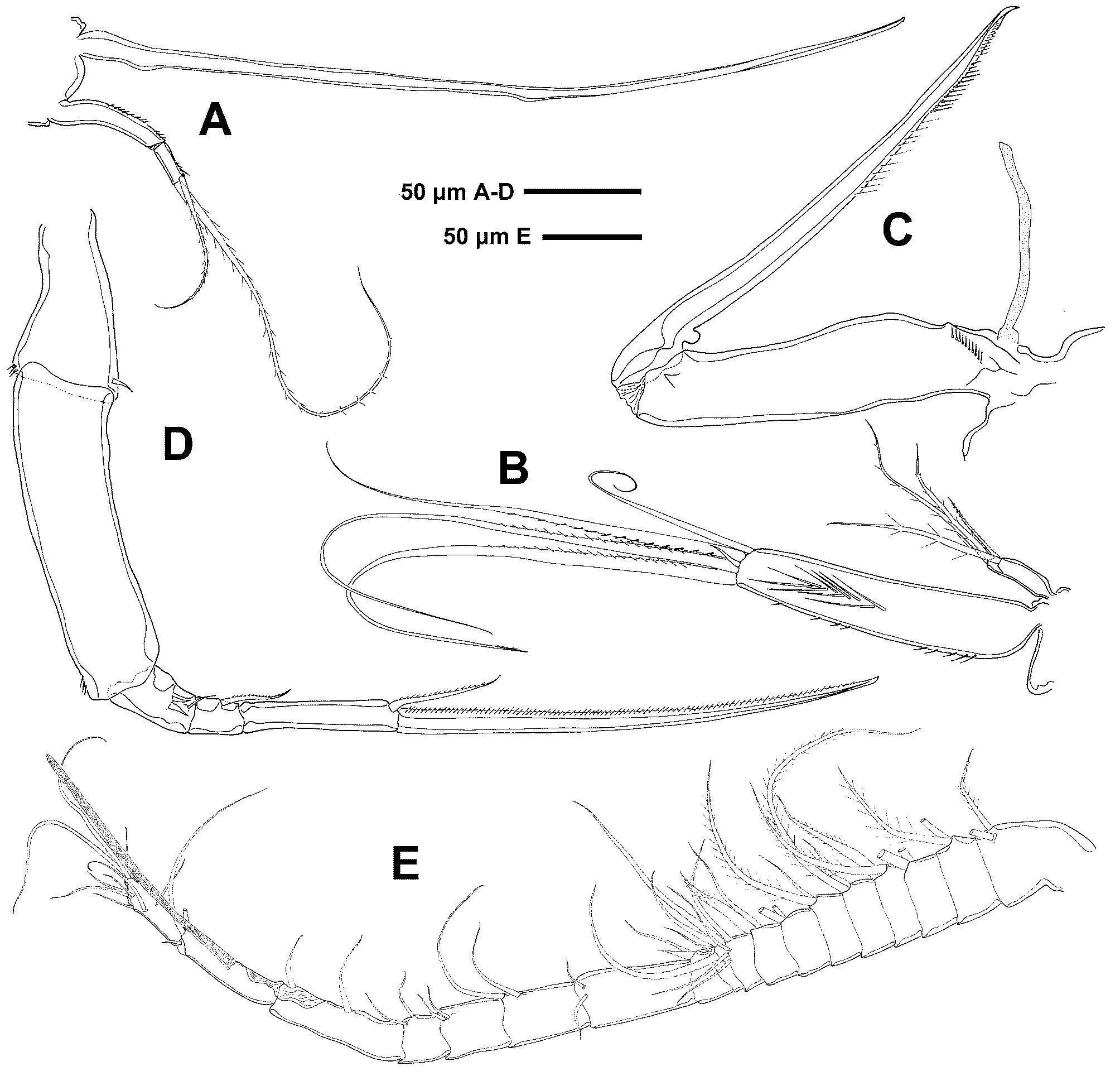
Siphon long and slender, 370 µm long, reaching the intercoxal plate of leg 2. Mandible ( Fig. 3 View FIGURE 3 A) composed of stylet-like gnathobase (356 µm long) and 2-segmented palp. First segment of palp ornamented with an incomplete row of spinules; second segment with spinules along distal margin and armed apically with 2 unequal, pinnate setae. Maxillule ( Fig. 3 View FIGURE 3 B) bilobed, with inner lobe 4 times longer than outer lobe. Outer lobe armed distally with 4 barbed setae. Inner lobe ornamented with spinules laterally and long setules along midline, and armed with 5 distal setae, one of them very short.
Maxilla ( Fig. 3 View FIGURE 3 C) 2-segmented, with partial transverse suture on syncoxa, possibly marking plane of praecoxa-coxa fusion; praecoxal region bearing long flaccid element medially, representing tubular extension of external opening of maxillary gland; coxal region unarmed, ornamented with row of spinules proximally. Claw-like basis bearing small hyaline process proximally and row of spinules along distal margin. Maxilliped ( Fig. 3 View FIGURE 3 D) 5-segmented, comprising short syncoxa, long basis and 3 free endopodal segments. Syncoxa with short seta distomedially and few spinules distolaterally; basis with few spinules distolaterally. First endopodal segment with thin distal seta and 2 robust medial setae. Second endopodal segment with medial barbed seta. Third endopodal segment bearing long spinulate claw and apical spinulate seta.
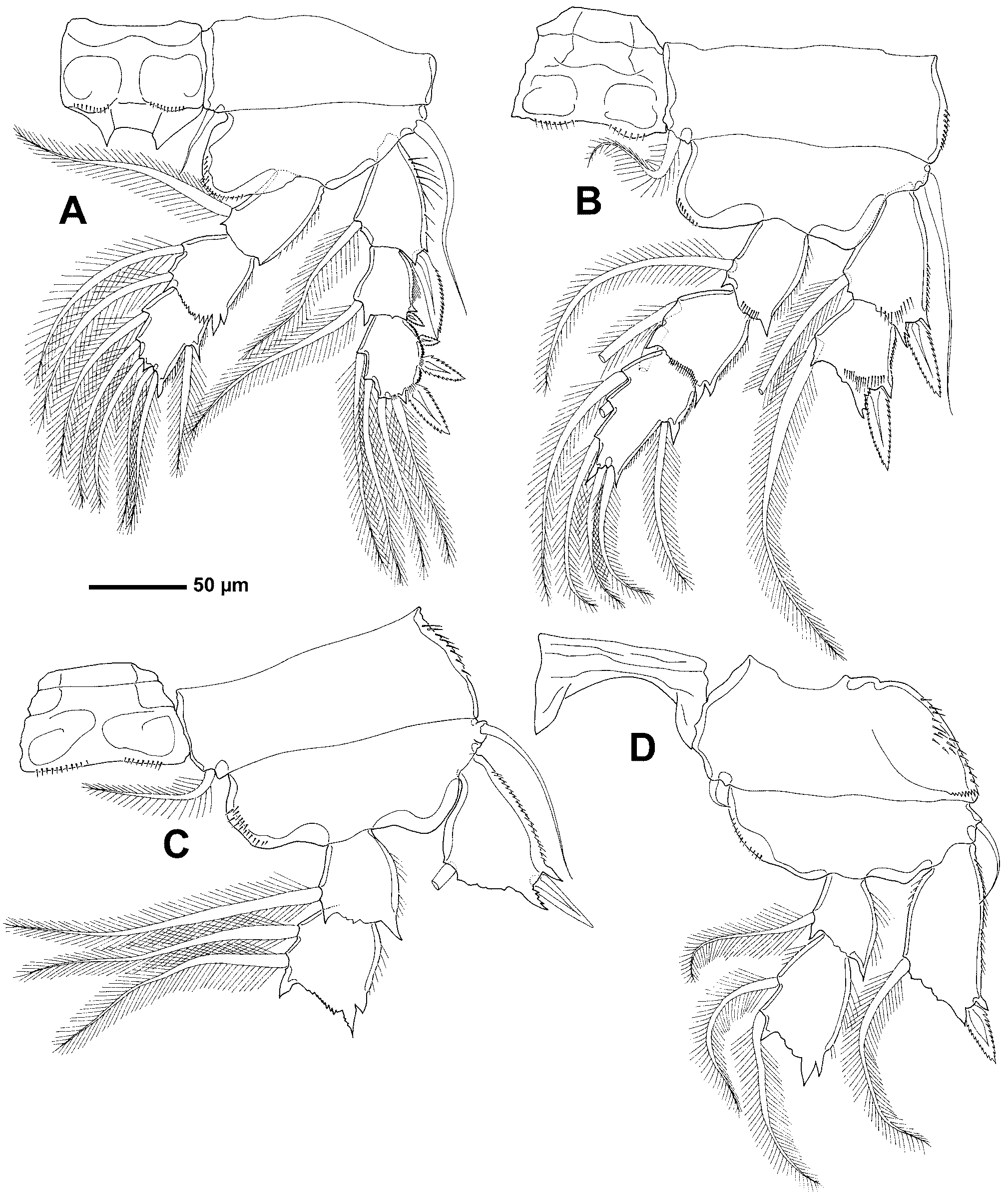
Swimming legs 1–4 ( Figs 4 View FIGURE 4 A–D) biramous, with only leg 1 complete. Spine and seta formula as follows: Intercoxal sclerite of legs 1–3 ornamented with rows of spinules along posterior margin. Coxae of legs 2–4 ornamented with rows of spinules laterally; coxal seta naked in legs 1 and 4, plumose in legs 2 and 3; outer basal seta of all legs naked. Outer spine of first exopodal segment of leg 3 smooth. Lateral margins of exopodal segments with minute spinules; lateral margins of endopodal segments with row of setules.
Leg 5 ( Fig. 2 View FIGURE 2 B) with protopodal segment incorporated into somite; protopodal seta located laterally; free segment slender, armed with 3 distal plumose setae and ornamented with medial spinular row. Leg 6 ( Fig. 2 View FIGURE 2 B) represented by paired opercular plates closing off gonopores on genital double-somite; armed with 2 smooth setae, one of them minute.
Adult male: Body cyclopiform, with oval cephalothorax and cylindrical urosome. Total length 890 µm (n = 1); maximum width 460 µm. Prosome comprising cephalothorax fully incorporating first pedigerous somite and 3 free pedigerous somites. Urosome 5-segmented, comprising pedigerous somite 5, genital somite and 3 free abdominal somites. Caudal ramus armed as in female.
Appendages as in female, except for the following. Antennule ( Fig. 3 View FIGURE 3 E) 18-segmented, geniculate; segmental homologies (expressed segment given first followed by ancestral segment(s) in brackets) and setation pattern as follows: 1(I)-2, 2(II)-2, 3(III)-2, 4(IV)-2, 5(V)-2, 6(VI)-2, 7(VII)-2, 8(VIII)-2, 9(IX-XII)-7, 10(XIII)-1+spine, 11(XIV)-1+spine, 12(XV)-2, 13(XVI)-2, 14(XVII)-2, 15(XVIII)-2, 16(XIX–XX)-2, 17(XXI–XXIII)-3+ae, 18(XXIV–XXVIII)-9. Geniculation located between segments 16 (XIX–XX) and 17 (XXI–XXIII). Segment 10(XIII) reduced, partly overlapped by distal expansion of compound segment 9 (IX–XII). Maxilliped with small tooth-like process in proximal-half of basis. Leg 6 forming large opercular plates closing off genital apertures, armed with 2 smooth setae.
Hosts. Unknown.
Distribution. Atlantic Ocean ( Hansen, 1923).
Remarks. A detailed re-examination of Asterocheres tenerus type material has revealed the following differences between our observations and the original description by Hansen (1923): 1) the highly reduced tenth (XII) antennulary segment of the female was overlooked by Hansen; 2) the antennary exopod bears 3 instead of one element, and the proximal margin of the distal endopodal segment articulates on the lateral side of the preceding segment; 3) the oral siphon extends to the intercoxal plate of leg 2, a feature that is unclear in the original description (according to Hansen, the siphon reaches beyond the insertion of leg 1 or beyond that of leg 2); 4) the inner lobe of the maxillule possesses 5 rather than 4 terminal setae; 5) the aesthetasc-like extension on the proximal part of the maxillary syncoxa was overlooked by Hansen; 6) the maxilliped is composed of 5 instead of 4 segments; 7) the swimming legs, which were not described by Hansen, are indeed present in the type specimens; 8) the free segment of leg 5 bears 3 instead of 2 distal setae; and 9) the male antennule is comprised of 18 rather than 17 segments.
Based on our redescription, A. tenerus belongs to the group of Asterocheres species having a 21-segmented antennule, which is currently composed of 20 species total as mentioned in the Remarks section of A. intermedius . Asterocheres tenerus differs from A. bacescui , A. madeirensis , A. minutus , and A. violaceus by having a 2-segmented rather than 1-segmented mandibular palp. Asterocheres tenerus can be distinguished from A. lilljeborgi , A. echinicola , A. uncinatus , A. tenuicornis , A. simulans , A.suberitis , A. jeanyeatmanae , A. reginae , A. flustrae and A. lunatus by having an oral siphon that reaches the insertion of leg 2 rather than only to the insertion of the maxillipeds.
Asterocheres tenerus differs from A. ellisi by lacking a dorsoventrally flattened prosome and from A. intermedius by having a genital double-somite that is as long as wide rather than longer than wide. The length of caudal rami separates A. tenerus from A. hirsutus . Asterocheres tenerus has caudal rami that are slightly longer than wide, while A. hirsutus possesses caudal rami that are 2.5 times longer than wide. Asterocheres tenerus can be differentiated from the remaining 2 species, A. astroidicola and A. urabensis , by having considerably slimmer and longer claws on the antenna, maxilla and maxilliped.
| ZMUC |
Zoological Museum, University of Copenhagen |
No known copyright restrictions apply. See Agosti, D., Egloff, W., 2009. Taxonomic information exchange and copyright: the Plazi approach. BMC Research Notes 2009, 2:53 for further explanation.
|
Kingdom |
|
|
Phylum |
|
|
Class |
|
|
Order |
|
|
Family |
|
|
Genus |
Asterocheres tenerus ( Hansen, 1923 )
| Bandera, Eugenia & Conradi, Mercedes 2009 |
Asterocheres tenerus:
| Johnsson 1998: 92 |
| Stock 1966: 152 |
Ascomyzon tenerum
| Hansen 1923: 7 |