
Philactinoposthia novaecaledoniae, Nilsson, Karin Sara, Wallberg, Andreas & Jondelius, Ulf, 2011
|
publication ID |
https://doi.org/ 10.5281/zenodo.277458 |
|
DOI |
https://doi.org/10.5281/zenodo.5683937 |
|
persistent identifier |
https://treatment.plazi.org/id/03A6456D-FFBF-AE4D-FF4B-96EAFCE4FBA9 |
|
treatment provided by |
Plazi |
|
scientific name |
Philactinoposthia novaecaledoniae |
| status |
sp. nov. |
Philactinoposthia novaecaledoniae sp.nov.
( Figs. 3 View FIGURE 3 , 10 View FIGURE 10 , 11 View FIGURE 11 , 12 View FIGURE 12 )
Type Material: Holotype: SMNH Type-8055. Paratype 1: SMNH Type-8056, Paratype 2: SMNH Type-8057.
Type Locality. Poe beach outside inner coral reef, New Caledonia, (21° 37' 31" S, 165° 23' 47" E), at 1.5 m water depth in clean fine sand, and Amedée Island, (22° 28' 40" S, 166° 28' 22" E), at 15 m water depth in fine sand.
Other Material examined. Living specimens in squeeze preparations; 7 sets of 4–5 μm serial sagittal sections of paraffin-embedded specimens.
Etymology. The species epithet refers to the type locality, Nova Caledonia = New Caledonia in Latin.
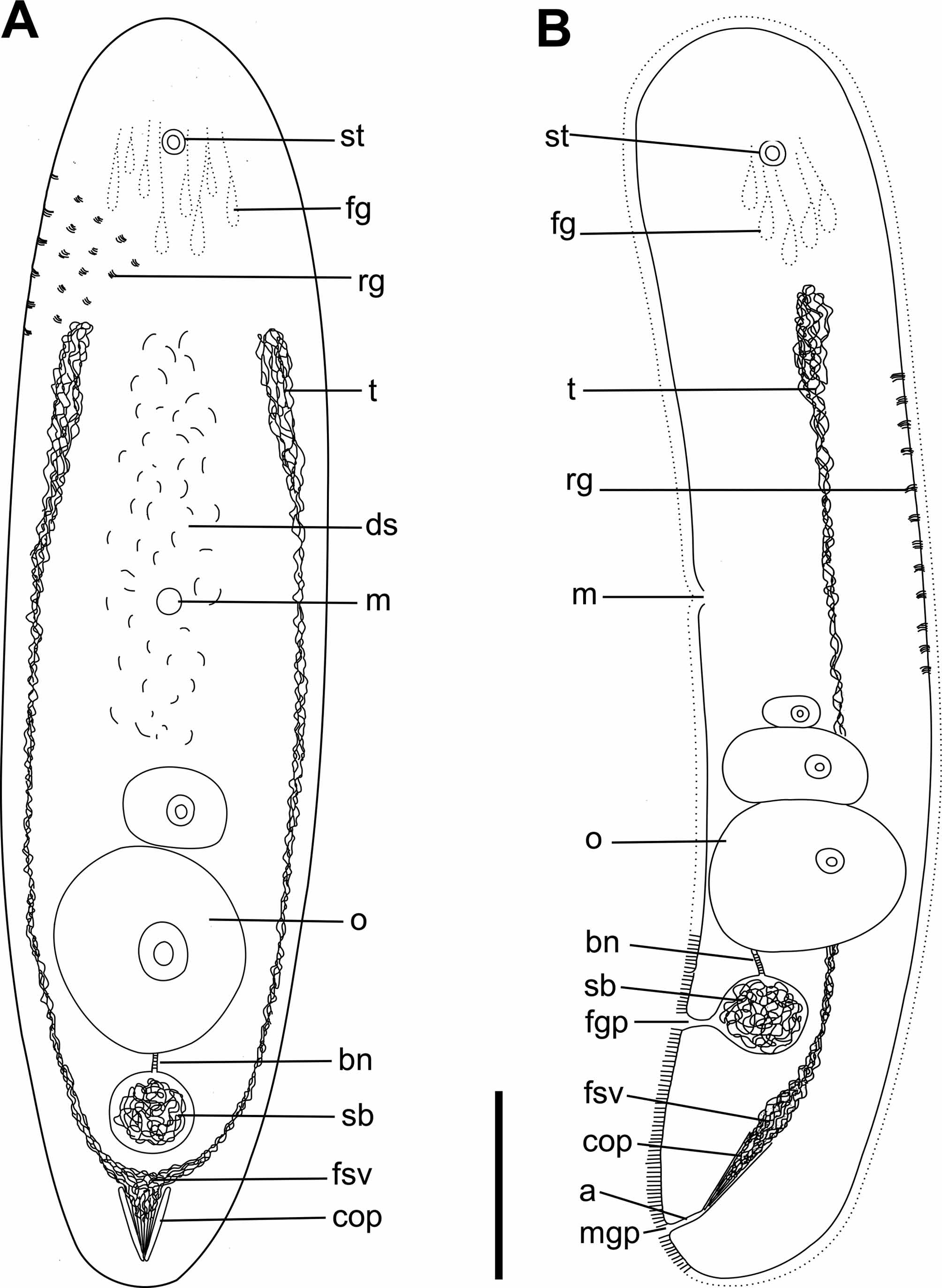
Description. Living specimens 500–600 μm long and ~130 μm wide (L/W=4). Sectioned specimens contracted, 200 μm long and 100 μm wide. Body shape cylindrical, both ends rounded bluntly. Epidermis completely covered with 5 μm cilia. Numerous red to brown colored rhabdoid glands in body wall, scattered uniformly across body ( Fig. 10 View FIGURE 10 AC). Each rhabdoid gland 15 μm long with up to 12 strokes. Epidermis uncolored by transmitted light. Brownish coloration of digestive syncytium. Statocyst, 7,5 μm in diameter, located 25 μm from anterior end in sections, at level U10. Frontal organ present, cell bodies of frontal organ located posterior to statocyst, extending from U11 to U20. Mouth ventral, located in anterior half of body, at level U55. Digestive central syncytium extends from level U25 to U60.
Ovary unpaired, ventral, with up to 3 oocytes, extending from posterior level of mouth to bursal nozzle, U60 to U80. Large ventro-median oocyte positioned close to bursal nozzle ( Fig. 10 View FIGURE 10 C). Female gonopore slightly anterior to seminal vesicle, at U87, opens to globular and very distinct seminal bursa, about 30 μm in diameter. Seminal bursa connects anteriorly to bursal nozzle. Bursal nozzle, 10 μm long, small in relationship to the size of seminal bursa ( Fig. 10 View FIGURE 10 C).
Testes paired, lateral to ovary, extending from anterior to mouth, posteriorly to male copulatory organ, U25 to U90. Male gonopore subterminal on ventral side, at level U97, opens to male copulatory organ with sclerotized vshaped penis-like structure ( Fig. 10 View FIGURE 10 AC). The characteristic v-shaped stylet consist of two groups containing a few needles each, ~20 μm long. Additional thin needles also present surrounded by spermatozoa. False seminal vesicle obscure, located at proximal end of penis-like structure, at U85.
Remarks. The presence of a a male copulatory organ consisting of sclerotized stylets, which are not invaginated into the seminal vesicle, the “normal” arrangement of the body-wall musculture with distal cicular muscle fibres, and the abscence of any symbiotic algae identifies all four Philactinoposthia species as members of the family Actinoposthiidae . The female accessory organs, the well developed frontal glands and distinct rhabdoid glands identify them as members of Philactinoposthia . The Bayesian analysis of the combined 18S, 28S and COI nucleotide data place P. brevis , P. ischiae and P. novaecaledoniae in a clade composed of Philactinoposthia species with maximum support ( Fig. 3 View FIGURE 3 ). P. multipunctata was not included in the phylogenetic analysis as there was no sequence data available for this species.
Philactinoposthia brevis sp.nov. can be distinguished from other species of the genus Philactinoposthia by its small arrow-shaped copulatory organ with at least four needles, ~30 μm long bursal nozzle with zigzag shape and ovary with one large oocyte visible in live specimens.
Philactinoposthia helgolandica Dörjes, 1968 is the most similar species, but has a longer and wider body (0.8- 1 mm long, 0.1–0.2 mm wide) which is dorsoventrally flattened, denser teardrop-shaped dark green rhabdoid glands that together with dark grey coloration of the digestive parenchyma gives a characteristic pattern, subterminal male gonopore, larger copulatory organ in relationship to body size, penis-like structure centrally located inside the inner bladder (penis anchored in the proximal wall of the vesicle), testes extending to frontal glands, bursal nozzle without a zigzag shape and more numerous eggs.
P. brevis can appear similar to Microposthia listensis Faubel, 1974 View in CoL due to the arrow-like copulatory organ, ovary with one large oocyte, similar body size and rhabdoid glands. However, P. b re vi s differs from M. listensis View in CoL in having a seminal bursa with bursal nozzle, longer body in relationship to width and not a dorso-ventrally flattened body and smaller rhabdoid glands.
Paraproporus xanthus Marcus, 1950 View in CoL has a similar combination of stylet and body size, but the copulatory organ is located terminally and a seminal bursa and bursal nozzle are absent.
Philactinoposthia ischiae sp.nov. can be distinguished from other Philactinoposthia species by its long curved posterior stylet (to the left from a dorsal view), male gonopore located subterminally slightly to the right, seminal vesicle at proximal end of stylet, well-defined bursal nozzle (thick in proportion to length), seminal bursa located at level of proximal part of stylet (i.e. not anterior to the stylet), paired ovary in posterior half of body, and the small uncolored rhabdoid glands that appear white in incident light. The rhabdoid glands were probably dissolved by the fixation and therefore not detectable in the sections. P. ischiae is similar to Philactinoposthia stylifera ( Westblad, 1946) due to the curved stylet-like structure and the dorsal position of the seminal bursa. However, P. stylifera has larger rhabdoid glands (20 μm), wider bursal nozzle with zig-zag appearance in sections, a female gonopore, and the ovary is more anterior. Philactinoposthia stylifera brasiliensis was reported by Hooge & Rocha (2006) and classified by them as a subspecies of P. stylifera , although it differs from Westblad’s description in the structure and size of rhabdoid glands, the absence of a female gonopore and a true seminal vesicle. The presence of a true seminal vesicle and the unpigmented digestive syncytium distinguishes P. ischiae from P. stylifera brasiliensis . P. ischiae can appear similar to Philocelis robrochai Hooge & Rocha, 2006 , but the position of the stylet, which is more curved and shorter, is different as it reaches the posterior end in P. i s c h i a e but not in P. robrochai . The bursal nozzle in P. ischiae is directed dorsally to the anterior rather than dorsally slightly to the posterior. Rhabdoid glands are scattered across the body rather than positioned in distinct rows. As in other species currently classified in Philocelis Dörjes,1968 , the vagina in P. robrochai is posterior to the male copulatory organ.
P. ischiae groups within a clade consisting of five Philactinoposthia species in the Bayesian analysis of the nucleotide dataset. A sister group relationship between the Philactinoposthia clade and a clade composed of four Philocelis species + Philactinoposthia saliens ( Graff, 1882) + Daku woorimensis Hooge, 2003 is supported by the Bayesian analysis ( Fig. 3 View FIGURE 3 ). It should be noted that several species of Philactinoposthia have male copulatory organs with sclerotized elements similar to those in Philocelis .
This species was only found in sand from the volcanic carbon dioxide vents. Samples from outside the vent area contained no specimens.
Philactinoposthia multipunctata sp.nov. is similar to Philactinoposthia coneyi Hooge & Rocha, 2006 , that shares with P. multipunctata an unpaired ovary, paired testes, red-colored rhabdoid glands and long and slender body shape. P. multipunctata is distinguished from other species of Philactinoposthia by its large body size with tapering ends, numerous red-colored rhabdoid glands, seminal bursa with fibrous disc in distal end, long and slender bursal nozzle with stack of hats appearance, v-shaped sclerotized penis-like structure filled with spermatozoa.
The body length of P. multipunctata is 2–3 mm, which is at least the double of P. coneyi , which is 700 μm. Body proportions are also different, P. coneyi is longer in relationship to its width compared to P. multipunctata (8:1, 6:1). Furthermore, P. coneyi has a rounded posterior end with vacuole, while P. multipunctata has a tapering posterior end without a vacuole.
The bursal nozzle in P. multipunctata is longer (~13 “hats”, 60 μm long) and with less distinctive “stack of hats” than P. coneyi ; especially discernible in sections. P. coneyi has a more muscular seminal vesicle with wellaligned sperm, shorter penis-like structure and not as numerous rhabdoid glands.
Philactinoposthia novaecaledoniae sp.nov. is distinguished from other Philactinoposthia species by its large seminal bursa in relation to body size (30 μm in diameter), the small bursal nozzle (10 μm long), and the posterior large copulatory organ (in relation to body size) with sclerotized elements forming a V-shaped stylet. P. novaecaledoniae can appear similar to P. viridorhabditis , P. helgolandica and P. coneyi due to unpaired ovaries, seminal bursa with bursal nozzle, posterior male copulatory organ and similar rhabdoid glands. P. novaecaledoniae has red to brown rhabdoid glands and a dark digestive syncytium whereas P. viridorhabditis is unpigmented with green-brown rhabdoid glands forming a species-specific pattern. The number of rhabdoid glands in P. novaecaledoniae is also higher than in P. viridorhabditis . Furthermore, the testes in P. viridorhabditis only extend anteriorly to the mid-body, the seminal vesicle is more massive, distinctive and separated from the male copulatory organ and the bursal nozzle is larger (~25 μm) than in P. novaecaledoniae . P. helgolandica has dark-green colored rhabdoid glands with dark grey coloration of digestive parenchyma, and much more massive seminal vesicle with well-aligned sperm and not as distinct stylet needles surrounded by seminal vesicle. P. coneyi is much longer in relation to body width, has a vacuole posteroterminally, shorter penis-like structure, more massive and muscular seminal vesicle and a welldefined bursal nozzle with the appearance of a stack of hats.


| SMNH |
Saskatchewan Museum of Natural History |
No known copyright restrictions apply. See Agosti, D., Egloff, W., 2009. Taxonomic information exchange and copyright: the Plazi approach. BMC Research Notes 2009, 2:53 for further explanation.
|
Kingdom |
|
|
Phylum |
|
|
Class |
|
|
Order |
|
|
Family |
|
|
Genus |
Philactinoposthia novaecaledoniae
| Nilsson, Karin Sara, Wallberg, Andreas & Jondelius, Ulf 2011 |
Philocelis robrochai
| Hooge & Rocha 2006 |
Philactinoposthia coneyi
| Hooge & Rocha 2006 |
Microposthia listensis
| Faubel 1974 |
Philactinoposthia helgolandica Dörjes, 1968
| Dorjes 1968 |
Philocelis Dörjes,1968
| Dorjes 1968 |
Paraproporus xanthus
| Marcus 1950 |
Philactinoposthia stylifera (
| Westblad 1946 |
Philactinoposthia saliens (
| Graff 1882 |