
Asterocheres hoi, Bandera & Conradi, 2013
|
publication ID |
https://doi.org/ 10.1080/00222933.2012.742588 |
|
publication LSID |
lsid:zoobank.org:pub:1507EC09-372A-4C75-9DD3-6AE64A90DF70 |
|
persistent identifier |
https://treatment.plazi.org/id/03C27E20-FFDB-6563-FDCF-02CCFC82FE41 |
|
treatment provided by |
Felipe |
|
scientific name |
Asterocheres hoi |
| status |
sp. nov. |
Asterocheres hoi sp. nov.
( Figures 3 View Figure 3 and 4 View Figure 4 )
Material examined
Holotype female (ZMA-Co.201.521) and one paratype female (ZMA- Co.201.521) associated with Lytechinus variegatus (Lamarck, 1816) in Piscadera Bay ( Curaçao) at 3 m depth collected 17 November 1958 by J.H.Stock.
Description
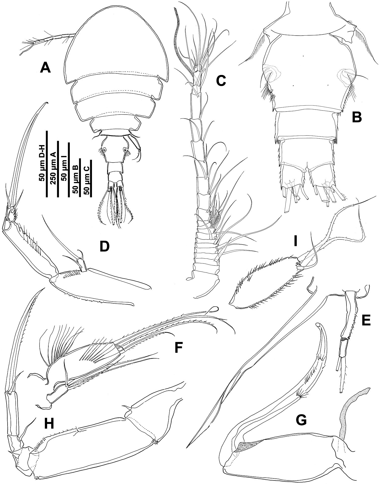
Female. Body cyclopiform with oval cephalothorax and short, cylindrical urosome ( Figure 3A View Figure 3 ). Mean body length 780 µm (750–810 µm) and maximum width 430 µm (410–450 µm), based on two specimens. Prosome comprising cephalothorax (fully incorporating first pedigerous somite) and three free pedigerous somites. Cephalothorax and free pedigerous somites with rounded posterolateral angles. Rostrum triangular. Dorsal cephalothoracic shield and free pedigerous somites with integumental pores and sensilla. Urosome four-segmented, comprising leg-5 bearing somite, genital double-somite, and two free abdominal somites ( Figure 3B View Figure 3 ). Genital double-somite slightly wider than long; paired genital apertures bipartite, each comprising lateroventral copulatory pore and dorsolateral gonopore; lateral margins with long spinules in middle third, posterior to genital apertures ( Figure 3B View Figure 3 ). Each genital area with smooth seta.
Caudal rami ( Figure 3A,B View Figure 3 ) slightly longer than wide (measured along outer margin), armed with six setae; seta I absent, setae III–VI plumose and setae II and VII slightly displaced onto dorsal surface and smooth.
Antennule 21-segmented ( Figure 3C View Figure 3 ), about 320 µm long. Segmental fusion pattern as follows: 1(I), 2(II), 3(III)-1, 4(IV)-1, 5(V)-2, 6(VI)-2, 7(VII)-1, 8(VIII)-2, 9(IX-XII)-7, 10(XIII)-1, 11(XIV)-1+1 spine, 12(XV)-1, 13(XVI)-2, 14(XVII)-2, 15(XVIII)-2, 16(XIX)-2, 17(XX)-1, 18(XXI)-1+1 aesthetasc, 19(XXII)-2, 20(XXIII-XXIV)-4 and 21(XXV-XXVIII)-6. Segment 10(XIII) reduced and partly overlapped by distal expansion of compound segment 9(IX-XII).
Antenna biramous ( Figure 3D View Figure 3 ), about 300 µm long (excluding coxa and including terminal claw). Coxa lost in dissection. Basis unarmed, with fine spinule rows on margins. Exopod one-segmented, about twice as long as wide; with one lateral seta, one subterminal seta and one long terminal seta, all naked. Endopod three-segmented; proximal segment elongate with spinular rows as figured; middle segment produced distally on medial side but articulating with distal segment proximally on lateral side, bearing one subterminal smooth seta; distal segment with spinules on inner margin and two smooth subterminal setae, and distal claw.
Siphon slender, about 350 µm long, reaching to intercoxal sclerite of leg 1.
Mandible ( Figure 3E View Figure 3 ) comprising stylet-like gnathobase and two-segmented palp. Proximal segment of palp longer, with row of spinules on lateral margin; distal segment with two unequal apical setae. Stylet located in oral cone, formed by anterior labrum and posterior labium, with denticulate margin subapically.
Maxillule bilobed ( Figure 3F View Figure 3 ); inner lobe 3.5 times longer than outer, with row of very long setules on lateral margin and medially, and with five terminal setae, four of them long and plumose and one minute and naked. Outer lobe with four terminal setae, three of them long and pinnate and one shorter and naked.
Maxilla ( Figure 3G View Figure 3 ) two-segmented but with partial transverse surface suture on syncoxa (proximal segment) possibly marking plane of praecoxa–coxa fusion; praecoxal portion bearing flaccid aesthetasc-like element medially, representing tubular extension of external opening of maxillary gland. Coxal portion unarmed. Basis claw-like with fan-like tufts of spinules medially and distally, and rows of spinules on distal half; recurved tip.
Maxilliped five-segmented ( Figure 3H View Figure 3 ), comprising short syncoxa, long basis and three-segmented endopod. Syncoxa with row of spinules and one short seta distally. Basis with rows of spinules on inner margin and distally and one minute seta medially. First endopodal segment compound, partial suture marking original separation of two ancestral segments, with (1,0) armature formula; second endopodal segment bearing one naked seta; third endopodal segment with recurved terminal claw plus additional apical seta. Inner margin of claw provided with row of minute spinules.
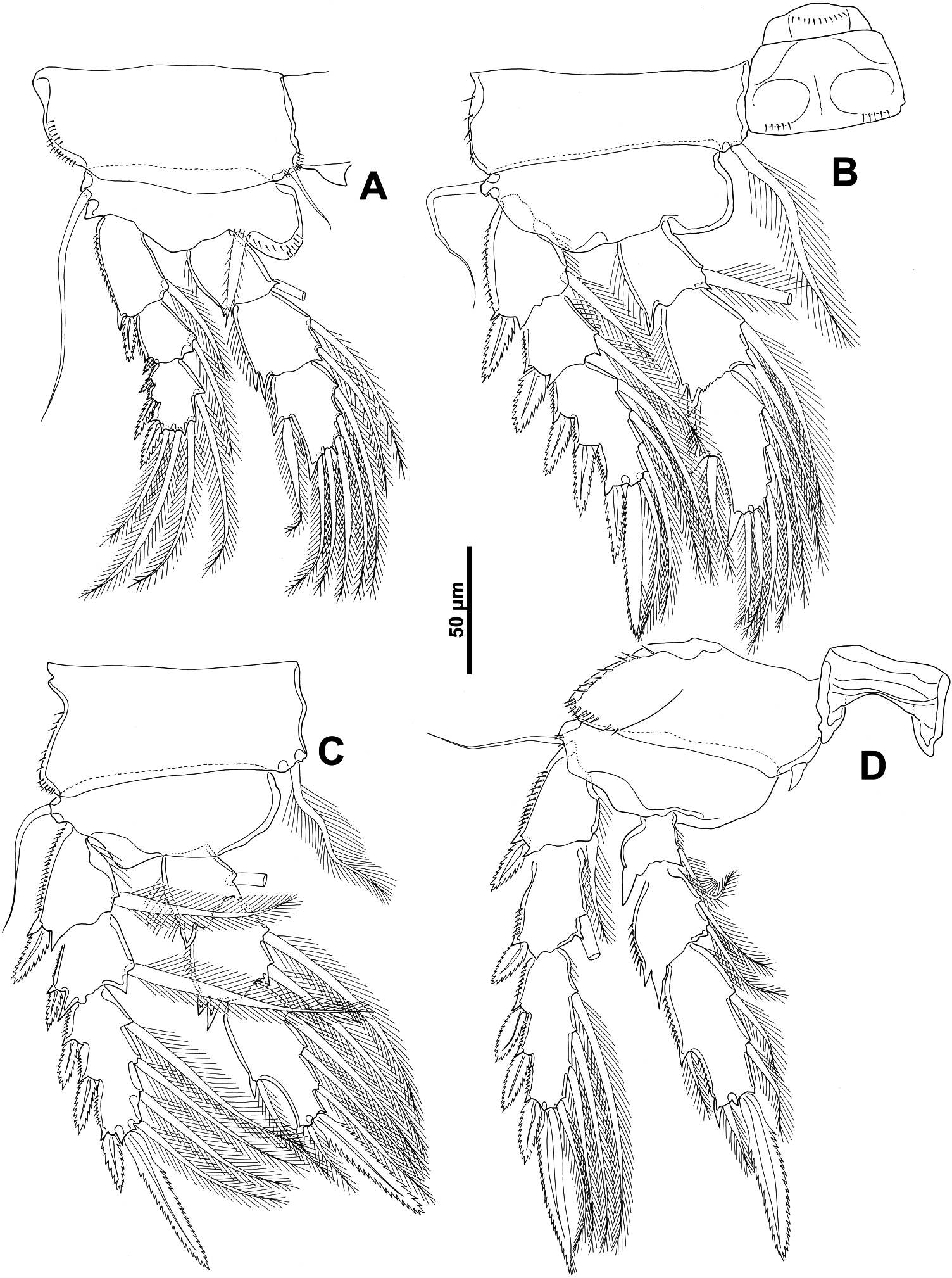
Swimming legs 1–4 biramous ( Figure 4A–D View Figure 4 ), with three-segmented rami and each with intercoxal sclerite. Spine and seta formula ( Table 2):
Coxae ornamented with spinule rows around outer margin; inner coxal seta short and naked in leg 1, long and plumose in legs 2–3, and reduced in leg 4 ( Figure 4A–D View Figure 4 ). Basis of leg 1 with spinules around inner margin; outer seta long and naked in legs 1–4. Outer spines of exopodal segments in legs 1–4 bilaterally serrate. Lateral margins of exopodal segments with minute serrations or spinular rows; those of endopodal segments with rows of setules.
Fifth leg ( Figure 3I View Figure 3 ) with protopod incorporated into somite. Free segment almost three times longer than wide, elongate-oval, with two long pinnate terminal setae and one shorter smooth subterminal seta; outer and inner margins with spinules.
Sixth leg ( Figure 3B View Figure 3 ) represented by paired opercular plates closing off gonopores on genital double-somite; each armed with one smooth seta.
Male. Unknown.
Etymology
The species is named in honour of Prof. Ju-shey Ho.
Remarks
This species was found in a vial labelled as “ Asterocheres cf. simulans (Th. Scott, 1898) ” which turned out to contain a new species. This species was collected by Stock in Curaçao (Piscadera Bay) in 1958 and lives associated with Lytechinus variegatus (Lamarck, 1816) . The most striking features of this species are: (1) the antennules are 21-segmented; (2) the antenna has a three-segmented endopod and a one-segmented exopod with three setae; (3) the mandible has two-segmented palp with two terminal setae and stylet with denticulate margin subapically; (4) the siphon reaches approximately to the insertion of leg 1; (5) the outer lobe of maxillule has four distal setae and the inner lobe has five distal setae (one minute and naked) and rows of long setules in the lateral margin and medially; (6) the maxilla bears a flaccid element medially, representing tubular extension of external opening of maxillary gland and claw with rows of spinules in the second half; (7) the maxilliped is five-segmented with terminal claw; (8) legs 1–4 biramous, as usual in the genus; (9) the free segment of fifth leg bears three terminal setae; (10) the caudal rami are slightly longer than wide with six terminal setae.
This species belongs to the group of species with a 21-segmented antennule in the female and a two-segmented mandibular palp. This group consists of 18 species named above.
The oral cone of A. hoi possesses an elongate siphon reaching to the intercoxal plate of legs 1. In contrast, the siphon of A. genodon , A. astroidicola , A. ellisi and A. tenerus reaches to the intercoxal plate of leg 2; in A. flustrae , A. reginae , A. simulans , A. suberitis , A. jeanyeatmanae , A. tarifensis , A. kervillei and A. tubiporae , the siphon reaches the insertion of maxillipeds; and in A. lilljeborgi the siphon extends only to the maxilla ( Ivanenko and Ferrari 2003).
The shape of the caudal rami separates A. hoi from A. hirsutus , A. tenuicornis , (according to the illustration in Eiselt 1965) and A. latus , as the new species has caudal rami only slightly longer than wide, but in A. hirsutus the caudal rami are 2.5 times longer than wide, in A. latus 2.6 times longer than wide and in A. tenuicornis six times longer than wide.
The ornamentation of the antenna and the maxillule serve to differentiate A. hoi from A. peniculatus . The new species has the antennal claw longer than the first endopodal segment and a row of very long setules on the lateral margin of the inner lobe of the maxillule. Asterocheres peniculatus has an antennal claw shorter than the first endopodal segment and the basis has a longitudinal row of bifurcate or trifurcate spinules or scales near base of the exopod; the inner lobe of the maxillule has a row of short setules, as usual in the genus ( Kim 2010).
Finally, the exopod of leg 5 is 2.5 times longer than wide and the two terminal barbed setae are much longer than the entire segment in A. hoi . In contrast, in A. urabensis the exopod of leg 5 is 3.8 times longer than wide and the two terminal smooth setae are shorter than the free segment ( Kim 2004a).
No known copyright restrictions apply. See Agosti, D., Egloff, W., 2009. Taxonomic information exchange and copyright: the Plazi approach. BMC Research Notes 2009, 2:53 for further explanation.
|
Kingdom |
|
|
Phylum |
|
|
Class |
|
|
Order |
|
|
Family |
|
|
Genus |