
Xetadrilus fabryi, Schmelz, Rüdiger M., Collado, Rut & Römbke, Jörg, 2011
|
publication ID |
https://doi.org/ 10.5281/zenodo.203260 |
|
DOI |
https://doi.org/10.5281/zenodo.5611835 |
|
persistent identifier |
https://treatment.plazi.org/id/520EAA7D-D65C-4512-FF40-8509FDC0FD4E |
|
treatment provided by |
Plazi |
|
scientific name |
Xetadrilus fabryi |
| status |
sp. nov. |
Xetadrilus fabryi View in CoL sp. nov.
( Figs 7 View FIGURE 7 , 8 View FIGURE 8 G, Table 3 View TABLE 3 )
Holotype. UFPR OL-23, adult specimen with 22 segments, posterior end regenerated, stained whole mount, Antonina, Rio Pequeno, 25°15'56.8''S, 48°44'23.5''W, 25 m a.s.l., agroforestry system [site 48], Jan 2008, leg. J. Römbke, R. M. Schmelz.
Paratypes. MZUSP, 4 specimens, stained whole mounts, Antonina, Cachoeira :
MZUSP 1215, 3 specimens, 25°15'2''S, 48°40'24'' and 25°15'13''S, 48°40'34''W, 30 and 40 m a.s.l., respectively, pasture, partly abandoned, on Cambisol [sites 1, 4], May 2003, leg. J Römbke, R. M. Schmelz.
MZUSP 1216, 1 specimen, 25°18'25"S, 48°40'24"W, 70 m a.s.l., pasture on Cambisol [site 2], March 2004, leg. B. Förster, R. M. Schmelz.
UFPR, 8 specimens, stained whole mounts:
UFPR OL-24, 5 specimens, Antonina, Rio Pequeno, 25°15'56.8''–16'23.4''S, 48°43'41.1''–44'23.5''W, 15 and 25 m a.s.l., respectively, agroforestry system [sites 47, 48], Jan 2008, leg. J. Römbke, R. M. Schmelz.
UFPR OL-25, 3 specimens, Curitiba, UFPR University Campus, Agrárias, 25°24'37''S, 49°14'56''W, 909 m a.s.l., grassland, Feb 2008, leg. R. M. Schmelz.
Additional material. One specimen from UFPR campus, Agrarias, examined in vivo, not preserved.
Etymology. Named in honour of Rainer Fabry, versatile German on-site coordinator of the SOLOBIOMAproject.
Xetadrilus maacki Xetadrilus aphanus Xetadrilus fabryi
Segment number 21–28 21–28 33
Description. Body dimensions. Living adult specimens ca. 3 mm long and 0.11–0.12 mm wide, whole mounts 2.5–3.5 mm long and 0.1–0.12 mm wide, up to 0.16 at XII. Segment number 30–33 (N=3), holotype 22 segments, with regenerated posterior end. Chaetae two per bundle, formula 2,0–0: 2–2. Lateral chaetae present in II–VII, absent from VIII on. Ventral chaetae from II on, absent in XII. Chaetae distally straight, or slightly bent to same side as proximal bend. Terminal chaetae enlarged, almost 2x as large as largest anterior chaetae, not sigmoid. Ventral anterior chaetae ca. 2 µm thick, in II 12 µm long, gradually increasing in size posteriad to about 28 µm in IX. Ventral chaetae in hindmost segments 45–55 µm long and 2.5–3 µm thick. Epidermal gland cells ( Fig. 7 View FIGURE 7 B) variable; entire epidermis glandular to varying degrees, gland cells pale and evenly distributed, or no epidermal gland cells distinguishable in living and preserved material. In specimens with glandular epidermis body surface often covered with foreign particles. Clitellum saddle-shaped, cells in ca. 25 transverse rows, separate (viv) or dense (fix). Hyalocytes on dorsal half, larger than granulocytes, isolated; in ventral half only granulocytes, mid-ventral interruption as wide as distance of male pores. Prostomium with head pore in mid-dorsal position.
Frontal prostomial epithelium ( Fig. 7 View FIGURE 7 A,E) thickened, with a vesicle-like recess or cleft at the frontal tip. Dorso-laterally several inner papillae in bilateral-symmetrical order, inner papillae also on peristomium. Prostomial musculature present. Body wall medium-thick, cuticle thin. Preclitellar septa not thickened. Brain ( Fig. 7 View FIGURE 7 E) in I–III, 2–2.5x as long as wide, incised posteriorly; prostomial ganglia conspicuous but small; ventral nerve cord with suboesophageal ganglion in II–IV and segmental ganglia from V. Oesophageal appendages absent. Pharyngeal glands ( Fig. 7 View FIGURE 7 A,B) as unpaired dorsal lobes in IV–VI, voluminous in IV and V, primary ventral lobes in V and VI, largest in VI; small and spherical secondary ventral lobes in V, VI and VII. Intestinal diverticula ( Fig. 7 View FIGURE 7 A,B) one pair in VII, elongate, oval, thick-walled, with distinct lumen; placed laterally of intestine, arranged in "V"- shape (dorsal view), slightly diverging anteriad; diverticula laterally flattened, higher than wide. Chloragocytes often black-grey in posterior segments. Dorsal blood vessel from 1/2 XIV in the two mature and complete specimens, from 1/4 XIV in holotype; the origin was difficult to see in living specimens. Preclitellar nephridia ( Fig. 7 View FIGURE 7 C) 3 pairs, from 7/8 to 9/10, postseptale without dorsal vesicle. Postclitellar nephridia elongate. Coelomocytes pale, not hyaline, without conspicuous texture.
Seminal vesicle absent. Spermatozoa distinct on top of sperm funnel, length not measured in vivo. Sperm funnel ( Fig. 7 View FIGURE 7 F) barrel-shaped, ca. 1/3 as long as body diameter (40–50 µm long, fix), collar not wider than funnel body. Vas deferens ( Fig. 7 View FIGURE 7 F) quite long, coiled densely in numerous loops in XII, sometimes filling entire dorsal half of segment XII; diameter 4 µm throughout except near sperm funnel, here ca. 5 µm (fix). Male copulatory organ ( Fig. 7 View FIGURE 7 F) small, circular in top view, not more than a body wall thickening in side view; bursa and bursal slit apparently absent; male pore on body surface, sometimes surrounded by folds. Glandular bulb tripartite, an eversible central bulb, diameter ca. 20 µm (fix), pierced centrally by vas deferens, and two smaller bulbs attached anteriorly and posteriorly. Copulatory muscles well-developed, encompassing bulbs and also extending dorsad as transverse copulatory muscles. Accessory glands absent. Spermathecae ( Fig. 7 View FIGURE 7 A) not attached to oesophagus, a simple tube. Ectal duct short (length 20 µm, diameter 10 µm, fix) with wide lumen, often filled with sperm, gradually widening into distal part of ampulla, here sperm arranged in parallel, or no ampulla distinguishable. The following tube narrow (diameter ca. 5 µm, fix), widening into thin-walled ental reservoir in VI or VII; reservoir not always developed. Sperm may be present in any part of the spermatheca. One mature egg at a time.
Habitat. X. fabryi was found in grazed and abandoned pastures and at agroforestry sites. It was not found at forest sites.
Remarks. This is the only species of the new genus with intestinal diverticula. Diverticula are similar in shape to the ones in Guaranidrilus joanae Christoffersen, 1977 and G. hoeferi sp. nov. (see above). The two Guaranidrilus species are much larger and with very large sperm-related sexual organs. X. fabryi has been assigned to Xetadrilus (and not to Guaranidrilus ) by virtue of the following characters: (1) Lateral chaeta absent from VIII, (2) epidermal gland cells oval, not transversely elongate, (3) prostomium with inner papillae and ganglia, (4) nephridia without dorsal vesicle, (5) pharyngeal glands with secondary lobes not only in V, VI, but also in VII, (6) post-clitellar origin of dorsal blood vessel.
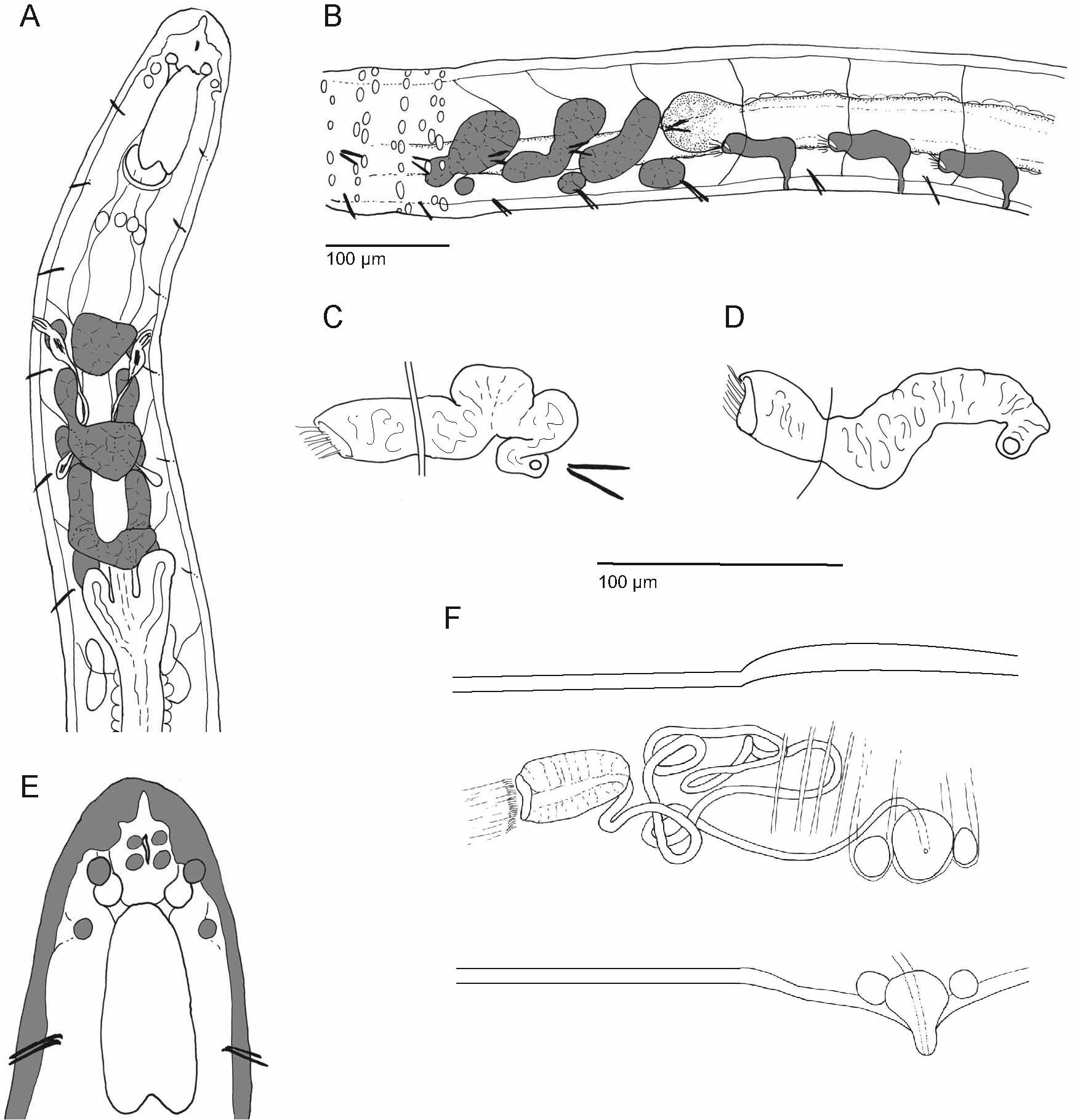
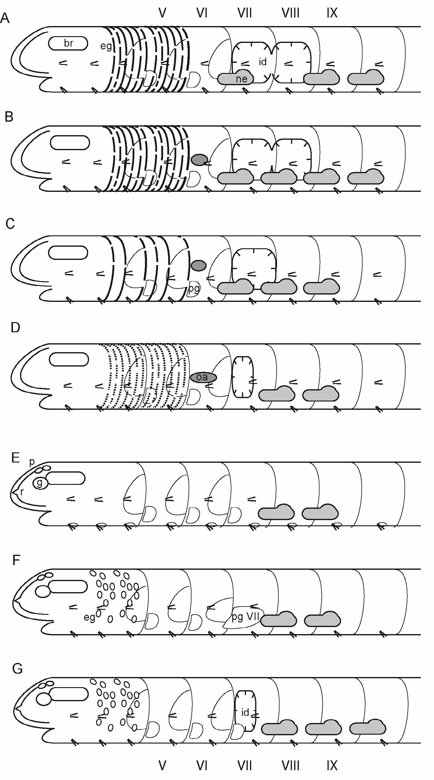
TABLE 3. Differences betweeen the three new species of Xetadrilus gen. nov. Idiosyncrasies underlined.
| Posterior chaetae | sigmoid, 2x as large as anterior | straight, 1.5–2x as large as anterior | straight, c. 1.8 x as large as anterior |
|---|---|---|---|
| Epidermal gland cells | mid-ventrally only | absent or all epidermis glandular | absent or all epidermis glandular |
| Prostomial ganglia | large | large | medium |
| Preclitellar nephridia | 2 pairs, 7/8, 8/9 | 2 pairs, 7/8, 8/9 | 3 pairs, 7/8 – 9/10 |
| Pharyngeal glands, dorsal lobes in VI | united | separate | united |
| Pharyngeal glands, ventral secondary lobes | at V–VII, increasing in size posteriad | at V–VII | V–VII, all small and spheri- cal |
| Intestine in VII | widened | widened | diverticula |
| Dorsal blood vessel | from XIII – XIV | from 1/2 XII – XIII | from XIV |
| Coelomocytes | distinctly brown, with refractile vesicles | with pale vesicles, aggre- gations pale or brownish | with pale vesicles, aggregations rarely with brownish tint |
| Further idiosyncrasies | Pharyngeal glands in VI– VII "Z"-shaped |
| MZUSP |
Museu de Zoologia da Universidade de Sao Paulo |
No known copyright restrictions apply. See Agosti, D., Egloff, W., 2009. Taxonomic information exchange and copyright: the Plazi approach. BMC Research Notes 2009, 2:53 for further explanation.