
Frontonia didieri, Long, Hongan, Song, Weibo, Al-Rasheid, Khaled A. S. & Wang, Yangang, 2008
|
publication ID |
https://doi.org/10.5281/zenodo.180528 |
|
DOI |
https://doi.org/10.5281/zenodo.5661178 |
|
persistent identifier |
https://treatment.plazi.org/id/C44C3C09-FFC9-5374-FF1B-92A6DF7E4F58 |
|
treatment provided by |
Plazi |
|
scientific name |
Frontonia didieri |
| status |
sp. nov. |
Frontonia didieri n. sp.
( Figs. 1–3 View FIGURE 1 View FIGURE 2 View FIGURE 3 ; Table 1 View TABLE 1 )
Diagnosis: Marine Frontonia in vivo ca. 100–150 × 45–80 μm, body dorsoventrally slightly flattened. 61–71 somatic, consistently 3 vestibular while 3–5 postoral kineties. Both peniculus 1 and 2 consisting of 4 kinety rows; peniculus 3 three-rowed, extremely different in lengths. One oval macronucleus. Single contractile vacuole centrally-located, with about eight conspicuous collecting canals.
Type location: A mesotrophic sandy beach near Qingdao, salinity ca. 12‰.
Type slides: One holotype with protargol impregnated specimens (slide number: 2007:5:17:1) is deposited in the Natural History Museum, London, UK, and one paratype with silver nitrate impregnated specimens (slide number: 2005110701-2) is deposited in the Laboratory of Protozoology, Ocean University of China.
Etymology: We dedicate this species to Dr. Pierre Didier, a famous French protozoologist, who has greatly contributed to the ciliate taxonomy and systematics.
Description: Size in vivo mostly about 120 × 60 µm, with ratio of length: width about 2:1. Body shape rather constant, elliptical in outline with both anterior and posterior ends slightly narrow; dorsoventrally flattened about 5:4 ( Figs. 1 View FIGURE 1 A, 1E; 3A). Extrusomes spindle-shaped, about 4 µm long, densely arranged ( Figs. 1 View FIGURE 1 A; 3B). Somatic cilia about 7 µm long. Cytoplasm transparent and colourless, usually filled with many large diatoms (up to 50 µm long) ( Figs. 1 View FIGURE 1 A, 1E; 3E). Macronucleus ellipsoidal, centrally positioned ( Figs. 1 View FIGURE 1 C, 1E, 1F; 3H). One contractile vacuole (CV), about 15 µm in diameter, positioned equatorially, with ca. eight long and conspicuous collecting canals ( Figs. 1 View FIGURE 1 A; 3J). CV-pores (CVP) mid-dorsally positioned ( Fig. 2 View FIGURE 2 A–D).
Movement mostly by gliding back and forth on substrate; when swimming, moderately fast with rotation about the long axis of the cell.
Buccal cavity shallow and small, triangular in outline, occupying about 1/6 of body length ( Figs. 1 View FIGURE 1 A; 3A– C, 3G). Buccal apparatus as shown in Figs. 1 View FIGURE 1 C, 1G and 3F, 3K: consistently 3 long vestibular kineties (VK) with densely arranged kinetosomes, extending from anterior level of buccal cavity to about middle level of cell. 3 peniculi (P1–3) located on left wall of cavity: P1 and 2 about equally long, positioned close to each other, and each composed of 4 rows of kinetosomes, whereas the posterior ends of those rows in P1 are conspicuously shortened; peniculus 3 (P3) consisting of only 3 kineties, of which only the rightmost one is complete, the middle row is about half length and the leftmost one is extremely shortened, i.e. about 1/10 length of rightmost one. The paroral membrane (PM) double-rowed, located on right edge of the buccal cavity ( Figs. 1 View FIGURE 1 G; 3F).
On average 66 somatic kineties. Both anterior and postoral sutures conspicuously long and extending onto dorsal side ( Figs. 1 View FIGURE 1 C, 1F; 2A–D). 3–5 postoral kineties (PK) locating posterior to the buccal cavity and ending at the postoral suture (PS) ( Fig. 1 View FIGURE 1 C, 1D, 1G).
Silverline system as in other congeners: quadrangular cortical meshes after silver nitrate impregnation ( Fig. 3 View FIGURE 3 D).
to be continued.
Comparison: Currently, over 40 morphotypes have been included in the genus Frontonia ( Bullington 1939; Carey 1992; Dragesco 1960; Dragesco 1972; Dragesco & Dragesco-Kernéis 1986; Foissner et al. 1994; Kahl 1931; Long et al. 2005; Petz et al. 1995; Roque 1961; Roque & Puytorac 1972). Among those, about 27 were reported from fresh water biotopes, whereas F. didieri n. sp. is diagnosed by the unique, conspicuous contractile vacuole collecting canals from living cells ( Alekperov 2005; Burkovsky 1970a, b; Foissner et al. 1994; Long et al. 2005; Petz et al. 1995; Roque 1961; Roque and Puytorac 1972).
Considering the body shape, size and the marine habitat, Frontonia didieri n. sp. is similar to F. caneti Dragesco, 1960 and F. vacuolata Dragesco, 1960 though in both forms the detailed structure of the oral apparatus is lacking (Table 2; Dragesco 1960). Nevertheless, F. d i d i e r i n. sp. can be clearly distinguished in vivo from the latter two by its centrally located contractile vacuole with the prominent collecting canals (vs. no collecting canals, CV left and located subcaudally in F. c a n e t i, or located caudally in F. vacuolata ) ( Fig. 10 View FIGURE 10 D and 10Q).
Frontonia lynni and F. salmastra Dragesco and Dragesco-Kernéis, 1986 also resemble F. didieri n. sp. with reference to the general morphology (i.e. living cells) and the infraciliature of the buccal apparatus ( Fig. 10 View FIGURE 10 I, 10J, 10 O, 10P). The new species can be recognized, however, in the presence of the collecting canals (vs. no collecting canals in the latter species), lower number of somatic kineties (61–71 vs. 71–83 in F. lynni , 90–100 in F. salmastra ) and fewer kinety rows in peniculus 3 (3 vs. 4 in F. lynni and F. s a l m a s t r a) (Table 2).
In addition, the dissimilarity of both forms is firmly supported by 18S rRNA gene sequence data as the sequence of F. didieri differs significantly in 143 nucleotides from that of F. l y n n i (structural similarity 91.8%) ( Fig. 11 View FIGURE 11 ).
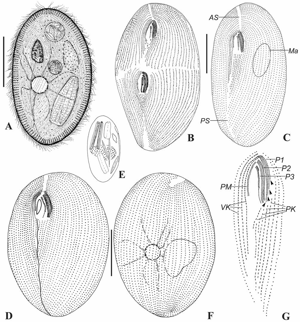
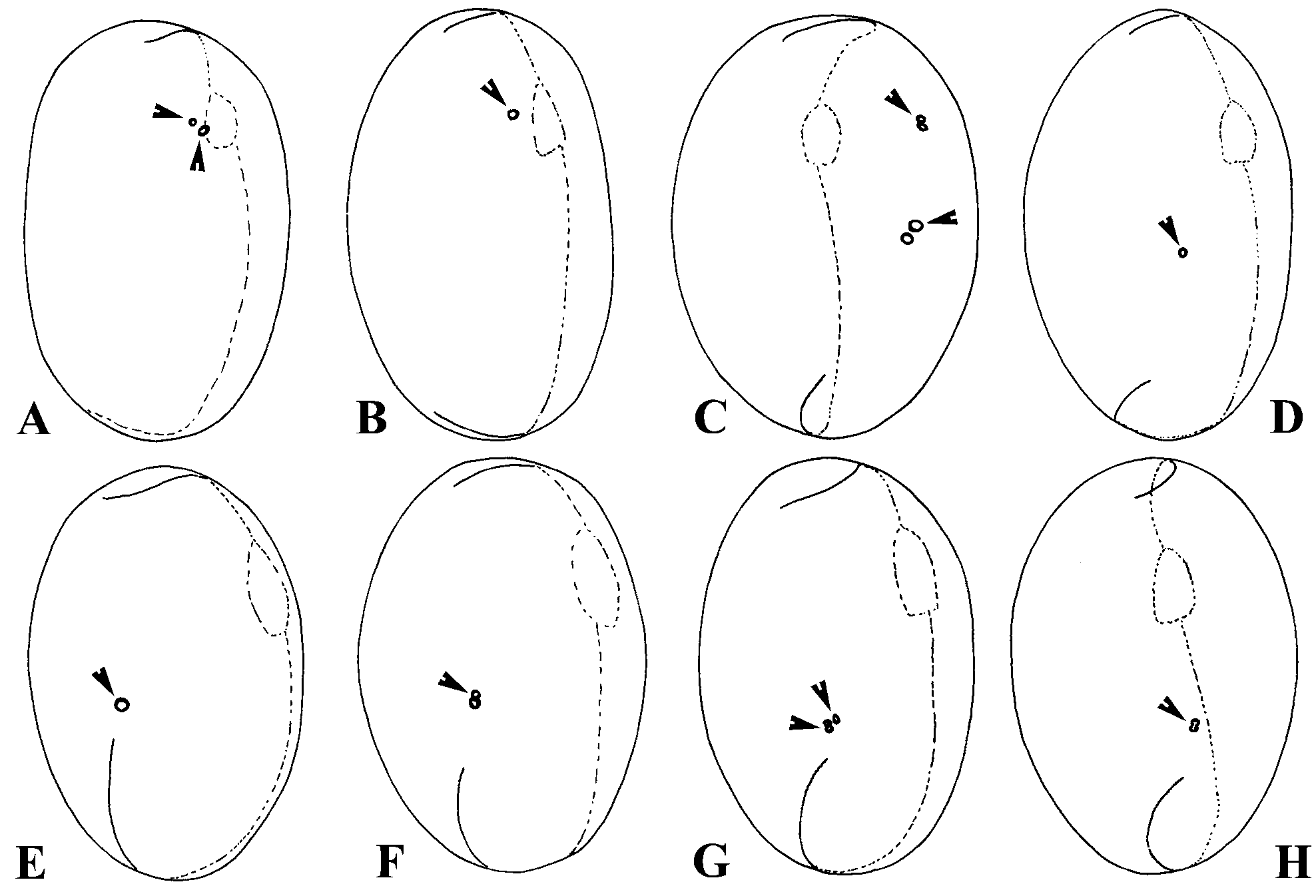
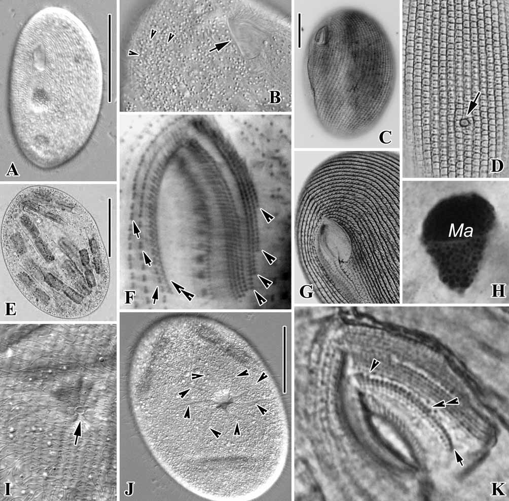

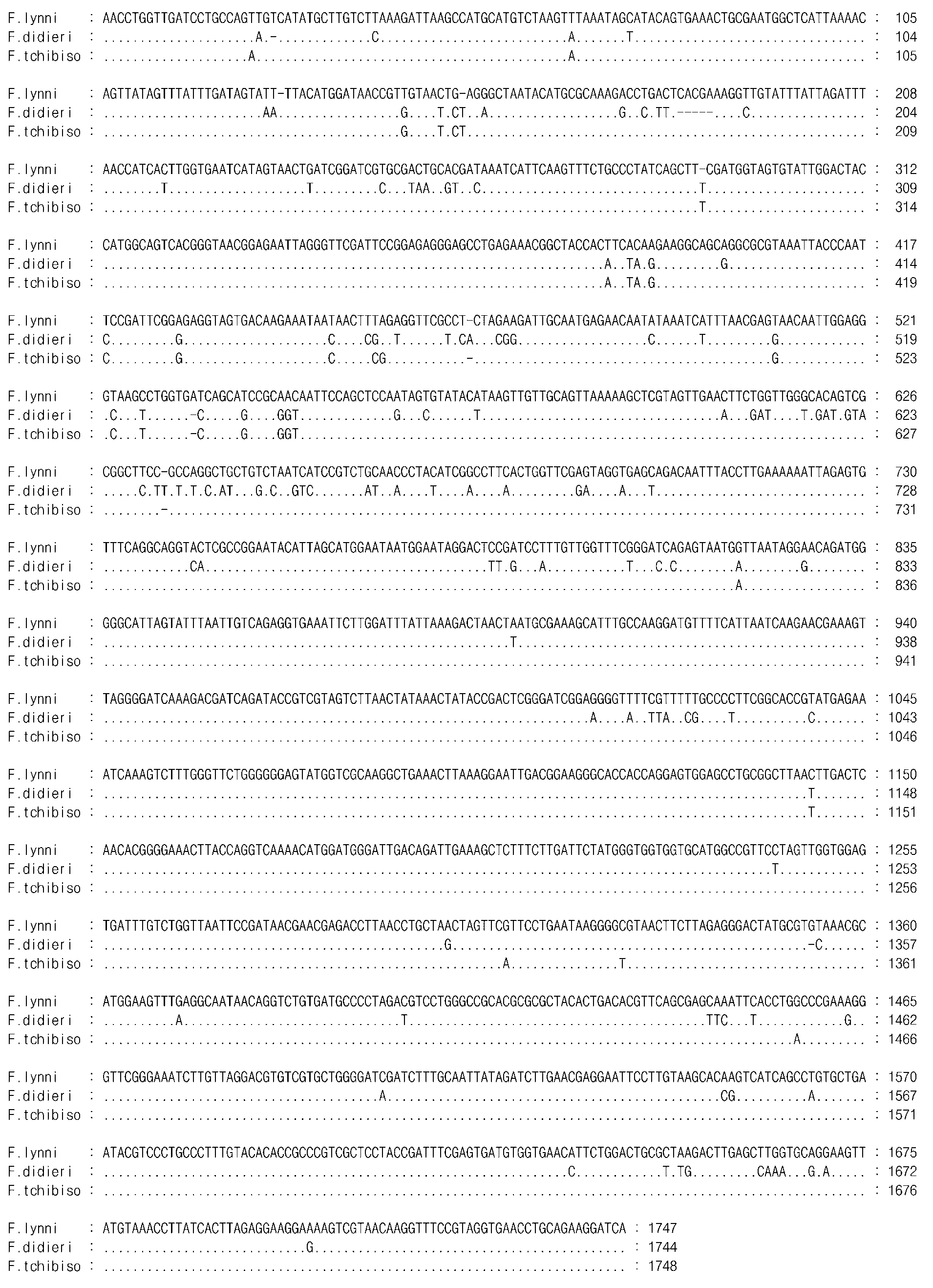
TABLE 1. Morphometric data of Frontonia didieri n. sp., F. multinucleata n. sp. and F. tchibisovae Burkovsky, 1970. Data are based on silver carbonate and silver nitrate impregnated specimens. All measurements in µm. CV = coefficient of variation in %; Max = maximum; Mean = arithmetic mean; Min = minimum; n = number of cells measured; P 1 – 3 = peniculus 1 – 3; SD = standard deviation.
| Characters | Min | Max | Mean | SD | CV | n |
|---|---|---|---|---|---|---|
| Body length | ||||||
| F. d i d i e r i n.sp. | 113 | 148 | 130.10 | 9.85 | 7.57 | 22 |
| F. multinucleata n.sp. | 72 | 112 | 94.20 | 10.42 | 11.06 | 20 |
| F. tchibisovae | 132 | 240 | 180.56 | 26.20 | 14.51 | 36 |
| Body width | ||||||
| F. d i d i e r i n.sp. | 48 | 73 | 61.32 | 6.97 | 11.37 | 22 |
| F. multinucleata n.sp. | 44 | 72 | 57.40 | 7.82 | 13.62 | 20 |
| F. tchibisovae | 88 | 186 | 135.50 | 22.93 | 16.92 | 36 |
| Number of somatic kineties | ||||||
| F. d i d i e r i n.sp. | 61 | 71 | 65.89 | 2.64 | 4.01 | 19 |
| F. multinucleata n.sp. | 58 | 67 | 63.39 | 2.28 | 3.60 | 18 |
| F. tchibisovae | 127 | 149 | 139.89 | 7.49 | 5.35 | 9 |
| Number of postoral kineties | ||||||
| F. d i d i e r i n.sp. | 3 | 5 | 4.76 | 0.56 | 11.74 | 17 |
| F. multinucleata n.sp. | 4 | 5 | 4.24 | 0.44 | 10.38 | 17 |
| F. tchibisovae | 5 | 7 | 5.82 | 0.67 | 11.51 | 28 |
| Number of vestibular kineties | ||||||
| F. d i d i e r i n.sp. | 3 | 3 | 3.0 0 | 0 | 0 | 18 |
| F. multinucleata n.sp. | 3 | 3 | 3.0 0 | 0 | 0 | 21 |
| F. tchibisovae | 3 | 4 | 3.07 | 0.27 | 8.79 | 27 |
| Number of macronuclei/us | ||||||
| F. d i d i e r i n.sp. | 1 | 1 | 1.0 0 | 0 | 0 | 15 |
| F. multinucleata n.sp. | 2 | 4 | 3.76 | 0.66 | 17.55 | 17 |
| F. tchibisovae | 1 | 1 | 1.0 0 | 0 | 0 | 20 |
| Number of ciliary rows in P1 | ||||||
| F. d i d i e r i n.sp. | 4 | 4 | 4.0 0 | 0 | 0 | 18 |
| F. multinucleata n.sp. | 4 | 4 | 4.0 0 | 0 | 0 | 15 |
| F. tchibisovae | 4 | 4 | 4.0 0 | 0 | 0 | 20 |
| Number of ciliary rows in P2 | ||||||
| F. d i d i e r i n.sp. | 4 | 4 | 4.0 0 | 0 | 0 | 18 |
| F. multinucleata n.sp. | 4 | 4 | 4.0 0 | 0 | 0 | 15 |
| F. tchibisovae | 4 | 4 | 4.0 0 | 0 | 0 | 20 |
| Number of ciliary rows in P3 | ||||||
| F. d i d i e r i n.sp. | 3 | 3 | 3.0 0 | 0 | 0 | 17 |
| F. multinucleata n.sp. | 4 | 4 | 4.0 0 | 0 | 0 | 15 |
| F. tchibisovae | 4 | 4 | 4.0 0 | 0 | 0 | 20 |
| Ratio of buccal/body length | ||||||
| F. d i d i e r i n.sp. | 0.14 | 0.21 | 0.17 | 0.02 | 11.76 | 17 |
No known copyright restrictions apply. See Agosti, D., Egloff, W., 2009. Taxonomic information exchange and copyright: the Plazi approach. BMC Research Notes 2009, 2:53 for further explanation.
|
Kingdom |
|
|
Phylum |
|
|
Class |
|
|
Order |
|
|
Family |
|
|
Genus |
Frontonia didieri
| Long, Hongan, Song, Weibo, Al-Rasheid, Khaled A. S. & Wang, Yangang 2008 |
F. salmastra Dragesco and Dragesco-Kernéis, 1986
| Dragesco and Dragesco-Kerneis 1986 |